ECG – stage 1

ECG of heart block

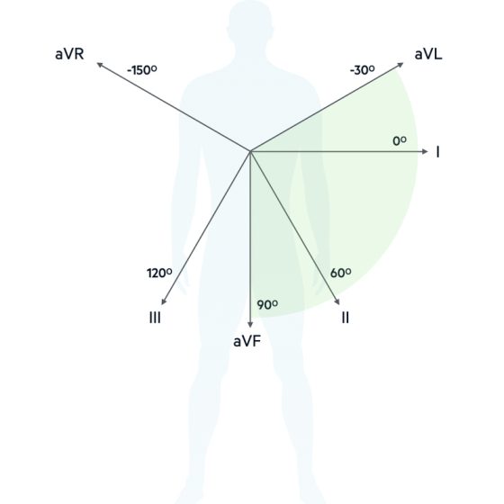
https://youtu.be/LhAOb9O8LGo ECG of heart block ECG training course Date of show : 18 May 2024, 09:00 PM CAI Introduction to ECG – 1st part 20:19 Heart axis deviation and alfa angle 23:13 Normal ECG values and heart rate calculation 37:53 ECG of atrial and ventricular hypertrophy 31:37 ECG changes in Myocardial Infarction MI 18:56 ECG of heart rate disorders 17:21 Arrhythmia, Flutters and Fibrillations 22:50 Extrasystole arrhythmias 14:49 ECG of heart block 27:19 PDF materials of lesson : Heart block

Extrasystole arrhythmias

https://youtu.be/95HV64rPXZA Extrasystole arrhythmias ECG training course Date of show : 16 May 2024, 09:00 PM CAI Introduction to ECG – 1st part 20:19 Heart axis deviation and alfa angle 23:13 Normal ECG values and heart rate calculation 37:53 ECG of atrial and ventricular hypertrophy 31:37 ECG changes in Myocardial Infarction MI 18:56 ECG of heart rate disorders 17:21 Arrhythmia, Flutters and Fibrillations 22:50 Extrasystole arrhythmias 14:49 ECG of heart block 27:19 PDF materials of lesson : Extrasystole arrhythmias

Arrhythmia, Flutters and Fibrillations

https://youtu.be/ZTEZGhaQ740 Arrhythmia, Flutters and Fibrillations ECG training course Date of show : 14 May 2024, 09:00 PM CAI Introduction to ECG – 1st part 20:19 Heart axis deviation and alfa angle 23:13 Normal ECG values and heart rate calculation 37:53 ECG of atrial and ventricular hypertrophy 31:37 ECG changes in Myocardial Infarction MI 18:56 ECG of heart rate disorders 17:21 Arrhythmia, Flutters and Fibrillations 22:50 Extrasystole arrhythmias 14:49 ECG of heart block 27:19 PDF materials of lesson : Arrhythmia, Flutters & Fibrilations

ECG of heart rate disorders

https://youtu.be/6AJzzA_5NDk ECG in heart rate disorders ECG training course Date of show : 12 May 2024, 09:00 PM CAI Introduction to ECG – 1st part 20:19 Heart axis deviation and alfa angle 23:13 Normal ECG values and heart rate calculation 37:53 ECG of atrial and ventricular hypertrophy 31:37 ECG changes in Myocardial Infarction MI 18:56 ECG of heart rate disorders 17:21 Arrhythmia, Flutters and Fibrillations 22:50 Extrasystole arrhythmias 14:49 ECG of heart block 27:19 PDF materials of lesson : Heart rate disorders

ECG changes in Myocardial Infarction MI

https://youtu.be/a1aWjGqhmIY ECG changes in Myocardial Infarction MI ECG training course Date of show : 10 May 2024, 09:00 PM CAI Introduction to ECG – 1st part 20:19 Heart axis deviation and alfa angle 23:13 Normal ECG values and heart rate calculation 37:53 ECG of atrial and ventricular hypertrophy 31:37 ECG changes in Myocardial Infarction MI 18:56 ECG of heart rate disorders 17:21 Arrhythmia, Flutters and Fibrillations 22:50 Extrasystole arrhythmias 14:49 ECG of heart block 27:19 PDF materials of lesson : ECG changes in Myocardial Infarction

ECG of Atrial and ventricular hypertrophy

https://youtu.be/wGz1jQD9YLU ECG of atrial and ventricular hypertrophy ECG training course Date of show : 08 May 2024, 09:00 PM CAI Introduction to ECG – 1st part 20:19 Heart axis deviation and alfa angle 23:13 Normal ECG values and heart rate calculation 37:53 ECG of atrial and ventricular hypertrophy 31:37 ECG changes in Myocardial Infarction MI 18:56 ECG of heart rate disorders 17:21 Arrhythmia, Flutters and Fibrillations 22:50 Extrasystole arrhythmias 14:49 ECG of heart block 27:19

Normal ECG values and heart rate calculation

https://youtu.be/eW6B1K6dZuo Normal ECG values and heart rate calculation ECG training course Date of show : 06 May 2024, 09:00 PM CAI Introduction to ECG – 1st part 20:19 Heart axis deviation and alfa angle 23:13 Normal ECG values and heart rate calculation 37:53 ECG of atrial and ventricular hypertrophy 31:37 ECG changes in Myocardial Infarction MI 18:56 ECG of heart rate disorders 17:21 Arrhythmia, Flutters and Fibrillations 22:50 Extrasystole arrhythmias 14:49 ECG of heart block 27:19 PDF materials of lesson : Normal ECG and Heart Rate calculation

Heart axis deviation and alfa angle

https://youtu.be/Qr-G4Sw71zk Heart axis deviation and alfa angle ECG training course Date of show : 04 May 2024, 09:00 PM CAI Introduction to ECG – 1st part 20:19 Heart axis deviation and alfa angle 23:13 Normal ECG values and heart rate calculation 37:53 ECG of atrial and ventricular hypertrophy 31:37 ECG changes in Myocardial Infarction MI 18:56 ECG of heart rate disorders 17:21 Arrhythmia, Flutters and Fibrillations 22:50 Extrasystole arrhythmias 14:49 ECG of heart block 27:19 PDF materials of lesson : Heart axis deviation

Introduction to ECG – 1st part

https://youtu.be/wEgCgh4LI7E Introduction to ECG – 1st part ECG training course Date of show : 02 May 2024, 09:00 PM CAI Introduction to ECG – 1st part 20:19 Heart axis deviation and alfa angle 23:13 Normal ECG values and heart rate calculation 37:53 ECG of atrial and ventricular hypertrophy 31:37 ECG changes in Myocardial Infarction MI 18:56 ECG of heart rate disorders 17:21 Arrhythmia, Flutters and Fibrillations 22:50 Extrasystole arrhythmias 14:49 ECG of heart block 27:19 PDF materials of lesson : Introduction to ECG for beginners

Heart block

  Sinus blockade The normal heart beat starts in the sinus node that contains the fastest pacemaker cells. If no impulse should reach the atrium, and no atrial rescue impulse appears, there will be a pause without any P wave. The pause – sinus arrest – may be brief or long. When a pause exceeds 3-5 seconds, the patient may experience dizziness or a fainting spell. Usually, the normal rhythm soon recovers, and the patient immediately feels well. Important notes : » If the speed is 50 mm/sec;      1 mm = 0.02 sec» If the speed is 25

Extrasystole arrhythmias

  Supraventricular extrasystoles   Sinus extrasystoles » Sinus node reentry» Normal sinus form P, unchanged ventricular complex» The post-extrasystolic pause is equal to the length of the normal sinus cycle   Atrial extrasystoles » Extraordinary altered P wave, narrow QRS, supraventricular type, incomplete compensatory pause » With lower atrial extrasystole, the P wave can be negative in II, III, aVF   AV extrasystoles » The extrasystolic impulse arising in the AV node propagates in two directions: retrograde to the atria and the usual way to the ventricles. Therefore, the QRS complex has an unchanged appearance, and the excitation of the

Arrhythmia, Flutters & Fibrillations

  Sinus arrhythmia » If the difference between the maximum and minimum P-P (R-R) is more than 0.1 sec (or more than 10% compared to the mean), then this is sinus arrhythmia» Respiratory arrhythmia : sinus arrhythmia is associated with the phases of breathing » No difference in P-P values – rigid sinus node (damage to sinus node cells) Normal rhythm : » Must be sinus, that’s means each P wave is followed by QRS complex » Must be regular – the difference between the longest and the shortest R-R interval does not exceed 0.1 sec » Must be equality of

Heart rate disorders

  Sinus rate disorders » Sinus rate means this impulses that are produced by sinoatrial node “SA-node”» Normal sinus rate : 60 : 100 beats per minute   Sinus bradycardia • Sinus rhythm with a frequency of less than 60 bpm• PQ can be extended up to 0.21 s, other intervals are normal• ST is sometimes raised above the isoline, concave, the T wave is widened at the base and increased in amplitude   Sinus tachycardia • Frequent sinus rhythm with more than 100 bpm• The maximum rate of the rhythm of the atria and ventricles is the same and

ECG changes in Myocardial Infarction

Our goal : To detect » Stage of myocardial infarction» Area of heart wall which affected» Coronary branch which occluded        To detect stage of MI   Stages of Myocardial Infarction – [ MI ]   Pre-acute stage   Evolving period   » ST elevation   Acute stage   Attack period » Pathological Q formation » ST elevation Pathological Q: depth more than ¼ of R amplitude in the same lead, its width (duration) more 0.03 sec   Sub-acute stage   Healing period » Pathological Q» ST at isoline» Coronary T (negative symmetric)   Post Infarction   Healed

Atrial and ventricular hypertrophy

Normal ECG values : Prong P < 2.5 mm height, 0.05 – 0.11 sec. duration.» P is positive in lead II.» The height of P should not exceed the height of T in the same lead.» P can be biphasic in III, aVL, V1 Galery Important notes : » If the speed is 50 mm/sec;      1 mm = 0.02 sec» If the speed is 25 mm/sec;      1 mm = 0.04 sec   Atrial hypertrophy characters   Right atrium hypertrophy Overload by pressure or volume • Prong P is high (>2.5 mm) and pointed in II, III,

Normal ECG and Heart Rate calculation

  ECG cycle components Atrial depolarization (P) axis : it is the general direction in which atrial depolarization conducts PR segments : From the end point of P wave to the start point QRS complex PR interval : Summation of P wave and PR segment , From the start point of P wave to the start point of QRS complex Ventricular depolarization (QRS) axis : it is the general direction in which depolarization conducts ST segment : from the end point of S wave to the start point of T wave Ventricular repolarization (T) axis : it is the general

Heart axis deviation

 Main ECG waves Atrial depolarization (P) axis : it is the general direction in which atrial depolarization conducts Ventricular depolarization (QRS) axis : it is the general direction in which depolarization conducts Ventricular repolarization (T) axis : it is the general direction in which ventricular repolarization conducts Rt : RightLt : Leftmv : Milli voltaVR : Augmented Voltage on Right armaVL : Augmented Voltage on Left armaVF : Augmented Voltage on FootRA : Right AtriumLA : Left AtriumRV : Right VentricleLV : Left VentricleLI : Lead 1LII : Lead 2LIII : Lead 3 Definition Of Mean QRS Axis : The

Introduction to ECG

Clinical values of ECG By the end of this course you will learn how to detect many diseases by ECG which is important, easy & inexpensive clinical test as following : Heart rate : Normal, bradycardia or tachycardia How to detect Heart axis and measure α angle Hypertrophy of both aria & ventricles on both sides right and left Myocardial Infarction, type, localization & stage Arrhythmia types, localization & stages Heart blocks, types & stages unusual syndromes of arrhythmia Features of Time voltage chart The P-QRS-T sequence is usually recorded on special ECG graph paper that is divided into grid-like