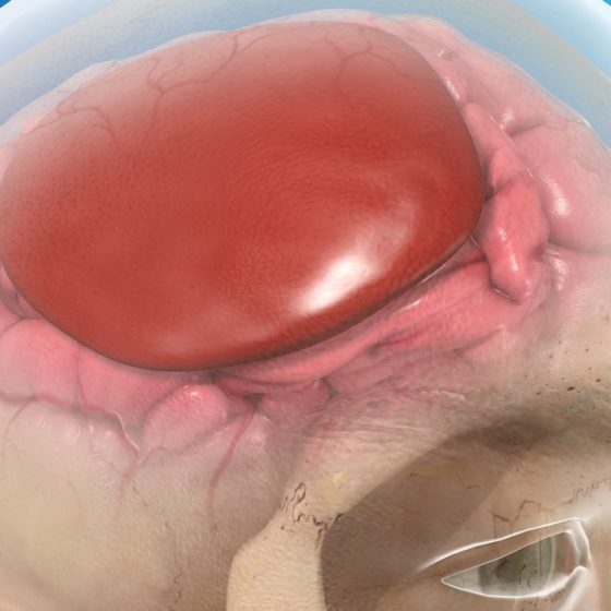
Subarachnoid haemorrhage
Definition Subarachnoid haemorrhage (SAH) is bleeding in the subarachnoid space between the arachnoid and pia mater meningeal layers. Aetiology SAH can be a life-threatening emergency and it is estimated that 10-15% of patients die before they reach hospital. SAH can be divided into traumatic or spontaneous Traumatic (tSAH): most common cause of SAH. Usually in setting of a head injury (e.g. fall, assault, road traffic collision) Spontaneous: commonly due to rupture of a cerebral aneurysm (aSAH) Traumatic Trauma is the most common cause of SAH. There is usually evidence of trauma in the clinical history (e.g. road traffic accident). An isolated SAH in