Day: March 31, 2024

Radiculopathies

Overview Radiculopathy refers to symptoms or impairments related to the involvement of a spinal nerve root. The spinal nerve roots serve as the main communication between the central nervous system (i.e. the spinal cord) and the peripheral nerves. When a spinal nerve root is affected, it is known as radiculopathy. Radiculopathies may be single or multiple. When multiple nerve roots are affected it is known as polyradiculopathy. The symptoms of radiculopathy are usually characteristic because each root supplies a specific area of cutaneous tissue, known as the dermatome, and a functional group of muscles, known as a myotome. Therefore, patients typically

Extradural haematoma

Definition An extradural haematoma (EDH) is a collection of blood in the extradural space, above the dura mater. Extradural haematoma (EDH), also known as epidural haematoma, refers to a collection of blood in the extradural space between dura and skull. It is most often due to trauma and commonly associated with a skull fracture (75-95% of cases). Aetiology EDH is most commonly due to trauma from a direct head injury. EDH is often life-threatening and patients may require emergency neurosurgery depending on the patient’s neurological status and size of the lesion. It is worth noting that the majority of patients with an

Chronic subdural haematoma

Definition Subdural haematoma is a collection of blood in the subdural space. Subdural haematoma (SDH) refers to a collection of blood in the subdural space. This is situated underneath the dura and above the arachnoid mater meningeal layers. There are several classifications of SDH: Acute (ASDH): bleeding occurring in the last 1-3 days. Chronic (CSDH): blood that has usually been present for > 3 weeks. Subacute: bleeding that occurs between 4 days and 2-3 weeks. Acute on chronic: chronic haematoma that may expand secondary to recurrent bleeding. Aetiology The most common cause of CSDH is trauma. The most common cause of CSDH is trauma in

Cauda equina syndrome

Overview Cauda equina syndrome is due to compression of the collection of nerves distal to the terminal part of the spinal cord known as the cauda equina. The cauda equina, which is Latin for ‘horses tail’, refers to the collection of spinal nerves that lie within the subarachnoid space distal to the last part of the spinal cord known as the conus medullaris. Compression of the cauda equina leads to a syndrome characterised by lower limb weakness, bladder and bowel dysfunction, and abnormal perianal sensation. It is considered a neurosurgical emergency. While not a true spinal cord lesion, cauda equina syndrome is usually

Brain tumours

Overview Brain tumour is a generic and broad term for a wide range of pathologies. A brain tumour is a broad term for an intracranial tumour, or mass, affecting structures such as the meninges, brain, glands, neurovascular structures and/or bone. Other words that may be used to describe an intracranial tumour include a mass, growth, space-occupying lesion (SOL) or central nervous system (CNS) lesion. The term ‘brain tumour’ itself is very generic and should generally be avoided as the management and prognostication for each type differ considerably. Where possible, it is important to use precise terminology. The clinical presentation, management, and prognostication

Acute subdural haematoma

Definition Subdural haematoma is a collection of blood in the subdural space. Subdural haematoma (SDH) refers to a collection of blood in the subdural space. This is situated underneath the dura and above the arachnoid mater meningeal layers. There are several classifications of SDH: Acute (ASDH): bleeding occurring in the last 1-3 days. Chronic (CSDH). blood that has usually been present for > 3 weeks. Subacute: bleeding that occurs between 4 days and 2-3 weeks Acute on chronic: chronic haematoma that may expand secondary to recurrent bleeding Aetiology The most common cause of ASDH is trauma. ASDH is often a life-threatening emergency and historically

Pancreatic adenocarcinoma

Overview Pancreatic adenocarcinoma is the most common form of pancreatic cancer and a major cause of cancer-related death. They arise from the ductal epithelium as the result of accumulated genetic mutations. Adenocarcinomas are the most common pancreatic neoplasm, accounting for around 85% of cases. Asymptomatic through much of its development, sadly this cancer is commonly diagnosed at a late stage when the chance of cure is faint. When caught early, surgery may offer a chance at curative therapy. Epidemiology There are approximately 10,500 cases of pancreatic cancer each year in the UK. Overall it is the tenth most common cancer in the UK,

Chronic pancreatitis

Overview Chronic pancreatitis refers to chronic, irreversible, inflammation and/or fibrosis of the pancreas. Chronic pancreatitis is traditionally considered as chronic, irreversible, inflammation and/or fibrosis of the pancreas. It is essentially a fibroinflammatory syndrome (evidence of fibrosis and inflammation) that occurs in patients with genetic and/or environmental risk factors for pancreatic injury. In the Western world, the leading cause of chronic pancreatitis is alcohol. Injury leads to structural and/or functional changes that include atrophy, calcification, strictures, exocrine dysfunction, endocrine dysfunction, and even increased risk of pancreatic cancer. Management centres on pain control and endoscopic or surgical treatment of complications. Exocrine and endocrine

Acute pancreatitis

Introduction Acute pancreatitis refers to an acute inflammatory process involving the pancreas. Pancreatitis occurs due to the uncontrolled release of activated pancreatic enzymes within the pancreas resulting in autodigestion. Patients may suffer a spectrum of disease from mild abdominal discomfort to multi-organ failure. Pancreatitis has an annual incidence of 13-45 cases per 100,000, with trends showing that it is becoming more common. It tends to occur more in men and is commonly secondary to gallstones or alcohol misuse. Aetiology Alcohol misuse and gallstones are the most common causes of acute pancreatitis. Alcohol misuse and gallstones are responsible for upwards of 75% of cases of

Peritonitis

Overview Peritonitis refers to inflammation of the peritoneum, which is the lining of the abdomen. Peritonitis is a really important clinical sign that refers to inflammation of the lining of the abdomen. The peritoneum is a serous membrane that essentially lines the abdominal cavity. It is composed of two layers and a potential space: Parietal peritoneum: lines the abdominal cavity. Boundaries are the pelvis inferiorly, retroperitoneal space posteriorly, diaphragm superiorly, and abdominal wall anteriorly Visceral peritoneum: wraps around the visceral (internal) organs located within the intraperitoneal space Peritoneal cavity: a potential space located between the parietal and visceral peritoneum. It contains

Perianal abscess

Overview Perianal abscesses are one of the most common complaints on the general surgery take. Perianal abscess present with an acute and tender perianal swelling. Systemic upset – with fevers and malaise – may be present. Management is typically with incision and drainage, though conservative management with antibiotics may be trialed. Risk factors There are a number of risk factors associated with perianal abscesses. Fistula-in-ano Inflammatory bowel disease Diabetes mellitus   Immunosuppression Clinical features Symptoms are typically pain and swelling in the peri-anal region. Most patients are systemically well at presentation, but a significant minority may present with features of systemic infection or

Inguinal hernias

Definition & classification A hernia refers to an organ or part of an organ that protrudes outside the walls of its usual cavity. Hernias may be described as: Reducible: the hernia may be completely returned into its original cavity. Irreducible: the hernia cannot be completely reduced, typically secondary to adhesions between the hernia and hernial sac (incarcerated). Strangulated: constriction of the hernia results in impaired circulation. These hernias represent a surgical emergency. Inguinal hernias refer to protrusions in the inguinal or scrotal region. They are a common surgical pathology, responsible for over 60,000 procedures in England in 2011/12. Men are nine times more likely to be affected than women. There

Haemorrhoids

Overview Haemorrhoids refer to abnormally swollen vascular cushions that are located in the anal canal. Haemorrhoids are essentially a cluster of vascular, smooth muscle, and connective tissue that lies along the anal canal in three columns. These are often referred to as mucosal anal cushions or haemorrhoidal cushions. These cushions are actually normal anorectal structures that are found universally in healthy individuals. In clinical practice, we use the term ‘haemorrhoids’ to refer to the symptomatic enlargement and displacement of the normal haemorrhoidal cushions that can lead to perineal irritation, anal itching, fecal soiling, and most commonly painless rectal bleeding. Haemorrhoids are extremely

Diverticulitis

Overview Diverticulitis is characterised by acute inflammation and infection of diverticula. Diverticulitis is a common presentation on any surgical take. It typically presents with abdominal pain and fevers. Terminology is key to understanding diverticulitis and its associated diseases: Diverticula: Sac like protrusion of the colonic mucosa through the muscular wall. Diverticulosis: The presence of asymptomatic diverticula. Diverticular disease: Symptomatic diverticula (e.g. abdominal pain) in the absence of inflammation. Also used as an umbrella term. Diverticulitis: Symptomatic acute inflammation and infection of diverticula. Diverticulosis most commonly affects the sigmoid colon but may occur anywhere in the gastrointestinal tract. Right sided colonic disease is more common in those of Asian

Colorectal cancer

Introduction Colorectal cancer (CRC) is the fourth most common malignancy in the UK and a major cause of morbidity and mortality. It refers to malignancies that arise from the beginning of the colon, the caecum, through to the end of the rectum. It may be found in a myriad of ways including with screening, incidentally on imaging or endoscopy, or following presentation with change in bowel habit, iron deficiency anaemia or bowel obstruction. Management depends on staging, patient factors and patient wishes. Treatment modalities include surgical resection, metastasectomy, chemotherapy and radiotherapy. Epidemiology In the UK there are around 42,300 cases of colorectal cancer

Colonic ischaemia

Introduction Colonic ischaemia refers to insufficient blood supply to the large bowel. Intestinal ischaemia occurs when blood flow to the intestines (small and large bowel) is reduced and thereby insufficient for the needs of the intestines. The cause of insufficient blood flow varies and can be occlusive (thrombosis, embolus) or non-occlusive (vasoconstriction, hypoperfusion). The terminology surrounding intestinal ischaemia can be confusing at times with certain terms used interchangeably. For the purposes of this note we define the following terminology (in line with the general consensus): Colonic ischaemia: refers to ischaemia affecting the colon (the focus of this note). Mesenteric ischaemia: this

Cholelithiasis

Overview Cholelithiasis (gallstones) refers to the development of a solid deposit or ‘stone’ within the gallbladder. Though largely asymptomatic in a significant proportion of patients they become problematic. In the UK around 60,000 cholecystectomies are performed each year. The terminology around gallstones can be confusing so is worth reviewing: Cholelithiasis: refers to gallstones – solid deposits that develop in the gallbladder. Choledocholithiasis: refers to gallstones within the biliary tree. Biliary colic: refers to a self-limiting pain in the RUQ/epigastrum associated with gallstones. Acute cholecystitis: refers to the acute inflammation of the gallbladder, most commonly caused by gallstones. Acute cholangitis: refers to infection of the biliary tree, commonly

Bowel obstruction

Definition & classification Bowel obstruction refers to complete or partial disruption of the normal flow of gastrointestinal content. It may occur in the small or large intestines, and is secondary to mechanical obstruction and/or peristaltic failure (non-mechanical). Classifying bowel obstruction depends on the location, segments of intestines involved, underlying aetiology and whether blood flow is compromised, which could lead to ischaemia and perforation. Complete obstruction: no fluid or gas is able to pass beyond the site of obstruction. Partial/incomplete obstruction: some fluid or gas is able to pass beyond the site of obstruction. Mechanical obstruction: physical blockage to the flow of gastrointestinal content.

Biliary colic

Overview Biliary colic refers to a pain in the RUQ/epigastrium caused by gallstones. It is the most common symptomatic manifestation of cholelithiasis (gallstones) affecting around 10-20% of patients. Though termed a ‘colic’ the pain is normally constant lasting from 30 minutes to 6 hours. The pain occurs when a stone impacts against the cystic duct during contraction of the gallbladder with increased pressures in the gallbladder itself. Biliary colic is generally considered an indication for elective laparoscopic cholecystectomy after appropriate investigation. See our Cholelithiasis notes for more about the aetiology, types and risk factors. Gallstones Cholelithiasis (gallstones) refers to the development of a solid

Appendicitis

Introduction Appendicitis may be defined as inflammation of the appendix. Acute appendicitis is a common surgical pathology that typically presents with acute abdominal pain. There are upwards of 50,000 cases in the UK each year. Appendicitis has a slight male preponderance and is uncommon at the extremes of age. The majority of cases occur in those aged 15-59 years old. Anatomy The appendix is a short appendage, normally 5-10 cm long, that opens onto the caecum. The appendix may also be referred to as the vermiform (to resemble a worm) appendix. It is a blind-ended tube that arises at the posteromedial aspect

Anterior abdominal wall

Introduction The abdominal wall refers to the layers of anterolateral structures including skin, muscles, nerves, vessels and connective tissues between the thorax and pelvis. The muscles of the abdominal wall have multiple functions. They act to protect and contain the abdominal viscera. They are able to contract, increasing intra-abdominal pressure, aiding expiration, coughing and vomiting. Together they help with truncal movements, particularly against resistance. They are organised into layers with the rectus abdominis found centrally and the (from superficial to deep) external oblique, internal oblique and transversus abdominis found anterolaterally. Surface anatomy The anterior abdominal wall may be divided into nine regions. The nine regions of the abdomen

Anal fissure

Overview An anal fissure refers to a tear in the lining of the anus or anal canal. An anal fissure is one of the most common anorectal disorders that refers to a tear in the lining of the anal canal. This classically leads to pain, particularly on defaecation, and can lead to PR bleeding. They may be acute or chronic: Acute: present for < 6 weeks Chronic: present for > 6 weeks Anal fissures are common with a peak incidence between 15-40 years old, although they can occur at any age and have an equal sex prevalence. Aetiology Anal fissures are most

Acute mesenteric ischaemia

Introduction Mesenteric ischaemia refers to insufficient blood supply to the small intestines leading to ischaemic and inflammatory changes. The terminology surrounding intestinal ischaemia can be confusing at times with certain terms used interchangeably. For the purposes of this note we define the following terminology (in line with the general consensus): Colonic ischaemia: refers to ischaemia affecting the colon. Mesenteric ischaemia: this term tends to be reserved to describe ischaemia affecting the small intestines. Mesenteric ischaemia can be divided into two major categories: acute and chronic. Acute mesenteric ischaemia (AMI) results from the acute insufficiency of blood supply to the small intestines. It

Acute cholecystitis

Overview Acute cholecystitis refers to inflammation of the gallbladder most commonly occurring due to impacted gallstones. After biliary colic, acute cholecystitis is the second most common complication of gallstones (cholelithiasis) affecting an estimated 0.3-0.4% of patients with asymptomatic gallstones each year. Relatively rarely acute cholecystitis occurs in the absence of gallstones (acalculous cholecystitis). Management aims to treat the infection and symptoms (antibiotics, fluids, analgesia) and prevent recurrence (laparoscopic cholecystectomy – ‘hot’ or interval). See our Cholelithiasis notes for more about the aetiology, types and risk factors. Calculous cholecystitis Cholelithiasis (gallstones) are by far the most common cause of acute cholecystitis. Cholelithiasis Gallstones affect up

Acute cholangitis

Overview Acute cholangitis refers to infection of the biliary tree characteristically resulting in pain, jaundice and fevers. Acute cholangitis almost always occurs due to bacterial infection secondary to biliary obstruction. The terms acute and ascending cholangitis can be used interchangeably. Biliary obstruction is often secondary to choledocholithiasis (gallstones in the biliary tree) or biliary strictures (both benign and malignant). Management involves antibiotics, supportive care and urgent decompression of the obstructed biliary system. Epidemiology Acute cholangitis is a relatively uncommon condition. The exact incidence is unknown. The median presenting age is 50-60, affecting men and women equally. There appears to be greater incidence in

Abdominal incisions

Overview A surgical incision refers to a cut made through the skin to access deeper tissue or facilitate an operation. An abdominal incision refers to a surgical cut made anywhere on the abdomen. This enables access to deeper tissue to facilitate an operation by gaining access to the intra-abdominal or intra-pelvic cavities. There are a number of characteristic incisions that are completed on the abdomen to facilitate open surgery. Many of these have eponymous names. Knowledge of abdominal incisions is important for exams. Kocher This is a subcostal incision that is completed to gain access to the upper abdomen. A Kocher (subcostal) incision

Signs and symptoms of cancer

Spotting cancer at an early stage saves lives, so tell your doctor if you notice anything that isn’t normal for you. You don’t need to try and remember all the signs and symptoms of cancer – listen to your body and talk to your doctor if you notice anything that isn’t normal for you. Signs and symptoms are more often caused by something less serious than cancer – but if it is cancer, spotting it early can make a real difference. This page covers some of the key signs and symptoms of cancer, which could help you spot cancer early. We

X-rays

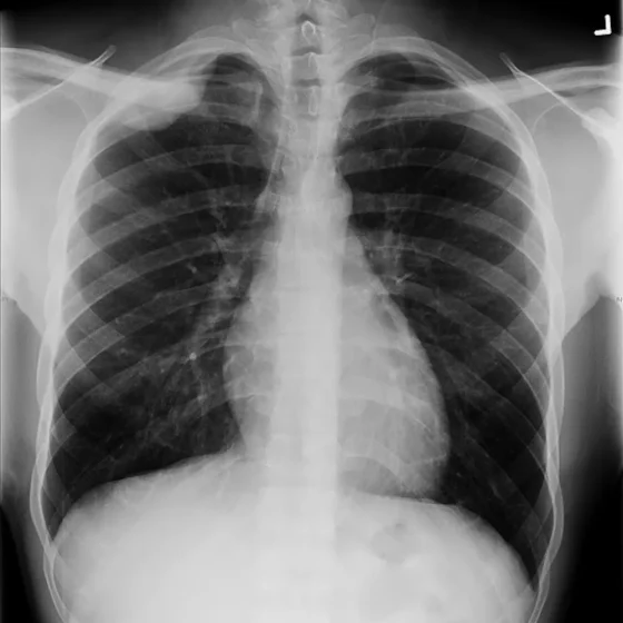
An x-ray is a test that uses small amounts (doses) of radiation to take pictures of the inside of your body. They are a good way to look at bones and can show changes caused by cancer or other medical conditions. X-rays can also show changes in other organs, such as the lungs. You usually have x-rays in the imaging department of the hospital, taken by a radiographer. But in an emergency they are sometimes done on the ward.  Types There are different types of tests using x-rays, including: chest x-rays to show fluid, signs of infection, an enlarged heart or tumours in the chest

Womb biopsy

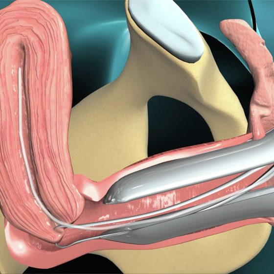
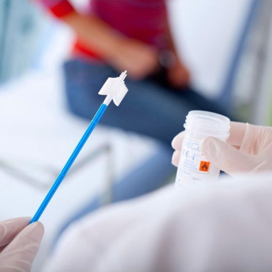
The only way to definitely diagnose womb cancer is to take a sample of the tissue lining the womb. This is called an endometrial biopsy. Your doctor sends the sample to the lab. This is where a pathologist checks it for abnormal or cancerous cells. There are different ways to take a biopsy of the womb lining. Aspiration biopsy To have this test you lie on your back on a couch with your knees up and feet apart. You’ll need to remove your underwear, but you will have a sheet to cover yourself with. Your doctor or nurse gently opens your vagina with a speculum. This is just the same as when

Vulval biopsy 

A vulval biopsy means removing a sample of tissue from the vulva. This is the best way to find out whether or not you have vulval cancer or another vulval condition. Preparing for your vulval biopsy You usually have this test as a day patient using a local anaesthetic. Rarely, you may have it under a general anaesthetic. This is when you are asleep and don’t feel anything. Your doctor or nurse will ask you to sign a consent form once they have given you information about the test. Eating and drinking You usually have a vulval biopsy under local anaesthetic,

Video of your vocal cords (videostroboscopy)

This test uses a long, thin, flexible tube called an endoscope to examine your voice box (larynx) and vocal cords while you speak. The endoscope has a camera and light and is connected to a video monitor and recorder. It helps your doctor to see the movement of your vocal cords while you speak. This test is also called a videostroboscopy, or videolaryngoscopy with stroboscopy. Why do you have a videostroboscopy? You have a video of your vocal cords to help your doctor see how they move when you speak. It also gives your doctor a chance to look for abnormalities on

Video of your throat and larynx (transnasal oesophagoscopy)

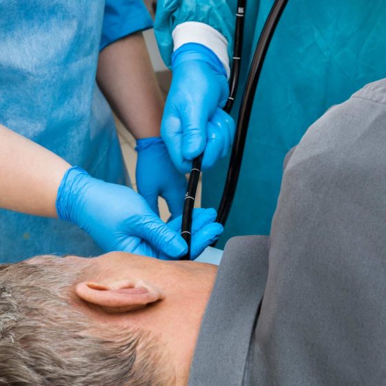
In this test the doctor uses a long, flexible telescope through your nose. This is to make a video of the inside of your: nose throat voice box (larynx) upper part of the food pipe (oesophagus) The test is called a transnasal oesophagoscopy. Why do you have a transnasal oesophagoscopy? This test helps your doctor to view your voice box (larynx) and food pipe (oesophagus). You might have it instead of having an endoscopy. This might happen if you aren’t well enough to have a general anaesthetic. Endoscopy instruments are bigger. You usually need a general anaesthetic for an endoscopy. How to prepare for

Vaginal biopsy

Your doctor may want you to have a biopsy. This means removing a sample of tissue from the vagina. You might have this done during a colposcopy or as a separate test. The doctor sends the sample to the laboratory where a pathologist  examines it.  You might have:  an excision biopsy a punch biopsy Why do you have this test? You have a vaginal biopsy to find out if you have pre cancerous changes (VAIN) or cancer of the vagina. If you have cancer, looking at the cells under a microscope will show which type of vaginal cancer it is. Your doctor may