Day: March 30, 2024

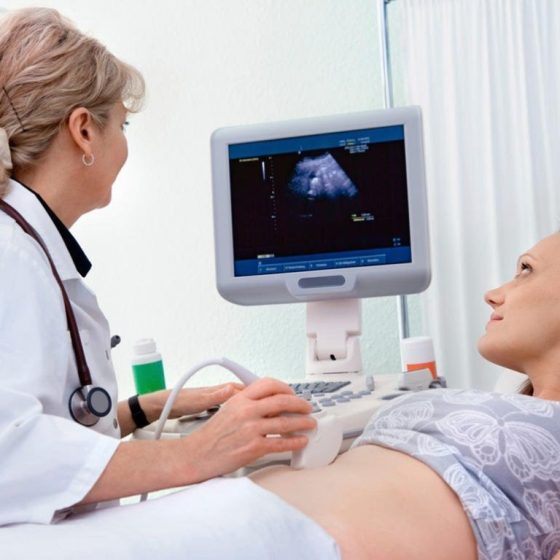
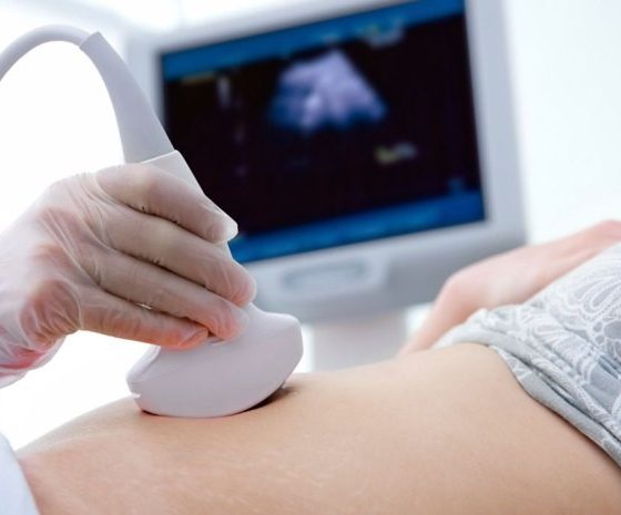
Ultrasound scan of the ovaries

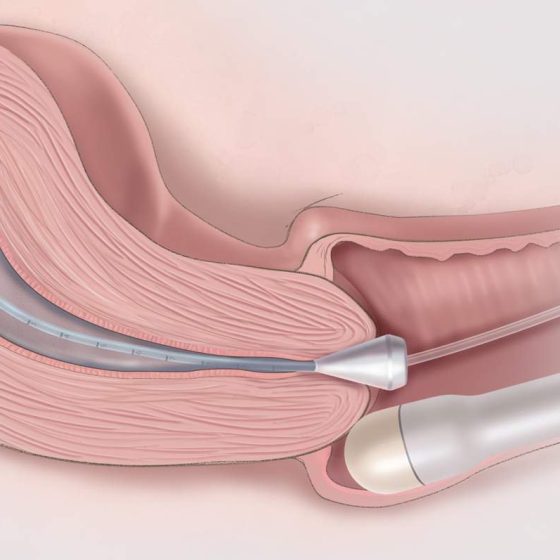
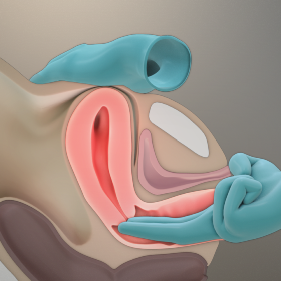
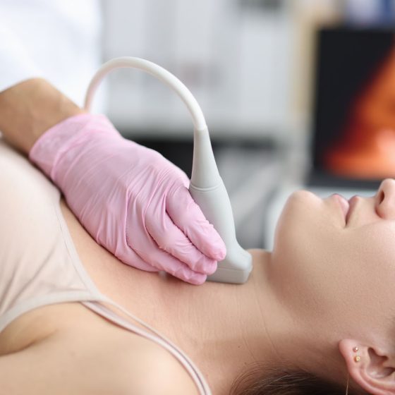
Ultrasound scans use high frequency sound waves to create a picture of a part of the body. It can show the ovaries, womb and surrounding structures. You might have this test to help diagnose ovarian cancer.  To have an ultrasound scan of the ovaries, your doctor or radiographer moves a probe over the lower part of your tummy. You may also have an internal vaginal ultrasound. This is when your doctor gently puts the probe into your vagina. This is also called a transvaginal ultrasound scan. We have separate information about what happens during a transvaginal ultrasound scan.  Why you have it

Ultrasound scan

An ultrasound scan is a procedure that uses high frequency sound waves to create a picture of a part of the inside of your body. The ultrasound scanner has a probe that gives off sound waves. The probe looks a bit like a microphone. The sound waves bounce off the organs inside your body, and the probe picks them up. The probe links to a computer that turns the sound waves into a picture on the screen. Ultrasound scans aren’t painful but can cause some discomfort. You might have it at one of the following: your GP surgery local community clinic in your

Transvaginal ultrasound scan

An ultrasound scan is a procedure that uses high frequency sound waves to create a picture of a part of the inside of your body. The ultrasound scanner has a probe that gives off sound waves. The probe looks a bit like a microphone. The sound waves bounce off the organs inside your body, and the probe picks them up. The probe links to a computer that turns the sound waves into a picture on the screen. Ultrasound scans aren’t painful but can cause some discomfort. You might have it at one of the following: your GP surgery local community clinic in your

Transrectal ultrasound scan (TRUS) and biopsy for prostate cancer

A transrectal ultrasound scan (TRUS) is an examination of the prostate gland using ultrasound. Your doctor might take samples of tissue from the prostate during this test. This is a TRUS guided biopsy. It can help to diagnose prostate cancer. You usually have an MRI scan before your TRUS guided biopsy. The MRI scan helps your doctor decide whether you need a biopsy and where to take the biopsies from. What is a TRUS? Ultrasound scans use high frequency sound waves to create a picture of a part of the body. A prostate ultrasound scan can show changes in your

Transperineal biopsy for prostate cancer

A transperineal biopsy is a needle biopsy to look for cancer cells in the prostate. This helps to diagnose prostate cancer. Your doctor puts a needle into the prostate through the skin behind the testicles (perineum). They take a number of samples, which are sent to the laboratory. In the laboratory, a specialist doctor looks at the samples under the microscope. Preparing for your transperineal biopsy You usually have this test in the outpatient department under local anaesthetic. Sometimes, you may have it in the operating theatre under a general anaesthetic. This is when you are asleep and don’t feel anything. Your doctor

Tissue typing

Tissue typing is a set of tests that are done with your blood and saliva. You might have it if your doctors think that you need a stem cell or bone marrow transplant  from someone else (donor transplant). It is to show how closely a possible stem cell or bone marrow donor’s tissue matches your own. You might also hear this test called other names such as: HLA tissue typing HLA typing Histocompatibility testing HLA crossmatching You usually have one or more of the following tests: a blood test a swab taken from the inside of your cheek (buccal swab)

Thoracoscopy and pleural biopsy

Thoracoscopy is a test to look at the space between your lung and chest wall. This is the pleural cavity. You might have a thoracoscopy to check for signs of cancer such as mesothelioma. It is a small operation. The doctor uses a flexible tube with a light and video camera attached. This is called a thoracoscope. They use it to take samples (biopsies) from the tissues that cover your lung (pleura). Why you might have it You have a thoracoscopy to look inside your chest to see possible areas of cancer. Doctors use a thoracoscopy to help diagnose cancers

Surgical biopsy for lung cancer

During a surgical biopsy for lung cancer, your surgeon takes a sample of the lung tissue by making small cuts in your chest. The surgeon carries out your biopsy in one of the following ways: keyhole surgery , also called video assisted thoracoscopy surgery (VATS) open surgery, through a small cut in the chest wall (small thoracotomy) You have this test under general anaesthetic . So, you will be asleep and won’t feel anything. Why do I need a surgical biopsy? A surgical biopsy of the lung can help diagnose lung cancer. You might have this test if: a CT

Percutaneous transhepatic cholangiography (PTC)

A percutaneous transhepatic cholangiography (per-kew-tay-nee-us trans-hep-attic col-an-jee-og-raf-ee) is a way of looking at your bile ducts using x-rays. This test is also called PTC.  Your doctor puts a long thin needle through the skin and into your liver and bile ducts. They take x-rays of the pancreas, gallbladder and bile ducts. They can also take samples (biopsies) of any abnormal looking areas. You usually have a PTC if you can’t have an ERCP for any reason or if you have had an ERCP but it didn’t work. For example, if doctors weren’t able to take samples of tissue or drain the bile ducts. Why

Penile dynamic sentinel lymph node biopsy

A dynamic sentinel lymph node biopsy (DSLNB) is a test to find out if there is cancer in the lymph nodes near your cancer. Not everyone with penile cancer needs a sentinel lymph node biopsy. You might have this test if there is no obvious sign that cancer cells have spread to the lymph nodes after: you have been examined by your specialist you have had an ultrasound scan It means that your doctor can avoid removing all the lymph nodes in the groin if this is not necessary. You may have to travel to a specialist hospital for this test. This is

Pelvic examination under anaesthetic

This is an internal examination under general anaesthetic . An examination under general anaesthetic is also called an EUA. The examination includes checking your: cervix, vagina and vulva womb bladder anus and rectum (back passage) Your doctor (gynaecological oncologist) can take samples of tissue (biopsies) during the test if necessary. Why you might have a pelvic examination under anaesthetic You might have this examination to check for signs of cancer. It can help your doctor diagnose a number of cancers such as: Preparing for a pelvic examination under anaesthetic Your doctor checks that you are fit and well enough for a general anaesthetic. Let

Pelvic examination

A pelvic examination is when a doctor or a specialist nurse examines your pelvis. Your pelvis is the area between your hip bones. It contains the pelvic organs, including the: bladder lower part of the bowel In women, it also contains the female reproductive system . This includes the: ovaries fallopian tubes womb cervix vagina You may also hear your doctor calling this test an internal or vaginal examination. During the pelvic examination, your doctor looks and feels for anything that doesn’t feel normal. They might refer you for more tests or treatment if they find anything abnormal. Your doctor

Panendoscopy

A panendoscopy is a test to look at your upper airway. This includes your: mouth nose throat voice box top of your food pipe A specialist doctor does the test using a panendoscope, which is a series of connected telescopes. They look through one end and there is a camera and light at the other.  Why you have it You might have a panendoscopy to: get a more detailed look at an abnormal area  take tissue samples from an abnormal area (a biopsy) This is a common test if you have symptoms that could be due to head and neck cancer.  Before your test You

PSA test

The PSA test is a blood test that measures the amount of prostate specific antigen (PSA) in your blood. It can help to diagnose prostate cancer.  Your doctor or nurse takes a sample of your blood. This can be done at your GP practice. They then send this off to a laboratory to check the amount of PSA.  When you have the test You might have a PSA test if you: have symptoms that could be caused by prostate cancer are aged 50 and over and have asked your GP for a test Doctors also check your PSA level as you

PET-MRI scan

A PET-MRI scan combines a PET scan and an MRI scan into one to give detailed information about your cancer.  PET stands for positron emission tomography. It uses a mildly radioactive liquid (radioactive tracer) to show up areas of your body where cells are more active than normal.  MRI stands for magnetic resonance imaging. It uses magnetism and radio waves to create cross section pictures of the body. It shows up soft tissues very clearly. You usually have a PET-MRI scan in the nuclear medicine or radiology department as an outpatient. These scanners tend to be only in the major

PET-CT scan

A PET-CT scan combines a CT scan and a PET scan. It gives detailed information about your cancer. The CT scan takes a series of x-rays from all around your body and puts them together to create a 3 dimensional (3D) picture. The PET scan uses a mildly radioactive liquid (radioactive tracer) to show up areas of your body where cells are more active than normal.  You usually have a PET-CT scan in the radiology department as an outpatient. A radiographer operates the scanner. It usually takes between 30 and 60 minutes. These scanners tend to be available only in the major cancer hospitals. So you

PET scan

A PET scan is a test that creates 3 dimensional (3D) pictures of the inside of your body. PET stands for positron emission tomography. The PET scan uses a mildly radioactive liquid (radioactive tracer) to show up areas of your body where cells are more active than normal. It’s used to help diagnose some conditions, including cancer. It can also help to find out where and whether cancer has spread.    You usually have a PET scan in the radiology or nuclear medicine department as an outpatient. These scanners tend to be only in the major cancer hospitals. So you might have to

Tonsillitis

Overview Tonsillitis refers to the acute inflammation of the palatine tonsils secondary to infection. Acute tonsilitis is very common, especially in children. It is most frequently viral and associated with an upper respiratory tract infection (URTI). It may also be caused by bacteria (e.g. Group A Streptococci). Management typically involves reassurance, fluids and analgaesia. Where a bacterial cause is suspected, antibiotics +/- steroids are given. The most common complication of acute tonsillitis is a peritonsillar abscess (see Quinsy). Recurrent severe tonsillitis results in considerable morbidity and time lost from school or work. Clinical features Clinical examination typically demonstrates enlarged and erythematous tonsils. Exudate is more uncommon and typically suggests

Thyroid cancer

Introduction Thyroid cancer accounts for around 1% of new malignancies in the UK each year. Thyroid cancer refers to a group of malignant conditions affecting the thyroid gland. They most commonly present with a thyroid lump and are diagnosed following triple assessment. Surgical resection (thyroidectomy and hemithyroidectomy) is the mainstay of treatment. Prognosis is related to the type, stage and age at diagnosis. This note will focus primarily on the diagnosis and management of papillary and follicular thyroid cancer (together often referred to as differentiated thyroid cancers). Epidemiology There are approximately 3,900 diagnoses of thyroid cancer in the UK each year. It is

Thyroid anatomy

Overview The thyroid is located in the mid-line of the neck, anterior to the trachea and inferior to the larynx. It is found within the pre-tracheal fascia, one of the fascial compartments of the neck. It is a thin fascia in the anterior part of the neck and is composed of two parts: Muscular part: contains the infrahyoid muscles Visceral part: contains the thyroid, trachea and oesophagus The thyroid gland is comprised of two lateral lobes connected by a central isthmus. It is surrounded by a fibrous capsule and located at the level of vertebrae C5-T1. The pyramidal lobe, an embryological

Ramsay Hunt syndrome

Overview Ramsay hunt syndrome refers to the reactivation of herpes zoster in the geniculate ganglion. The terminology can be confusing. Technically it is Ramsay Hunt syndrome type II (type I is a form of cerebellar degeneration). It can also be referred to as herpes zoster oticus. It is characterised by a facial nerve palsy associated with a vesicular rash affecting the ipsilateral ear, hard palate and anterior two-thirds of the tongue. Treatment is with antivirals, steroids (+/- gastro-protection) and appropriate eye care.   Facial nerve The facial nerve is divided into five branches, which collectively control the muscles of facial expression. The

Quinsy

Overview Quinsy, also termed peritonsillar abscess, is a collection of pus in the peritonsillar space. There are two types of infection that affect the peritonsillar regions: Peritonsillar cellulitis: refers to infection of the peritonsillar tissue in the absence of a discrete collection of pus (abscess). Peritonsillar abscess (quinsy): refers to a discrete collection of pus in the peritonsillar region. Quinsy occurs most commonly as a complication of bacterial tonsillitis. Unlike simple tonsillitis, it mandates surgical intervention to drain the collection. Group A beta-haemolytic streptococcus and haemophilus influenzae are commonly implicated. Severe complications are rare but can include sepsis, the spread of the infection through the deep

Pharyngeal pouch

Overview A pharyngeal pouch, also known as Zenker’s diverticulum, is a sac-like out-pouching that occurs within the hypopharynx. A Zenker diverticulum (ZD), more commonly known as a pharyngeal pouch, is a herniation of the posterior pharyngeal wall that occurs at an area of weakness within the inferior pharyngeal constrictor. It typically occurs in older adults and can lead to symptoms of dysphagia, regurgitation, and more seriously, aspiration pneumonia. Pharyngeal pouch seen at endoscopy with a transparent distal attachment. Evidence of previous endoscopic stapling Epidemiology A pharyngeal pouch is rare before the age of 40. A pharyngeal pouch is a relatively rare condition

Ménière’s disease

Introduction Ménière’s disease is a disorder of the inner ear that manifests itself with attacks of vertigo, tinnitus and hearing loss. The majority of cases of Ménière’s disease are idiopathic but the condition is thought to be related to abnormal production and absorption of endolymph (the fluid that fills portions of the labyrinthine system). Where an underlying cause is identified it is referred to as Ménière’s syndrome. The disease is characterised by episodes of vertigo, tinnitus and fluctuating hearing loss. As the condition develops, symptoms, in particular tinnitus and hearing loss, may become permanent and progressively worse. Management involves supportive care during acute

Malignant otitis externa

Overview Malignant otitis externa is a serious condition where infection spreads from the external auditory canal to the skull base. It most commonly affects the elderly, diabetic or immunocompromised. Pseudomonas aeruginosa is by far the most common causative pathogen. Treatment is primarily with intravenous antibiotics and supportive care. Risk factors The majority of cases of malignant otitis externa are associated with diabetes or glucose intolerance. Diabetes Old age Immunocompromise (e.g. HIV) Aetiology Pseudomonas aeruginosa is the most common cause of malignant otitis externa. Pseudomonas aeruginosa is a gram-negative rod. It normally causes infections in patients with deficient immune systems or poor glycemic control. Less commonly other pathogens are

Epistaxis

Introduction Epistaxis refers to bleeding from the blood vessels within the nasal mucosa. Nosebleeds are common. The vast majority resolve spontaneously or with basic first aid. However, it remains a relatively common cause of presentation to A&E departments, with up to 6% of the UK population seeking medical attention due to epistaxis at some point in time. On occasion, epistaxis requires intervention (e.g. nasal cautery or nasal packing) typically performed by an A&E or ENT doctor. In certain settings (discussed in more detail below), epistaxis should trigger an outpatient referral to ENT to exclude an underlying causative lesion (e.g. malignancy). Aetiology

Chronic rhinosinusitis

Overview Chronic rhinosinusitis refers to inflammation of the nasal cavities and paranasal sinuses lasting > 12 weeks. Chronic rhinosinusitis is broadly defined as inflammation of the nasal cavities and paranasal sinuses that lasts for longer than 12 weeks. However, it is actually a very heterogenous condition that is difficult to fully define and may be part of a wider systemic illness (e.g. granulomatosis with polyangiitis). There are various subtypes of chronic rhinosinusitis although the cardinal features remain the same, which include nasal congestion, mucopurulent nasal discharge, and facial pain or pressure. Treatment aims to reduce symptom burden and improve quality of

BPPV

Overview Benign paroxysmal positional vertigo (BPPV) is an inner ear disorder characterised by episodes of positional vertigo. BPPV is the most commonly encountered cause of vertigo seen in clinical practice. It is caused by otoconia (loose debris composed of calcium carbonate) within the semilunar canals of the inner ear. Attacks are triggered by head movements that result in movement of the otoconia, abnormal motion of endolymph and the feeling of vertigo. A careful history and examination are required to exclude other causes of vertigo. ‘Manoeuvres’ (explained in detail below) are used in both the diagnosis and treatment of BPPV. The Dix-Hallpike

Allergic rhinitis

Overview Allergic rhinitis is a common condition secondary to IgE-mediated inflammation of the nasal mucosa. Allergic rhinitis is a common condition characterised by sneezing, runny nose (i.e. rhinorrhoea), nasal obstruction and itching. It is more colloquially known as ‘Hayfever’. These symptoms are brought about by IgE-mediated inflammation secondary to antigens known as ‘allergens’ that can include grass, tree pollen, or house dust mites among many others. The diagnosis of allergic rhinitis is clinical and management aims to dampen the immune response to allergens principally with anti-histamines and intranasal glucocorticoids. It may be a very disabling condition and is commonly associated with

Acute rhinosinusitis

Overview Acute rhinosinusitis refers to acute inflammation of the nose and paranasal sinuses. Acute rhinosinusitis is a common condition that is usually caused by a viral pathogen. It leads to typical features of nasal congestion, nasal discharge, and facial pressure/pain that is worse on bending forward. These symptoms completely resolve within 4 weeks. Similar to an upper respiratory tract infection, symptoms will improve without intervention and antibiotics are rarely required. Terminology Rhinosinusitis is a better term than simply ‘sinusitis’ because inflammation of the nasal cavities almost always accompanies sinusitis. Sinusitis versus rhinosinusitis Sinusitis: symptomatic inflammation of the paranasal sinuses Rhinosinusitis: symptomatic inflammation

Acute otitis media

Overview Acute otitis media (AOM) refers to inflammation of the middle ear with effusion and clinical features of a middle ear infection. Normally AOM is a self-limiting condition, commonly occurring in children, though adults can also be affected. In general symptoms settle after 3 days, but can remain for a week. Bacteria or viruses are normally implicated – differentiating the two is challenging. The majority of cases do not require antibiotic therapy and will settle with time. Though rare, serious complications such as mastoiditis, meningitis, intracranial abscess, sinus thrombosis or facial nerve paralysis may occur. AOM may become persistent or recurrent. NICE

Acute otitis externa

Overview Otitis externa refers to inflammation of the external auditory canal. Though the inflammation can have many causes (e.g. allergic, dermatologic), infection, in particular by bacteria, is the most common cause. It may be categorised (definition from NICE CKS) as: Acute: lasts 3 weeks or less Chronic: lasts 3 months or longer Malignant otitis externa (MOE) is a rare life-threatening condition where the infection spreads to the surrounding bone, it is covered here. Epidemiology Up to 10% of people will experience acute otitis externa in their lifetime. Though otitis externa (both acute and chronic) can occur at any age, it is more common in children and

Acoustic neuroma

Overview An acoustic neuroma is a benign Schwann-cell derived tumour, which commonly arises from the eight cranial nerve. An acoustic neuroma, also known as a vestibular schwannoma, is a benign intracranial tumour that is derived from Schwann cells that are one of the major supporting nerve cells in the peripheral nervous system. It is typically a slow growing tumour that arises from the eighth cranial nerve supplying the inner ear. This cranial nerve, also known as the vestibulocochlear nerve, has important functions in both hearing and balance. An acoustic neuroma is typically unilateral and leads to unilateral sensorineural hearing loss (bilateral

Lactational mastitis

Introduction Lactational mastitis is a relatively common condition affecting post-partum women. Also termed puerperal mastitis, it can affect up 1/3 of women in the post-partum setting (although many studies put this figure closer to 10%). It can be complicated by the development of a breast abscess. These terms may be defined as: Mastitis: refers to inflammation of the breast tissue. Mastitis may be lactational (occurring in lactating women) or non-lactational. It can also be categorised as infectious or non-infectious. Breast abscess: refers to a localised collection of pus within the breast. Again these may be lactational (occurring in lactating women) or

Fibroadenoma

Introduction Fibroadenomas are the most common benign tumour of the breast. Traditionally referred to as ‘breast mice’, these discrete, mobile lumps often occur in younger women. It is important to distinguish and exclude breast cancer, as such patients should be referred for a triple assessment in line with NICE guidance. As a general rule, 50% regress spontaneously, 25% will remain unchanged and 25% will get bigger. Epidemiology Fibroadenomas most commonly occur in women aged 14 to 35. These benign tumours are typically seen in younger patients and often regress after menopause. There is up to a 10% lifetime incidence of fibroadenomas in women. Types Fibroadenoma may be split

Breast cancer

Introduction Breast cancer is the most common malignancy affecting women in the UK. It may be diagnosed during screening or patients may present with a breast (or axillary) lump. Pain, skin and nipple changes may also prompt presentation. On occasion, patients will present with symptoms of metastatic spread. Management is holistic, with input from members of the multi-disciplinary team (MDT) and centred around the individual patient’s thoughts and wishes. Breast cancer can occur in anyone. It can affect women, trans-women, trans-men, men and non-binary individuals. In men, it is less common and is not within the top 20 cancers affecting men in the

Vasculitis

Overview Vasculitis refers to inflammation of blood vessels. Vasculitis is a very broad term that in its simplest form refers to inflammation of blood vessels. There are many different causes of vasculitis and they usually present in a myriad of ways. The exact cause of vasculitis is unknown and the underlying mechanisms leading to blood vessel inflammation vary between conditions (e.g. antibody-mediated, T-cell mediated). There has been a lot of work on classifying vasculitides into different types. They are a very heterogenous group of conditions that can affect multiple organs and in some cases be life-threatening. Several important terms are used

Systemic sclerosis

Overview Systemic sclerosis is a chronic, multi-system disorder that is characterised by widespread vascular dysfunction and fibrosis. Systemic sclerosis (SSc) is a chronic multi-system disorder. The condition is heterogeneous and can present with a range of clinical manifestations involving multiple organs. One of the hallmark features of SSc is thickened, hardened skin known as scleroderma. The term scleroderma is often used synonymously with SSc. Disease subtypes SSc may be divided into several disease subtypes based on the extent of skin involvement and pattern of organs affected. Limited cutaneous systemic sclerosis (lcSSc): characterised by sclerosis (hardening) of the skin in the distal limbs. Some

Systemic lupus erythematosus

Overview Systemic lupus erythematosus (SLE) is a multi-system, inflammatory, autoimmune disorder. SLE, also shortened to lupus, is a multi-system condition and therefore may present in a myriad of ways. Typical manifestations include characteristic skin rashes, arthralgia and renal impairment. Management is complex but involves symptomatic relief, immunosuppressive agents and more novel biologics. Epidemiology SLE is thought to affect almost 1 in 1000 individuals in the UK. It often presents in women of reproductive age, with an average age at diagnosis of 48.9, but can manifest at any age. The condition is more common in women and those of Afro-Caribbean or South Asian

Spondyloarthropathies

Overview The spondyloarthropathies refer to a diverse group of conditions associated with the HLA-B27 gene. The spondyloarthropathies (SpAs) refer to a diverse group of conditions associated with the HLA-B27 gene. They are characterised by chronic inflammation affecting the axial skeleton (i.e. spine and sacroiliac joints) and/or peripheral joints. Due to the predominant axial skeletal involvement, they may be collectively termed ‘axial spondyloarthropathies’ (AxSpA). These conditions include: Ankylosing spondylitis (AS) Psoriatic arthritis (PsA) Reactive arthritis (ReA) IBD-associated spondyloarthropathy The SpAs are characterised by pain, stiffness, and loss of mobility. They can present with numerous musculoskeletal features: Spinal & sacroiliac joint inflammation Peripheral arthritis Dactylitis:

Sjögren syndrome

Overview Sjögren’s syndrome is a chronic autoimmune disease characterised by reduced lacrimal and salivary gland function. Sjögren’s syndrome (SS) is characterised by dry eyes and dry mouth due to reduced lacrimal and salivary gland function, respectively. It is a systemic condition associated with extraglandular clinical features and can affect almost any organ. Terminology A number of terms are used in SS. Sicca syndrome: old term that is synonymous with SS. Keratoconjunctivitis sicca: refers to the dry eye symptoms experienced in SS Xerostomia: refers to dry mouth Primary vs. secondary SS may be classified as primary or secondary: Primary: Sjögren’s syndrome in

Rheumatoid arthritis

Overview Rheumatoid arthritis is a chronic, systemic inflammatory disorder characterised by inflammatory polyarthritis. Rheumatoid arthritis (RA) is classified as an inflammatory arthropathy because it characteristically affects multiple joints leading to chronic joint pain, swelling and stiffness. However, RA can be a multi-system disorder associated with extra-articular manifestations and systemic features including myalgia, fatigue, low-grade fever, weight loss, and depression. Patients with RA are often described as seropositive or seronegative depending on the presence or absence antibodies (e.g. Anti-CCP, Rheumatoid factor – discussed below). Epidemiology RA is estimated to affect around 1% of the UK population. The annual incidence of RA is estimated at

Reactive arthritis

Overview Reactive arthritis is one of the spondyloarthropathies that is defined as arthritis occurring after an infection. Reactive arthritis (ReA) is one of the spondyloarthropathies that can cause peripheral arthritis (i.e. joint inflammation) or spondylitis (i.e. spinal inflammation) following an infection. It causes ’sterile’ inflammation because there is no actual infection in the joint. ReA commonly occurs several weeks following infection by a variety of organisms that usually infect the urogenital or gastrointestinal tract. ReA was historically referred to as ‘Reiter’s syndrome’, which describes a clinical triad of arthritis, urethritis, and conjunctivitis. However, only a small proportion of patients present with

Raynaud phenomenon

Overview Raynaud phenomenon refers to skin colour changes that occur in the fingers and toes from vasospasm. Raynaud phenomenon is an exaggerated response to cold temperatures or emotional stress. It is due to abnormal vasoconstriction of digital and cutaneous arterioles often referred to as ‘vasospasm’. Vasospasm refers to the sudden constriction of blood vessels. Raynaud phenomenon presents with classic skin changes that occur suddenly. These may be divided into three stages: White: development of white fingers with sharp demarcation of colour change due to vasoconstriction. Blue: vasoconstriction leads to tissue hypoxia and development of cyanosis. Red: as vasoconstriction improves, there is an increase

Pseudogout

Overview Pseudogout is a crystal arthropathy, which may cause an acute inflammatory arthritis from deposition of calcium pyrophosphate. The principle mechanism underlying pseudogout is deposition of calcium pyrophosphate dihyrate (CPP). In fact, pseudogout is an old term that refers to one of the clinical syndromes within a spectrum of conditions associated with CPP deposition (CPPD) in connective tissue. The term pseudogout is still used commonly in clinical practice. This is because of the close resemblance to gout in both pathophysiology (crystal arthropathy) and clinical features (acute inflammatory arthropathy). CPPD disease CPPD is an umbrella term that covers all clinical conditions linked to the

Polymyalgia rheumatica

Overview Polymyalgia rheumatica is a common rheumatological disorder, which is characterised by shoulder and hip girdle pain. Polymyalgia rheumatica (PMR) is a common systemic inflammatory disease that is one of the most common indications for long-term steroids. It is characterised by myalgia and muscles stiffness with preponderance to the neck, shoulder and pelvic girdle. Similar to giant cell arteritis (GCA), PMR is predominantly a disease of older adults and rarely presents before 50 years old. The peak prevalence is estimated between 70-80 years. Women are 2-3 times more likely to be affected than men. Aetiology The exact cause of PMR is currently

Osteoporosis

Overview Osteoporosis is characterised by low bone mass and micro-architectural deterioration of bone with resulting fragility and fracture risk. Osteoporosis can be defined as a bone mineral density (BMD) of 2.5 standard deviations below the mean peak mass (for an average young healthy adult). With the exception of fracture, osteoporosis is an asymptomatic condition. The weaker bone predisposes patients to what are termed ‘fragility fractures’. This refers to those that result from ‘low-energy’ trauma that would not normally result in fracture. Common fragility fractures include vertebral crush fractures and those of the distal wrist and proximal femur. A number of therapeutic options

Neuroendoscopy to take a sample of tissue

Neuroendoscopy is a way of taking samples of tissue from a brain tumour. Doctors take samples of tissue (biopsy) to look at it under a microscope. This helps your doctor decide the best treatment for you. You may also have a neuroendoscopy to: remove fluid from the fluid filled spaces of the brain (ventricles) remove all or part of the brain tumour You usually have a neuroendoscopy under general anaesthetic. This means that you will be asleep and won’t feel anything. Having a neuroendoscopy sounds like a frightening procedure, but it is quite safe. Doctors who carry out these procedures

Needle biopsy through the skin for lung cancer 

This test is also called a percutaneous lung biopsy. A doctor called a radiologist takes a sample of lung tissue by passing a needle into the lung. They use a CT scanner or ultrasound to ensure they know the right place to take the sample. You have this test with a local anaesthetic . It takes about 30 to 45 minutes. Why do I need a biopsy through the skin? You might have this test if your doctor has seen an abnormal looking area in your lung or airways using an x-ray or CT scan. What do I need to

Needle biopsy of the thyroid

You have this test to find out if the lump in your thyroid needs further treatment. Preparing for your test You are able to eat and drink normally before a biopsy. Take your medicines as normal. But if you are taking any blood thinning medicines you might need to stop them before the test. Your doctor or nurse will tell you when to stop. You will need to remove any clothing around your neck such as a tie or scarf. You will also be asked to remove any jewellery such as a necklace or chain. You will lie on your back on a

Neck lymph node ultrasound and biopsy

During a neck lymph node biopsy, your doctor uses an ultrasound scanner to help them take a small amount of lymph node tissue using a fine needle. Ultrasound scans use high frequency sound waves to create a picture of a part of the body. The ultrasound scanner has a probe that gives off sound waves. The sound waves bounce off the structures inside your body, and the probe picks them up. The probe links to a computer that turns the sound waves into a picture. You normally have this test as an outpatient procedure in the hospital’s imaging department. Why

Nasoendoscopy

A nasoendoscopy is part of a normal ear, nose and throat examination. It is a procedure to look at the inside of the: nose back of the tongue throat voice box Your doctor passes a thin rigid or flexible tube called a nasoendoscope through your nostril into your nose. The tube has a camera and a bright light at the end. Why you might have it You may have a nasoendoscopy so that your doctor can check for any abnormal looking areas. This is a common test if you have symptoms that could be due to head and neck cancer.  Preparing for

Multiparametric MRI (mpMRI) scan for prostate cancer

MRI stands for magnetic resonance imaging. It is a type of scan that creates pictures using magnetism and radio waves. MRI scans produce pictures from angles all around the body and shows up soft tissues very clearly.  The multiparametric MRI (mpMRI) is a special type of MRI scan that produces a more detailed picture of your prostate gland than a standard MRI scan does. You usually have an mpMRI scan if doctors suspect that you have prostate cancer. Why you might have an mpMRI It is important to know that an mpMRI scan alone cannot diagnose prostate cancer. But it can