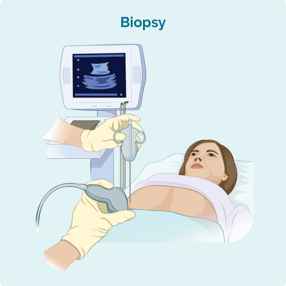
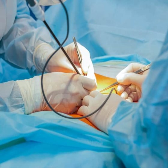
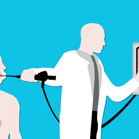
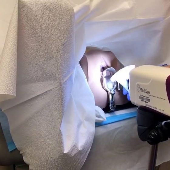
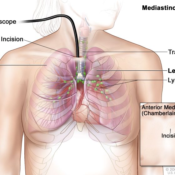
Day: March 29, 2024

Mediastinoscopy 

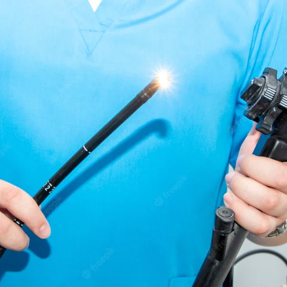
Mediastinoscopy is a test that examines the mediastinum. This is the centre of your chest and area between your lungs. It contains: the heart the main blood vessels lymph nodes (glands) the food pipe (oesophagus) The mediastinoscopy takes between 45 to 60 minutes. You have a general anaesthetic to have this test.  Why do I need a mediastinoscopy? You might have this test to see if cancer cells have spread into the lymph nodes around the windpipe.  Preparing for your mediastinoscopy You see a doctor before the mediastinoscopy. They’ll ask some general questions about your health. You will also need some other tests before

Mammogram

A mammogram is an x-ray of your breasts. X-rays use high energy rays to take pictures of the inside of your body. You might have mammograms: as part of the breast cancer screening programme to check for cancer if you have symptoms that could be due to breast cancer. This is called a diagnostic mammogram The health professionals who take mammograms are called mammographers. The mammogram itself usually only takes a few minutes, but the appointment may last about 30 minutes. Mammograms for breast screening Mammograms for breast screening can help to find breast cancer early when they are too

MRI scan

MRI (magnetic resonance imaging) is a type of scan that uses magnetism and radio waves to take pictures of inside the body. The scan takes between 15 and 90 minutes. You might have one to find out whether you have cancer and if you do to measure how big it is and whether it has spread. You might also have one to see how well treatment is working. It is a safe test. If you have an injection of contrast dye it can cause a headache, dizziness or a warm flushed feeling An MRI is a type of scan that creates

MRCP and MRI scan

MRCP stands for magnetic resonance cholangio pancreatography (col-an-jee-oh pan-kree-at-og-raf-ee). An MRCP scan is a type of MRI scan that you have in an MRI scanner. MRI stands for magnetic resonance imaging. An MRI scan produces pictures from angles all around the body and shows up soft tissues very clearly.  These scans create pictures using magnetism and radio waves to give detailed pictures of your: pancreas gallbladder bile ducts liver You might have an MRI scan of your tummy (abdomen) at the same time as the MRCP. You usually have these scans in the x-ray (radiology) department as an outpatient. It can

Radioactive scans for neuroendocrine tumours (NETs)

Radioactive scans can help your doctors diagnose some types of neuroendocrine tumours. You have an injection of a low dose radioactive substance, which can show up on a scan. Radioactive scans include: octreotide scans (or Octreoscans) – these are also called somatostatin receptor scintigraphy scans tektroyd scans gallium dotatate PET (positron emission tomography) scans You might also have an mIBG scan, but this is less common now. mIBG stands for metaiodobenzylguanidine. What are radioactive scans and why do I need one? Octreotide, dotatate and mIBG are substances that some neuroendocrine cells take up (absorb). Doctors can attach a radioactive substance

Lymph node ultrasound and biopsy – under the arm (axillary) 

Cancer can sometimes spread into the lymph nodes under the arm (axilla). This can happen with different types of cancer, including breast cancer. To check the lymph nodes under your arm for breast cancer cells, you have an ultrasound scan. Ultrasound scans use high frequency sound waves to create a picture of a part of the body. The ultrasound scanner has a microphone that gives off sound waves. The sound waves bounce off the organs inside your body and are picked up by the microphone. The microphone links to a computer and turns the sound waves into a picture. If

Lymph node ultrasound and biopsy – groin

Cancer can sometimes spread into the lymph nodes in the groin. Your groin is the area at the top of your legs, between the hips. This can happen with different types of cancer, such as anal and penile cancer.  To check the lymph nodes in your groin for cancer cells, your doctor checks the groin for swollen (enlarged) lymph nodes. You then might have an ultrasound scan to check the nodes. Ultrasound scans use high frequency sound waves to create a picture of a part of the body. If doctors find any abnormal lymph nodes, they use the ultrasound to

Lumbar puncture

A lumbar puncture is a test to check the fluid that circulates around the brain and spinal cord. This is called the cerebrospinal fluid or CSF. Why do I need a lumbar puncture? A lumbar puncture can check for cancer cells or for infection in the CSF. Doctors remove some of the CSF to test for cancer cells. They use a needle to take a sample of the CSF from your lower back. You normally have this test in the outpatient department under local anaesthetic. This means you are awake but the area is numb.  What Is a Lumbar puncture? A

Liver biopsy 

A biopsy means removing a sample of cells or tissue and looking at it under a microscope. You may have a liver biopsy to help diagnose liver cancer. But not everyone needs to have a liver biopsy. Doctors can often diagnose liver cancer just by looking at the scans. You usually won’t have a liver biopsy if your doctor thinks: they can remove the cancer you may be able to have a liver transplant This is because there is a small risk that a liver biopsy could spread the cancer along the path of the biopsy needle. It is important to

Laryngoscopy

A laryngoscopy is a test that an ear, nose and throat (ENT) specialist uses to look at the: back of your throat (pharynx) voice box (larynx) The ENT specialist uses a thin tube called a laryngoscope to do this test. They can see the inside of your throat very clearly. You have this test under general anaesthetic. This means that you are asleep and won’t feel anything. Your doctor might also call this test an examination under anaesthesia (EUA). They may take samples of tissue (biopsy) of any abnormal areas. Preparing for a laryngoscopy You might have a blood test 2 days beforehand to check

Laparoscopy

Laparoscopy is a small operation to look inside your tummy (abdomen) and the area between your hips (pelvis). Laparoscopy is also known as keyhole surgery or minimally invasive surgery. It can help diagnose certain types of cancer. It is also a very common procedure used to treat and diagnose many other non cancerous conditions. A laparoscopy involves your surgeon making several small cuts (incisions) instead of a large incision across your abdomen. The surgeon places hollow tubes inside the incisions. These are called ports. Your surgeon puts a thin tube with a light and a camera (laparoscope) through one of

LLETZ – Large Loop Excision of the Transformation Zone

LLETZ stands for large loop excision of the transformation zone. It’s also known as loop electrosurgical excision (LEEP) or loop diathermy. Your colposcopist uses a thin wire loop to remove the transformation zone of the cervix. The wire has an electrical current running through it, which cuts the tissue and seals the wound at the same time. The transformation zone is the area around the opening of the cervix. LLETZ is an outpatient treatment and takes up to 15 minutes. You usually have it under local anaesthetic. Why you might have LLETZ LLETZ is a treatment for abnormal cervical cells picked

Kidney biopsy

A kidney biopsy is a procedure to take a small piece of tissue from the kidney. A specialist doctor (pathologist) looks at it under a microscope and can tell if you have kidney cancer and which type it is. Knowing this helps your doctor decide on the best treatment for you.  Why you might have a kidney biopsy Your doctor will ask you to have a kidney biopsy to: find out if you have kidney cancer if other tests haven’t been clear help them decide if a small kidney cancer can be watched before being treated (active surveillance) tell them

IVU (Intravenous urogram) or IVP (Intravenous pyelogram)

An intravenous urogram (IVU) is a test that looks at the whole of your urinary system. It’s sometimes called an intravenous pyelogram (IVP). It looks at the: kidneys bladder tubes that connect the kidney with the bladder (ureters) The male urinary system The female urinary system The test uses a colourless dye, also called contrast medium. This shows up the soft tissues of the urinary system on a normal x-ray. Why do I need an IVU test? An IVU test can show if cancer is growing in any part of your urinary system. The cancer will show up as a

Gastroscopy for stomach cancers

A gastroscopy is a test that looks at the inside of your food pipe (oesophagus), stomach and the first part of your small intestine (small bowel). A doctor or specialist nurse (endoscopist) does the test. They use a long flexible tube which has a tiny camera and light at the end. This tube is called a gastroscope or endoscope.  You may also hear this test called an endoscopy or oesophago gastric duodenoscopy (OGD). Why do I need a gastroscopy? You might have a gastroscopy to find out the cause of symptoms such as: abnormal bleeding indigestion or heartburn that doesn’t go away with

Fluorescein angiogram of the eye

Your eye specialist might do a fluorescein angiogram (FA) to look at the blood vessels in your eye. You may also have a photograph taken of the surface of the inside of the eye (the fundus). Doctors call this colour fundus photography. What is it? A fluorescein angiogram looks at the blood vessels and the lining at the back of your eye (the retina). This test can pick up changes in the condition of your eyes. It can help your specialist make a diagnosis of eye cancer or tell them how well treatment is working. What happens You usually have the test in the

Flexible sigmoidoscopy

Flexible sigmoidoscopy is a test to look inside the lower part of your large bowel. It is also called bowel scope or flexi sig. The flexible sigmoidoscopy is a thin flexible tube called a colonoscope. This has a small light and camera at one end. The endoscopist (specially trained doctor or nurse) puts the tube into your back passage (anus). They then gently move it up into the lower part of your bowel. They can see the pictures of the inside of your bowel on a TV screen. You usually have this test in the endoscopy unit at the hospital. It may

FIT (Faecal Immunochemical Test)

FIT (Faecal Immunochemical Test) is a test that looks for blood in a sample of your poo. It looks for tiny traces of blood that you might not be able to see and which could be a sign of cancer. Traces of blood in your poo can be caused by other medical conditions and doesn’t necessarily mean you have cancer. But if it is cancer, finding it at an early stage means treatment is more likely to work. This information is about using the FIT test for people who have symptoms that could be caused by bowel cancer. We have separate

FISH (fluorescence in situ hybridisation)

FISH stands for fluorescence in situ hybridisation. It is a test that looks for gene changes in cells. Genes are made of DNA. They control everything the cell does, including when it grows and reproduces. FISH tests look for specific genes or parts of genes.  Changes in genes can make the cancer cell: produce particular proteins make far more of the protein than normal stop making a particular protein If a gene change occurs, the cell may produce too much of a protein or not enough. This can make the cancer cells grow and reproduce more than normal. Cancer treatment There are now

Eye examination

The specialist eye doctor (ophthalmologist) will examine your eyes using a few different instruments. They look at the different structures of your eye to check for changes or abnormalities. What happens before the eye examination? You usually have drops into your eyes before the examination. These enlarge the pupils to make it easier for the eye specialist to look in your eyes. The eye drops might sting a bit when they first go in. They can cause blurred vision, and your eyesight may be blurry for a few hours, so you shouldn’t drive yourself home. It might be helpful to take

Examination of your prostate

To examine your prostate your doctor puts a finger into your back passage (rectum). This procedure is also called a digital rectal exam (DRE). Why you might have this You might have a prostate examination to check for any problems in your prostate.  What happens before the examination? It’s normal to feel anxious about this test but it usually only takes a few minutes. You shouldn’t feel any pain but may be uncomfortable. Tell your doctor if you feel pain. You can ask for a man or woman doctor if you’d prefer. Or you can have someone else in the room,

Examination of anus and rectum (back passage)

This examination involves your doctor feeling inside the anus and rectum using their finger. This is also called a digital rectal exam (DRE). You might have a DRE if you have symptoms that could be due to bowel or anal cancer.  Why you might have an examination of the anus and rectum You might have this to help work out the cause of your symptoms. Preparing for the examination It’s normal to feel anxious about this test but it usually only takes a few minutes. It might be uncomfortable but you shouldn’t feel any pain. Tell your doctor if you

Endoscopy for cancers

An endoscopy is a test that looks inside the body. The endoscope is a long flexible tube which has a tiny camera and light on the end of it.  There are many types of endoscopes and the doctor uses these to look inside different parts of the body. The name of the test you have will depend on which part of the body the doctor is looking at. Why do I need an endoscopy? You might have an endoscopy to help find out what is causing your symptoms. During this test a doctor or specialist nurse (endoscopist) can also take

Endoscopic ultrasound for lung cancers

This test combines ultrasound and endoscopy. It helps your doctor to look through the wall of the food pipe at the surrounding tissue. The main airway (windpipe) is close to the food pipe. A doctor or a specialist nurse (endoscopist) does the test. To do the test the endoscopist uses a long flexible tube called an endoscope. They pass the endoscope down your throat into your food pipe. It has a tiny camera and light on the end and an ultrasound probe attached. The ultrasound scan uses high frequency sound waves to create a picture of the inside of your body. The

Endoscopic ultrasound (EUS)

This test combines an ultrasound and endoscopy. It can look at parts of your upper gastrointestinal tract. This includes the: food pipe (oesophagus) stomach pancreas bile ducts gall bladder first part of your small bowel (duodenum) lymph nodes  An endoscopy is a test to look inside your body. Your doctor uses a long flexible tube (endoscope) with a tiny camera and light on the end. The endoscope also has an ultrasound probe at its tip.  An ultrasound scan uses high frequency sound waves to create a picture of the inside of your body. The preparations for this test are the

Endobronchial ultrasound (EBUS)

Endobronchial ultrasound is also called Endobronchial Ultrasound-guided Transbronchial Needle Aspiration (EBUS-TBNA). It is a test that can show if: an abnormal looking area is cancer the size of the cancer the cancer has spread into other lung areas or outside the lung This test uses a narrow flexible tube to look at the inside of the breathing tubes (airways) in your lungs. The tube has an ultrasound probe. It uses high frequency sound waves to create pictures of the lungs and structures outside the airway walls, such as the lymph nodes. Your doctor can see any areas that look abnormal

Echo (echocardiogram)

An echo is an ultrasound scan of the heart. This scan is very similar to the scans women have during pregnancy. It uses high frequency sound waves to create a picture of your heart. Doctors can look at the structure of your heart and how well it is pumping, and the nearby blood vessels.  The ultrasound scanner has a microphone that gives off sound waves. The sound waves bounce off the heart and the microphone picks them up. The microphone links to a computer that turns the sound waves into a picture on the screen. This microphone device is called the probe.

ERCP (endoscopic retrograde cholangiopancreatography)

ERCP test ERCP stands for endoscopic retrograde cholangio pancreatography. It is a test to help diagnose conditions of the liver, bile ducts, pancreas or gallbladder. What is an ERCP? Your doctor uses a long flexible tube with a small camera and light at the end, called an endoscope. It’s also sometimes called a duodenoscope. They pass this tube through your mouth, throat, stomach and into the first part of your small bowel (duodenum). Your doctor can look down the endoscope or see pictures on an X-ray screen of the pancreas, gallbladder and bile ducts.   They can take samples (biopsies) of any abnormal looking areas. 

ECG (electrocardiogram)

An ECG is a test to check the electrical activity of your heart. It can show changes to your heart rate or rhythm and tell your doctor how well your heart is working. ECG stands for electrocardiogram. You might also hear this test called a heart trace. An ECG can give your doctor a lot of good information. It can help the doctor decide if you need further tests. To have the test a trained health care professional puts small sticky pads on your chest, arms and legs. Wires connect these to an ECG recording machine; these wires are ECG leads.

Draining fluid from your chest or tummy

Cancer cells can inflame the lung lining (pleura) or abdominal lining (peritoneum). This can cause fluid to build up. The fluid might contain cancer cells. Fluid around the lungs is called a pleural effusion. It can make it difficult to breathe. Draining fluid from your chest is called thoracocentesis or pleural aspiration. Fluid in the tummy (abdomen) is called peritoneal ascites. It can make the abdomen feel swollen, tight and uncomfortable. Draining fluid from your abdomen is called an abdoparacentesis or peritoneal aspiration.  Why you might have it The doctors might drain fluid from around your lungs or abdomen to: see if the fluid contains

Looking at your mole or skin (dermoscopy)

You will see a skin specialist (dermatologist). They will ask you questions about your mole or abnormal area of skin, such as how long you have had it and what changes you have noticed. They will look closely at the abnormal area, and will check the rest of your skin for any changes. They usually use a dermatoscope to do this.  What is a dermatoscope? A dermatoscope is a handheld instrument, a bit like a magnifying glass. It can make things bigger (magnify) by up to 10 times. Your specialist puts some oil or gel onto your skin. They then hold the dermatoscope

Cystoscopy to check for cancer

A cystoscopy is a test to look at the inside of your bladder and tube that carries urine from your bladder out of your body (urethra). It uses a thin tube called a cystoscope.  There are different types of cystoscopies: flexible cystoscopy rigid cystoscopy narrow band imaging (NBI), blue light cystoscopy or photodynamic diagnosis (PDD) Your doctor will discuss with you what type of cystoscopy you’re having. Flexible cystoscopy The flexible cystoscope has optic fibres inside it, a light and camera attached to it. Because it’s flexible it can bend around the tubes as it passes through your urethra. This is generally done under local

Colposcopy

A colposcopy is a test to look at the cervix in detail. A colposcope is a large magnifying glass that a doctor or specialist nurse (a colposcopist) uses to look closely at the skin-like covering of the cervix. By looking through it, the colposcopist can see changes that may be too small to see with the naked eye. They can take samples (biopsies) of any abnormal areas. You usually have a colposcopy in the hospital outpatient clinic. Why you might have a colposcopy You have a colposcopy if you’ve had an abnormal result after a cervical screening test. You may also have a colposcopy if you have symptoms

Colonoscopy

A colonoscopy looks at the whole of the inside of the large bowel. A doctor or nurse (endoscopist) uses a flexible tube called a colonoscope. The tube has a small light and camera at one end. The endoscopist puts the tube into your back passage and passes it along the bowel. They can see pictures of the inside of your bowel on a TV monitor.  Why might you have a colonoscopy You may need a colonoscopy: to help find the cause of bowel symptoms to look for early signs of bowel cancer as part of the national bowel cancer screening programme

Cholangiography

Cholangiography means looking at the structure of the bile ducts and gallbladder. It can help to find the size of a cancer and whether it has spread. What is cholangiography? Cholangiography means putting a dye called a contrast medium into the bile ducts and gallbladder to show them up clearly on x-ray. Preparing for your test You might have a blood test 2 days beforehand to check how well your blood clots. Tell your doctor if you’re having medicine that changes how your blood clots. This includes: aspirin clopidogrel arthritis medicines warfarin or heparin apixaban, rivaroxaban, dabigatran, edoxaban or betrixaban Your doctor will

Cervical biopsy (cone biopsy)

A cone biopsy is a small operation to remove a cone shaped piece of tissue from your cervix. You usually have it under general anaesthetic, which means you are asleep. The operation takes about 20 to 30 minutes. You will probably stay in hospital overnight.  Why you might have a cone biopsy You might have a cone biopsy if you have symptoms that could be caused by cervical cancer. It’s also a treatment for abnormal cervical cells picked up through cervical screening. The abnormal cells might be on the outer surface of the cervix (ectocervix) or the inner part of the cervix (endocervix).