What is an x-ray?
An x-ray is a type of radiation used to create a picture of the inside of the body. As x-ray beams pass through your body they are absorbed differently by various structures in the body, such as bones and soft tissues, and this is used to create an image. X-ray imaging is also known as radiography.
What are the types of x-rays?
There are several types of x-ray:
- plain radiography, or plain x-ray
- computed tomography, known as CT scanning
- fluoroscopy — which produces moving images of an organ
- mammography — an x-ray of the breasts
- angiography — an x-ray of the blood vessels
When is an x-ray done?
X-rays can be used to diagnose disease and injury, including:
- bone conditions — such as fractures, dislocations, bone infections or arthritis, and also osteoporosis and bone density
- lung conditions — such as pneumonia, collapsed lung and lung cancer
- congestive heart failure
- blood vessel problems, such as an aortic aneurysm — a bulge in the aorta
- cancer, e.g. lung cancer, bone cancer, breast cancer
- blockages of the bowel
- tooth decay
- detection of foreign objects, such as when a child accidentally swallows an item
- to check the position of wires, leads and tubes after surgical procedures
How do I prepare for an x-ray?
Preparing for an x-ray is simple:
- bring the referral that your doctor gave you
- bring along any x-rays you’ve had before of similar areas
- tell the radiographer if you might be pregnant
- tell the radiographer if you have kidney problems or have allergies to contrast material
- be prepared to remove your jewellery and change into a hospital gown if needed
- follow any instructions given to you by your doctor or radiographer
What should I tell my doctor before the x-ray?
You should tell the doctor if you are or may be pregnant. X-rays should be avoided in pregnancy.
Some types of x-ray use injected or swallowed contrast dye (contrast media) to improve the images so it is important that you tell the doctor if you have any kidney disease or if you have previously had any allergic reaction to contrast media. Also, tell the doctor if you have difficulty taking a deep breath and holding it.
What is contrast dye?
Contrast dye (also known as contrast medium) is a substance that is sometimes used during plain x-ray, CT scanning, angiography or other tests. It helps to improve the contrast in x-ray images, making it easier to distinguish between normal and diseased or damaged areas. It may be given to you orally (by mouth) or by injection.
Commonly used contrast dyes are iodine-containing contrast medium and gadolinium contrast medium.
Patients with kidney problems face greater risks when having contrast medium than other people. If you have kidney problems and need an x-ray with contrast medium, talk to your doctor first.
Some people have an allergic reaction to iodine-containing contrast dye. Reactions can be mild, moderate or severe. Anyone can have an allergic reaction, even if previously they have not had problems.
It’s normal to feel a warm feeling that spreads through the body for around 20 seconds after an injection of iodine-containing contrast medium. You may even feel like you have wet yourself, but you won’t have.
Gadolinium contrast medium is generally very safe and reactions are uncommon. You may be aware of a cold feeling in your arm during the injection.
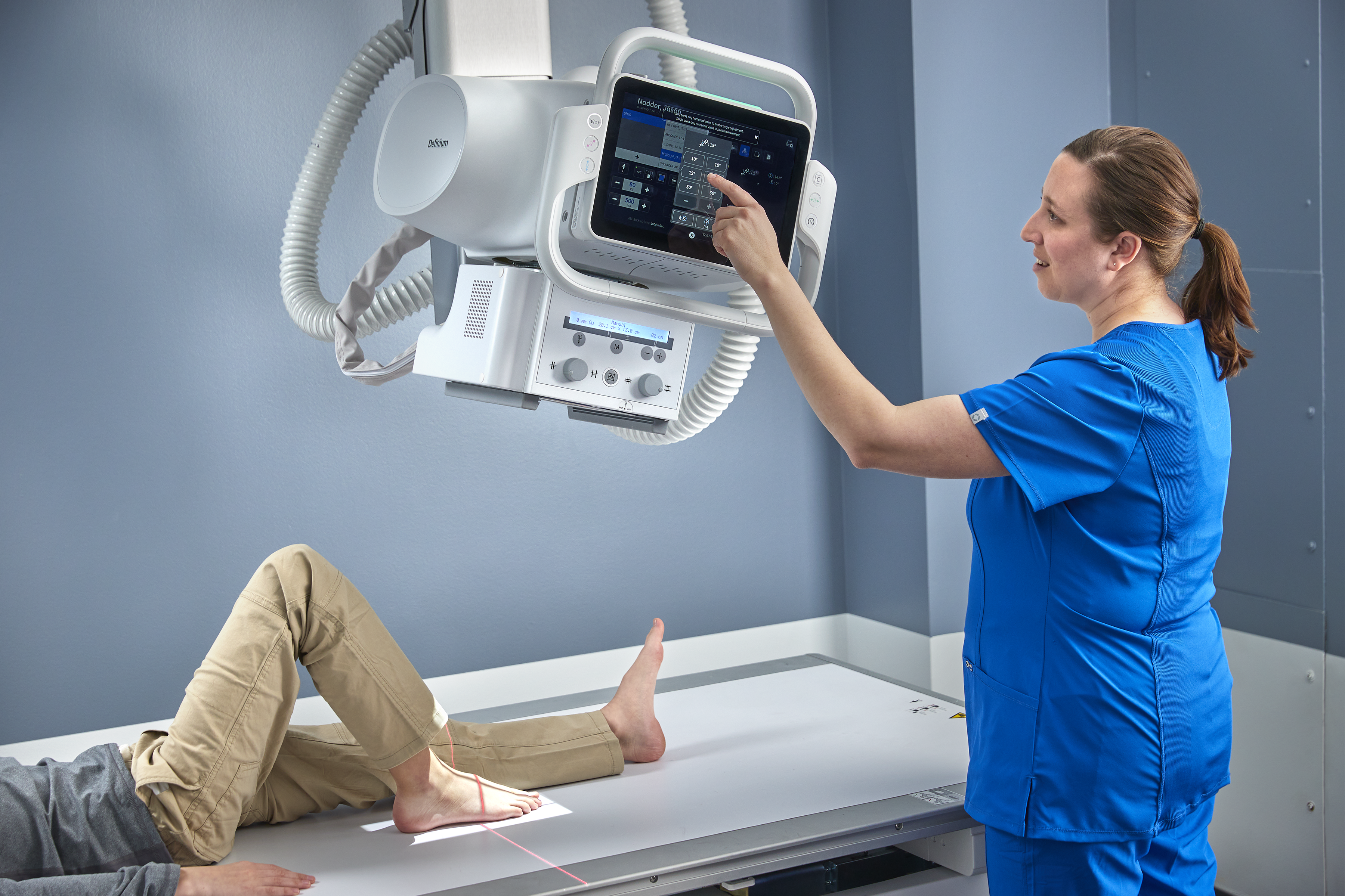
How is an x-ray done?
A plain x-ray is painless and usually takes less than 15 minutes. It can be done in a hospital or private radiology practice. X-rays are done by a radiographer or medical imaging technologist. The images are reviewed by a a specialist medical doctor called a radiologist.
Before the x-ray you may be asked to remove any metal objects or jewellery from your body. During the procedure you’ll be asked to lie, sit or stand, depending on the part of the body being x-rayed. A protective shield or apron may be used to protect parts of your body that are not being x-rayed. It is important not to move during the x-ray. You may be asked to take a deep breath and hold it while the images are taken.
eople who use a wheelchair may need to move or be moved onto a different chair to prevent the metal parts of the wheelchair affecting the image.
For children, a parent or carer can usually stay with a child during the x-ray to reassure them and help them keep still.
A radiologist will then assess the images and send a report to your doctor.
Are there risks with x-rays?
Yes. An x-ray uses a small amount of radiation to create an image. Some types of x-ray, such as CT scanning and angiography, use higher doses of radiation than plain x-rays. The amount of radiation used is unlikely to cause any serious problems, but if you are concerned you should talk to your doctor.
Generally, the benefit of the x-ray in diagnosing a health condition is greater than the risk of the radiation.
On the whole, it is wise to have x-rays that are necessary, but not ones that won’t help with treatment. For example, x-rays do not necessarily help in the diagnosis or treatment of low back pain.
For children, there may be alternatives to x-ray, such as an ultrasound, in some circumstances.
X-rays and pregnancy
In pregnant women, x-rays expose the foetus to small amounts of radiation. The dose used is so low that it is not usually a concern, however it is best to avoid exposing the mother’s abdomen to any radiation if possible.
A different test may need to be used.