Main ECG waves
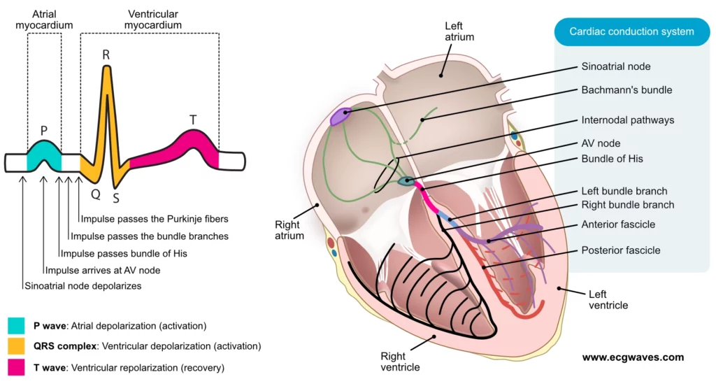
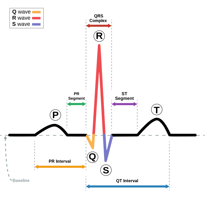
- Atrial depolarization (P) axis : it is the general direction in which atrial depolarization conducts
- Ventricular depolarization (QRS) axis : it is the general direction in which depolarization conducts
- Ventricular repolarization (T) axis : it is the general direction in which ventricular repolarization conducts
Rt : Right
Lt : Left
mv : Milli volt
aVR : Augmented Voltage on Right arm
aVL : Augmented Voltage on Left arm
aVF : Augmented Voltage on Foot
RA : Right Atrium
LA : Left Atrium
RV : Right Ventricle
LV : Left Ventricle
LI : Lead 1
LII : Lead 2
LIII : Lead 3
Definition Of Mean QRS Axis :
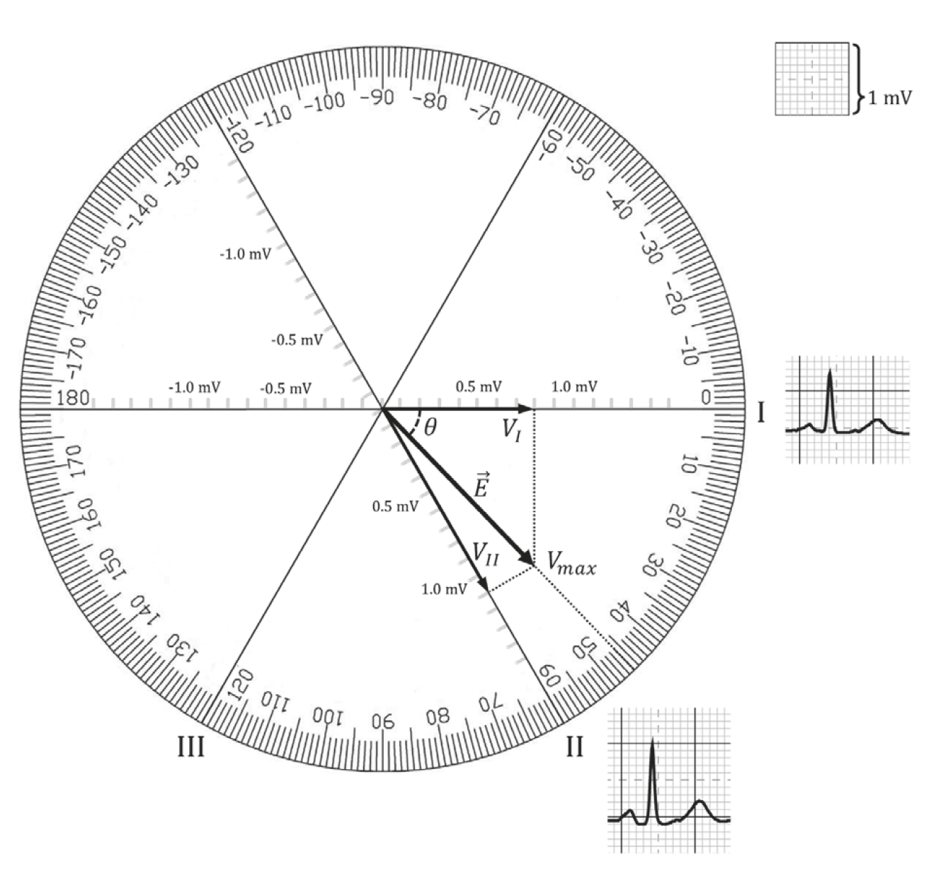
The term (mean QRS) axis describes the general direction in the frontal plane toward which the QRS complex is predominantly pointed.
- Einthoven’s triangle can easily be converted into a triaxial lead dia- gram by having the axes of the three standard limb leads (I, II, and Ill) intersect at a central point.
- Similarly the axes of the three augmented limb leads (aVR, aVL, and aVF) also form a triaxial lead diagram.
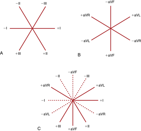
- These two triaxial lead diagrams can be combined to produce a hexaxial lead diagram, so we will be using this diagram to determine the mean QRS axis and describe axis deviation.
Some Explanation :
- Each lead has a positive and negative pole .
- As a wave of depolarization spreads toward the positive pole, an upward (positive) deflection occurs.
- As a wave spreads toward the negative pole, a downward (negative) deflection is occurs.
- Finally, a scale is needed to determine or calculate the mean QRS axis.
- By convention the positive pole of lead I is said to be at 0°.
- All points below the lead I axis are positive, and all points above that axis are negative . Thus, towards the positive pole of lead aVL (-30°), the scale becomes negative. Downward toward the positive poles of leads II, Ill, and
aVF, the scale becomes more positive (lead II at +60°, lead aVF at +90°, and lead Ill at +120°). - The completed hexaxial diagram used to measure the QRS axis.
- By convention again, an electrical axis that points toward lead aVL is termed leftward or horizontal.
- An axis that points toward leads II, III, and aVF is rightward or vertical.
How to detect Heart axis and prove axis deviation
In order to be able to determine the axis of the heart, we must understand how the ECG device determines the axis of the heart
Basically, As we described in the previous lesson ; Standard leads & Augmented leads which are 6 leads 3 for each.
We must combine these following axes into one axis
Bipolar leads ( Standard leads )
» Lead I : Rt arm → Lt arm
» Lead II : Rt arm → Lt leg ; Lead II = Lead I + Lead Ill
» Lead Ill : Lt arm → Lt leg

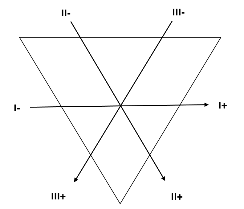
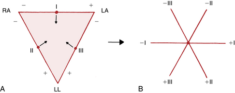
A : Einthoven’striangle.
B : The triangle is converted to a triaxial diagram by shifting leads I, II, and Ill so that they intersect at a common point
![]()
Unipolar leads ( Augmented voltage leads )
» aVF : (Rt/Lt arm) → Lt leg
» aVL : (Rt arm/Lt leg) → Lt arm
» aVR : (Lt arm/Lt leg) → Rt arm

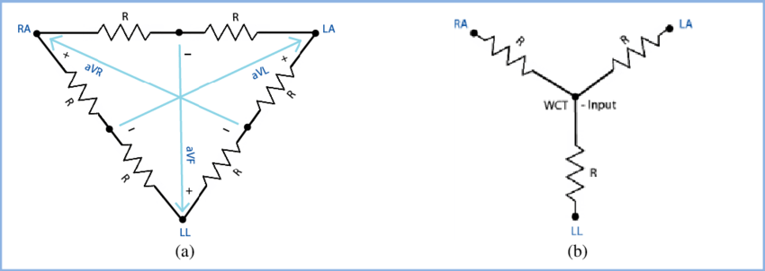

- Triaxial lead diagram showing the relation- ship of the three augmented (unipolar) leads.
- Notice that each lead is represented by an axis with a positive and negative pole.
NB : The term unipolar was used to mean that the leads record the voltage in one location relative to about zero potential, instead of relative to the voltage in one other extremity
After combination of these previous axes we can get this next Hexa-axial structure
Cardiac Hexa-axis
General rules :
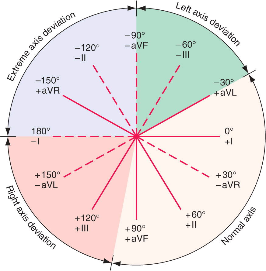
Normal axis : In the ECGs of most normal people this axis lies
between (- 30° and +90°) in some international textbooks it’s between (+30° and +75°)
Left axis deviation “LAD” : If an axis of – 30° or more negative.
Rt axis deviation : if an axis +90° or more positive.
Simple method for rapid determination of axis :
Look to the direction of QRS in In LI LII and LIII
– Normally QRS in LII is the highest
– If QRS in LI higher so → left axis deviated heart
– If QRS in LIII higher so → right axis deviated heart
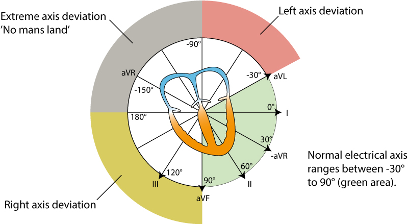
Values of detection of heart axis
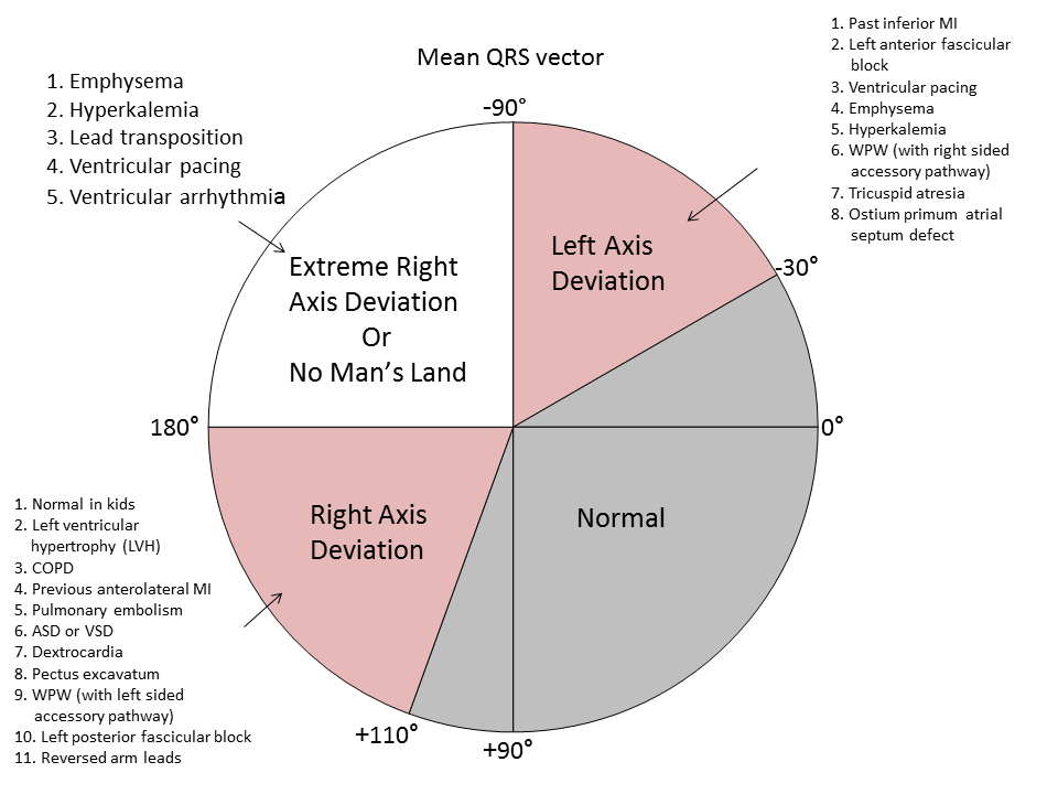
Practical work :
Method
Step 1.
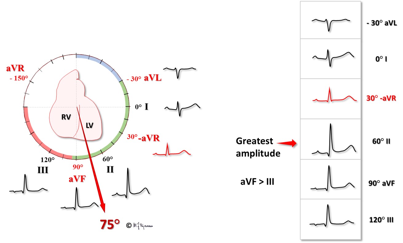
![]()
Step 2.
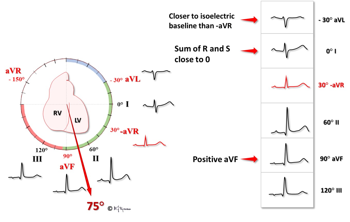
Practical tasks
Example 1.
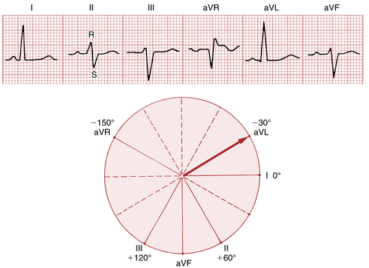
Example 2.
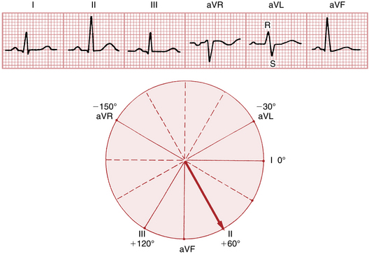
![]()
Example 3.
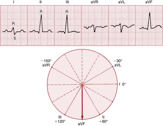
Try to train your self
Task 1.

Task 2.


