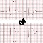
Our goal :
To detect
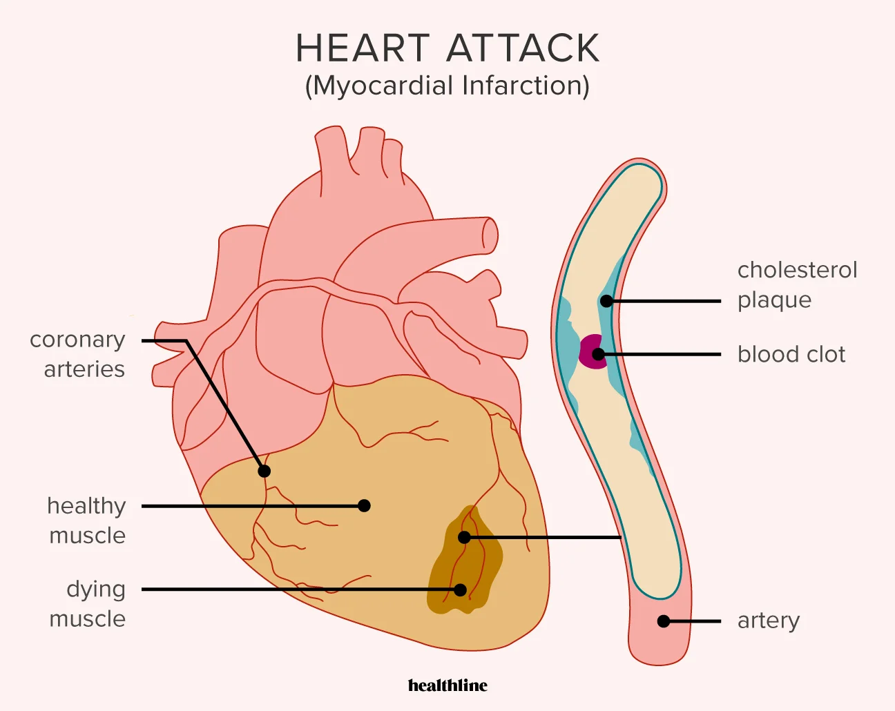
» Stage of myocardial infarction
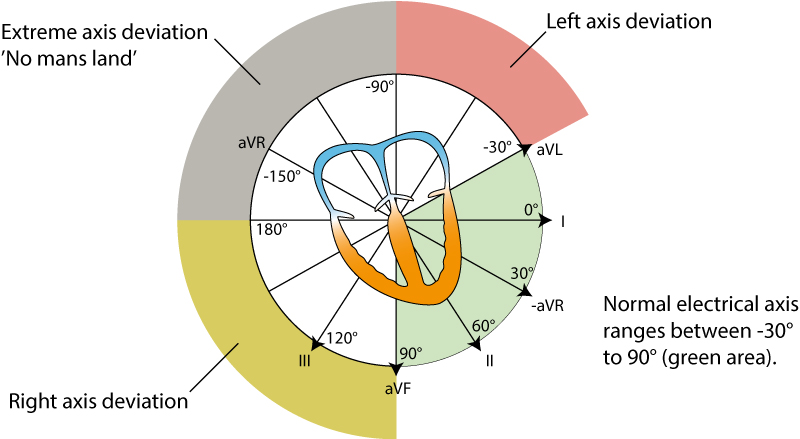
» Area of heart wall which affected
» Coronary branch which occluded
To detect stage of MI

Stages of Myocardial Infarction – [ MI ]
Pre-acute stage
Evolving period
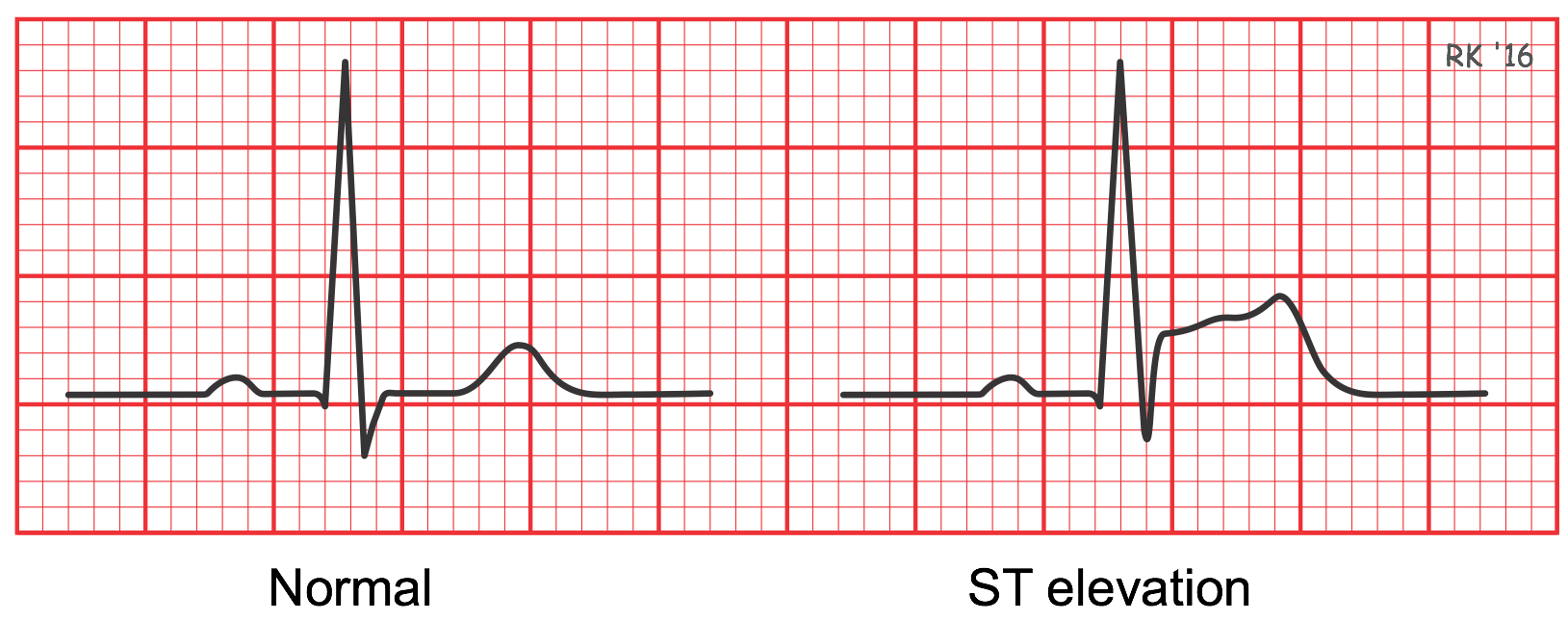
» ST elevation
Acute stage
Attack period
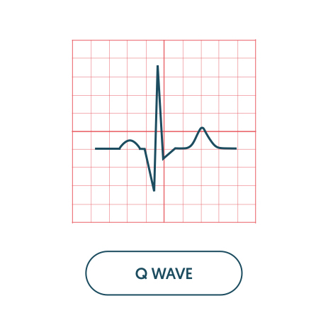
» Pathological Q formation
» ST elevation
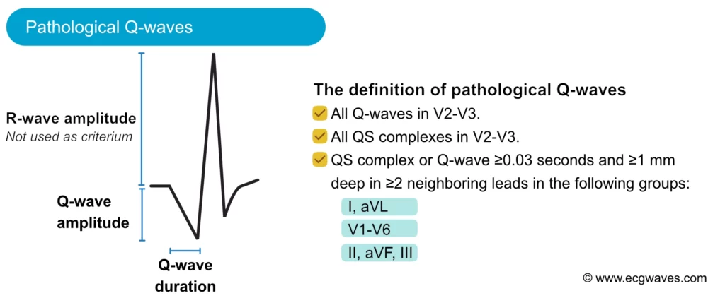
Pathological Q: depth more than ¼ of R amplitude in the same lead, its width (duration) more 0.03 sec
Sub-acute stage
Healing period
» Pathological Q
» ST at isoline
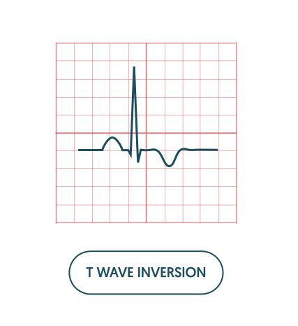
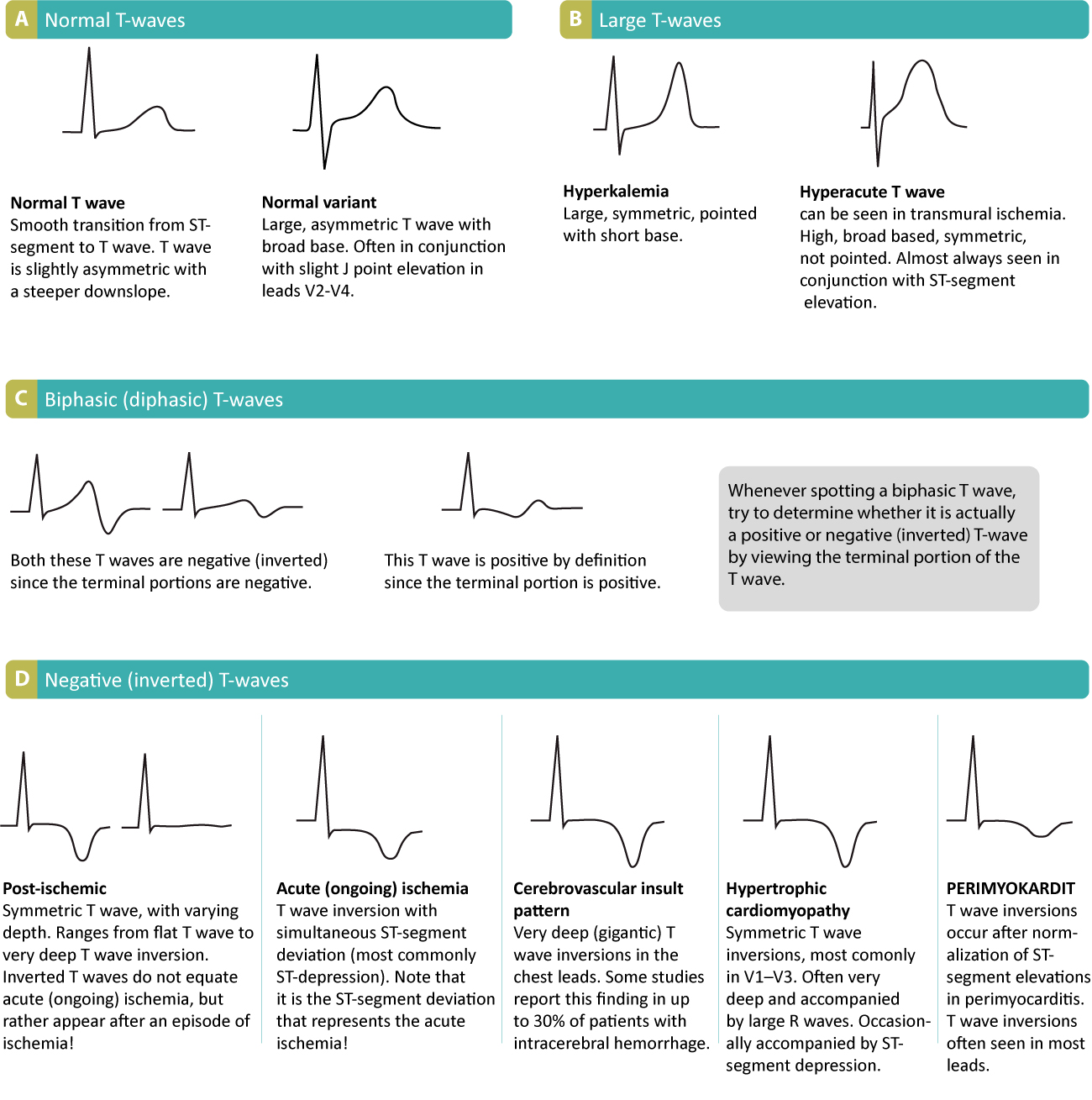
» Coronary T (negative symmetric)
Post Infarction
Healed period
» Pathological Q remains
» T becomes positive

Waves changes
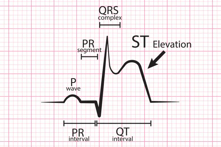
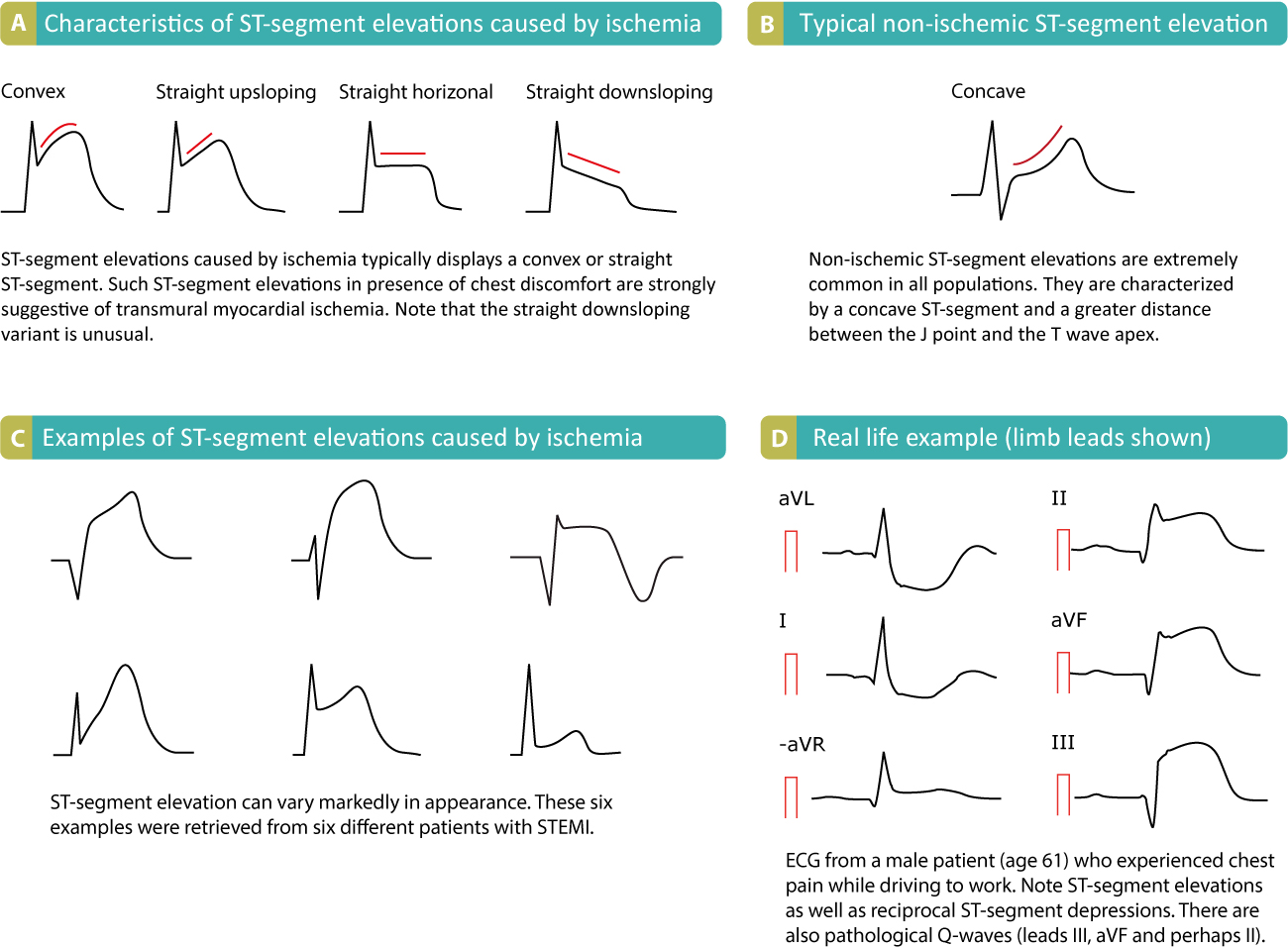
ST elevation characters
![]()
Pathological Q wave
Coronary T – Inverted T wave
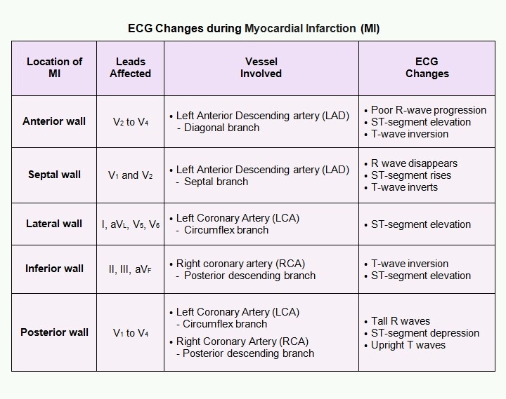
To detect area of heart wall which affected & occluded coronary branches
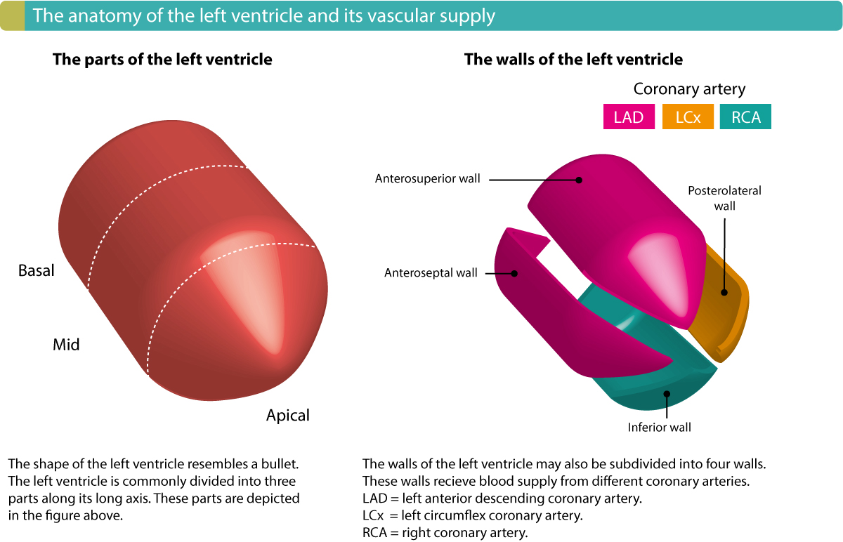

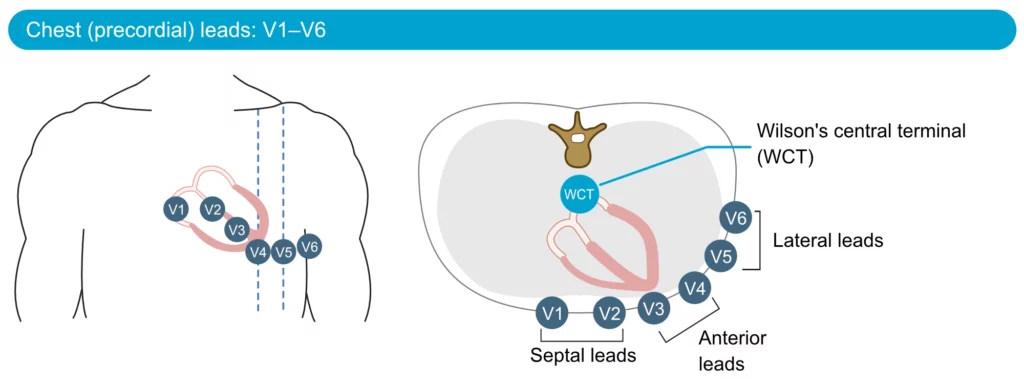
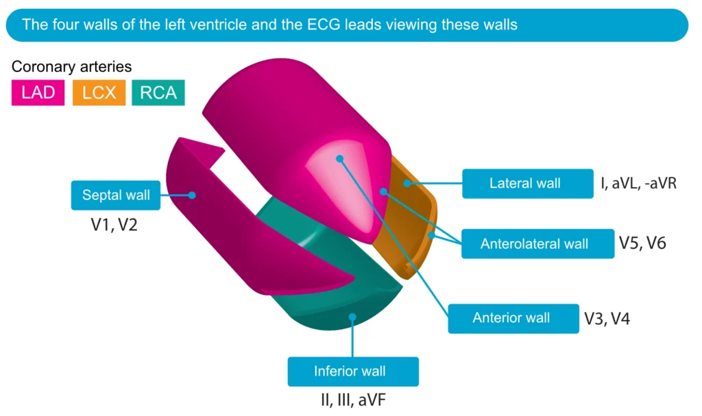
Anterior wall ischemia
Anterior wall contains septal area, lateral wall & heart apex
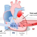
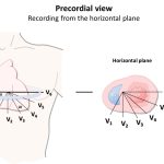
» Whole anterior wall → I, aVL, V1, V2, V3, V4, V5 & V6
» Septum → V1, V2
» Heart apex → V3, V4
» Lateral wall → V5, V6
Vessel involved :
» Whole anterior wall → Left Anterior Descending artery (LAD) [ Diagonal branch ]
» Septum →Left Anterior Descending artery (LAD) [ Septal branch ]
» Lateral wall →Left Coronary Artery (LCA) [ Circumflex branch ]
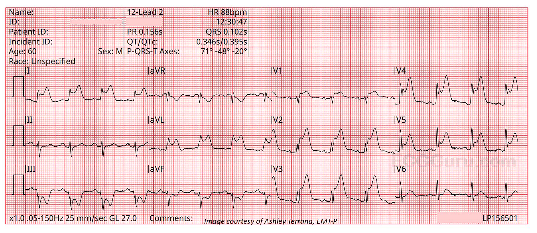
Inferior wall ischemia
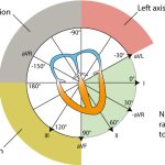
→ II, III, & aVF
→ Right Coronary artery (RCA) [ Posterior descending branch ]
![]()
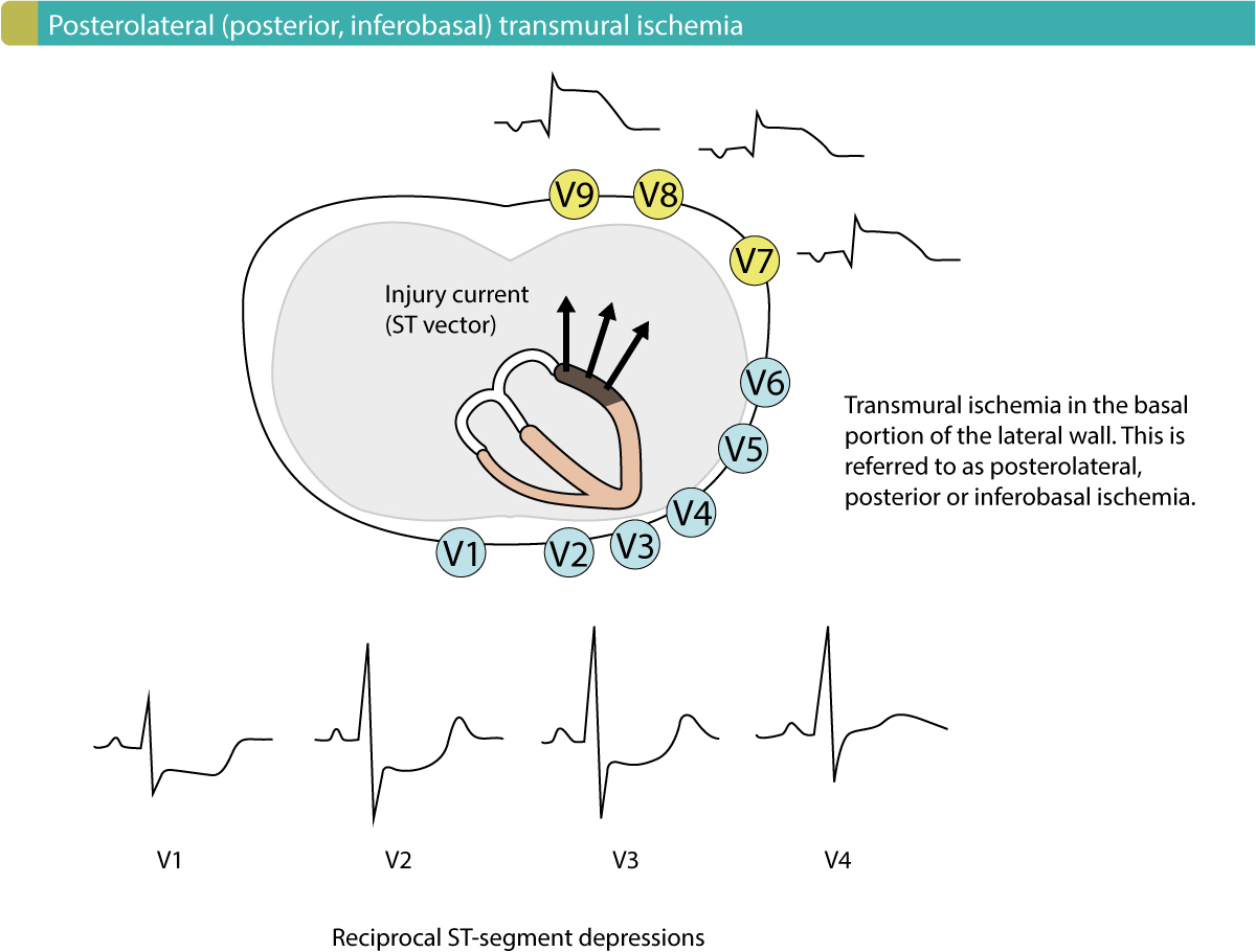
Posterior wall ischemia
» Reciprocal ST↓ → V1, V2 & V3
→ Left Coronary artery (LCA) [ Circumflex branch ]
→ Right Coronary Artery (RCA) [ Posterior descending branch ]
RA : Right Atrium
LA : Left Atrium
RV : Right Ventricle
LV : Left Ventricle
RCA : Right coronary artery
Rt : Right
Lt : Left
mv : Milli volt
MI : Myocardial Infarction
LI : Lead 1
LII : Lead 2
LIII : Lead 3
LAD : Left anterior descending coronary artery
aVR : Augmented Voltage on Right arm
aVL : Augmented Voltage on Left arm
aVF : Augmented Voltage on Foot
Lcx : Left circumflex coronary artery

