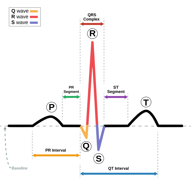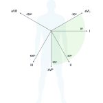
Normal ECG values :
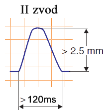
Prong P < 2.5 mm height, 0.05 – 0.11 sec. duration.
» P is positive in lead II.
» The height of P should not exceed the height of T in the same lead.
» P can be biphasic in III, aVL, V1
Atrial hypertrophy characters
Right atrium hypertrophy

Overload by pressure or volume
• Prong P is high (>2.5 mm) and pointed in II, III, aVF and low or isoelectric in the I lead
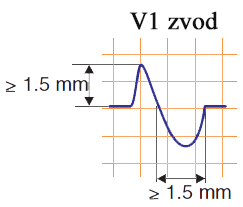
• In V1 and V2 prongs P are often biphasic with high first positive phase
(that demonstrates high electical activity of the right atrium and the deviation of its vector to the right)
• The width of P-prong remains normal
• Specific for right atrium hypertrophy P-prongs are called “P-pulmonale” (because they are usually registered


![]()
Left atrium hypertrophy

Overload by pressure or volume
• Prong P is wider (longer) than 0.12 sec.
P may be 0.14-0.16 sec
• P may be two-humped with the distance between prongs more than 0.04 sec
• These changes are more prominent in I, II, aVL, V4-V6
• Specific for left atrium hypertrophy and especially dilatation – negative or biphasic P in V1 and V2.
These changes are typical for mitral stenosis and are called “P-mitrale” (because of high end-diastolic pressure in left ventricle that causes left atrium overload)


Hypertrophy of both right and left atria
Wide and high P in II, split P in III and wide biphasic P in V1
• P may be changed in all the chest leads
• Called “P-cardiale”
Ventricular hypertrophy characters
Right ventricle hypertrophy
Direct signs
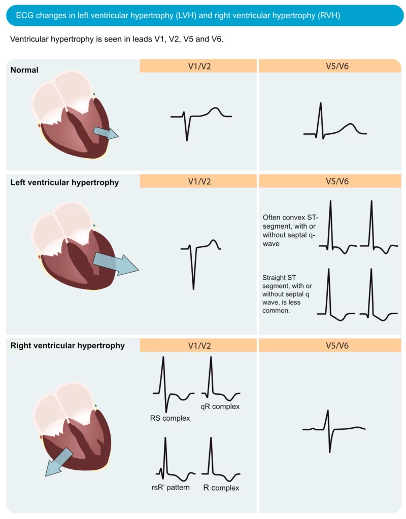
1. Increase in QRS complex voltage
• High R in III, aVF, V1 and V2
• Deep S in I and V4-6, small not deep S V1-2 (might even disappear)
• R/S V1>1, R/S V5<1
• RV1>7 mm, SV1<2 mm, RV1+SV6>10.5 mm
2. Changes of repolarization: ST segment deviation to the direction opposite to QRS:
• Deviation down of ST segment (to the direction opposite to the high R) in the leads V1, more seldom V2, III, aVF, converting to asymmetric, negative or biphasic T. Might be only negative T prong in right chest leads
• In the leads where there is deep S a slight elevation of ST may be observed
3. Impairment of repolarization:
• The change of QRS complexes form in V1 with different variants of prong R domination: rR, rsR’, RS, Rsr’, and without domination: rsr’ (very typical complex for pulmonary heart disease= cor pulmonale)
• Increase of internal deviation time (interval from the beginning of Q up to the perpendicular from the R) in V1 or V2 more than 0.035 sec – increase of right ventricle mass
Indirect signs
1. Vertical position or deviation of the electrical axis of the heart to the right (alpha angle +90 or >120)
2. Deviation of the transition zone to the left (lead V5-V6)
3. Deep S V5-V6 without increase of R in these leads

Left ventricle hypertrophy
Direct signs
1. Increase in QRS complex voltage
• High R in I, aVL, V4, V5, V6 (RV5>RV4 or even RV6>RV5)
• Deep S in III, aVF, V1 and V2
• RI >20 mm, SIII+RI >25 mm, RaVL >11-13 mm, RV5 or RV6 >25 mm, RV5+SV1 >35 mm (Sokolov-Layon criteria)
• SV3+RaVL > 28 mm for men, >20 mm for women (Cornella criteria)
2. Changes of repolarization: ST segment deviation to the direction opposite to QRS:
• Convex deviation down of ST segment, converting into asymmetric (with gentle slope and more steep climb), negative or more seldom biphasic (-+) prong T in those leads where there are high prongs R, that is in I, aVL, V5, V6 leads
• Slightly concave ST segment elevation, converting into positive prong T in leads III, aVF, V1-2 (discordant changes)
• TV1>TV6
3. Increase of internal deviation time (interval from the beginning of Q up to the perpendicular from the R or R’
Indirect signs
1. Horizontal position or deviation of the electrical axis of the heart to the left (seldom more than -30)
2. RI>RII>RIII, deep SIII
3. Deviation of the transition zone to the right (lead V2 or V2-V3)

Ventricular hypertrophy summarization
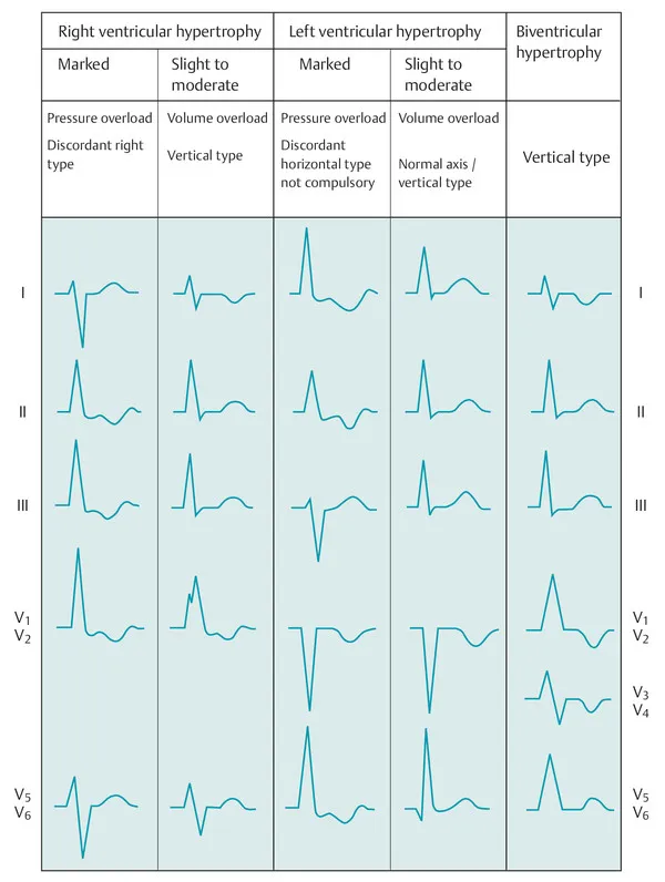
RA : Right Atrium
LA : Left Atrium
RV : Right Ventricle
LV : Left Ventricle
Rt : Right
Lt : Left
mv : Milli volt
LI : Lead 1
LII : Lead 2
LIII : Lead 3
aVR : Augmented Voltage on Right arm
aVL : Augmented Voltage on Left arm
aVF : Augmented Voltage on Foot

