Biopsy for pancreatic cancer 

0 0
Read Time:5 Minute, 11 Second

A biopsy means taking a sample of tissue and sending it to the laboratory so it can be looked at under a microscope.

There are different ways of taking biopsies to check for pancreatic cancer. But not everyone with a likely diagnosis of pancreatic cancer will have a biopsy.

Why do I need a biopsy?

The most sure way of diagnosing pancreatic cancer is by taking a biopsy and looking at it under a microscope.

Your doctor takes a biopsy by putting a needle into the area of suspected cancer.

Doctors don’t usually take biopsies from the pancreas if they think that the cancer could be removable with surgery (resectable). In that case, your likely diagnosis will be made by the doctor examining you and reviewing your scans and tests. A confirmed diagnosis can be made when the tumour is removed.

Your doctor might want to take a biopsy if you have a likely cancer that can’t be removed with surgery. Then they can find out exactly what kind of cancer it is and whether you can have treatment to slow down its growth.

Different ways of taking a biopsy

There are several different ways your doctor can take a biopsy. The type your doctor uses will depend on the position of the tumour in the pancreas.

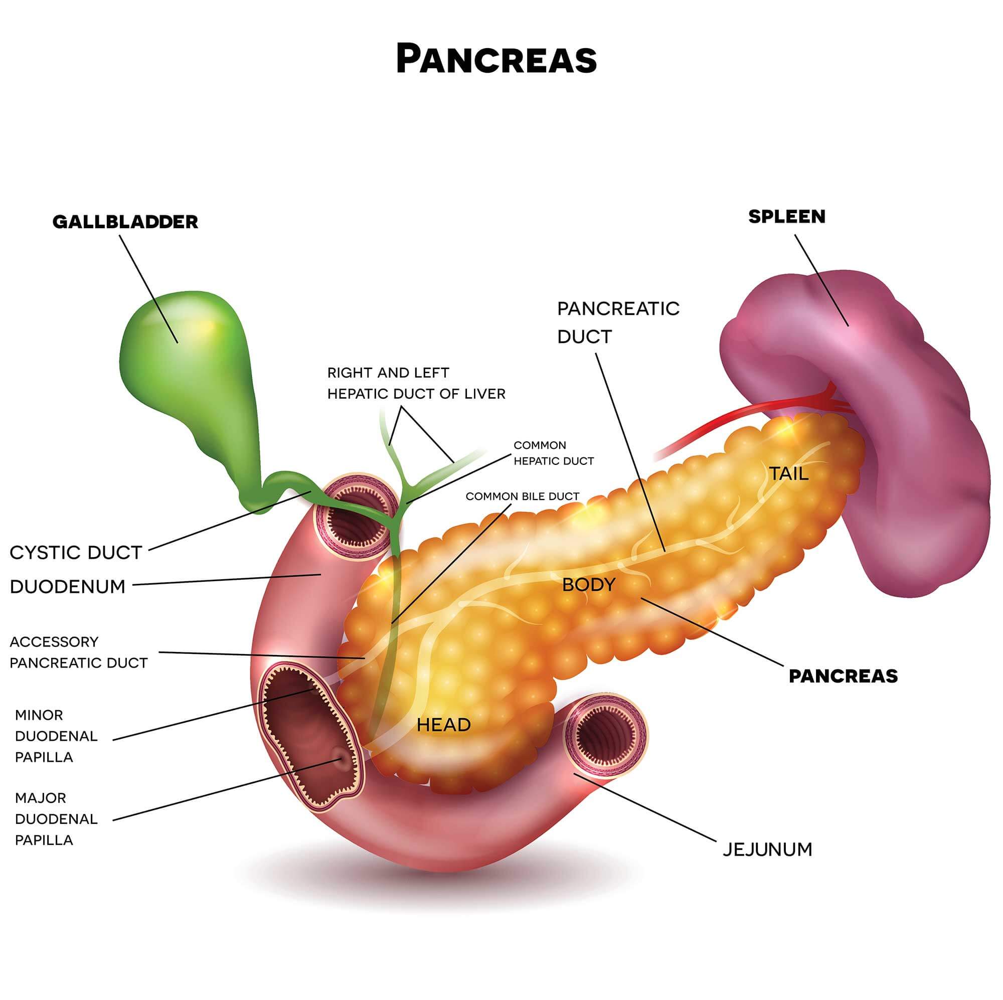
Endoscopic ultrasound combines an ultrasound scan and endoscopy Open a glossary item. It’s also called an endoluminal ultrasound or EUS.

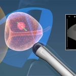
Your doctor or specialist nurse (endoscopist) does the test. They pass a flexible tube called an endoscope into your mouth and down your throat into the stomach. At the end of the endoscope is a small ultrasound probe. The ultrasound picture helps your doctor to see any areas that might be cancer. They can then guide a small needle into these areas to take the biopsy.

ERCP stands for endoscopic retrograde cholangio pancreatography. It uses an endoscopy and x-rays to show detailed pictures of the pancreas, bile ducts, and gallbladder.

Your doctor uses a long flexible tube called an endoscope that has a small camera and light at the end. They pass the tube through your mouth and then down your throat, food pipe, stomach and into the first part of your small bowel.

Your doctor can see if there are any growths or other abnormal looking areas in the pancreas or gallbladder and can take biopsies.

You might have a cholangioscopy at the same time as an ERCP. Your doctor puts a smaller tube down a channel in the endoscope. This smaller tube (cholangioscope) has a probe and camera attached. It can go directly into the bile ducts. Your doctor can take samples of any abnormal looking areas.

This way of taking a biopsy is useful for cancer that has spread to nearby parts of the body such as the liver or peritoneum Open a glossary item

Your doctor can use an ultrasound or CT scan to see exactly where the tumour or tumours are. You have a local anaesthetic Open a glossary item injection in the skin over the area so that it goes numb. Your doctor then puts a fine needle through the skin and into the tumour. They can take out a small piece of tissue.

You feel pressure when the needle goes in, but it shouldn’t be painful because of the local anaesthetic.

A laparoscopy is a small operation to look inside the tummy (abdomen). A laparoscopy involves your surgeon making several small cuts (incisions) instead of a large incision across your abdomen. This is also known as keyhole surgery or minimally invasive surgery.

You have this while your asleep, so you have a general anaesthetic. Your doctor passes a thin tube with a light and camera (laparoscope) through the incisions. They also insert some gas into your abdomen so they can see the organs more clearly.

There are several tools that your surgeon might use and this will depend on what your surgeons sees. So for example they may use an ultrasound probe to look at the deeper layers of the tissue inside your abdomen. Your surgeon can also take biopsies and fluid to send to the laboratory to check for cancer cells if they see any abnormal areas.

After this test is over, you have stitches or Steri-Strips holding the wounds closed. The cuts heal within a week or so.

A laparotomy is when your surgeon makes a cut in the skin of your tummy (abdomen) to take a tissue sample from the pancreas.

Today this is rare, and you are more likely to have a laparoscopy because it is a much smaller operation. You usually have to stay in hospital for a few days after a laparotomy.

Some surgeons send the biopsy to the laboratory while you are still under anaesthetic. If the laboratory confirms that you have pancreatic cancer your surgeon might carry on and remove the cancer.

Possible risks

Having a biopsy is generally safe but as with any medical procedure there are possible risks. These include a risk of infection or bleeding. Your doctor will go through this before you sign the consent form. They make sure the benefits of having the biopsy outweigh the possible risks.

Your nurse will tell you who to contact if you have any problems after the biopsy.

Getting your results

You should get your results within 1 or 2 weeks. 

Waiting for results can make you anxious. Ask your doctor or nurse how long it will take to get them. Contact the doctor who arranged the test if you haven’t heard anything after a couple of weeks.

You might have contact details for a specialist nurse who you can contact for information if you need to. It may help to talk to a close friend or relative about how you feel. You may want them to go with you to get the results for support.

Happy
Happy
0 %
Sad
Sad
0 %
Excited
Excited
0 %
Sleepy
Sleepy
0 %
Angry
Angry
0 %
Surprise
Surprise
0 %
Dr. Ahmed Hafez