0 0
Read Time:4 Minute, 48 Second

What is an ultrasound scan?

An ultrasound scan creates a real-time picture of the inside of the body using sound waves. Ultrasound is generally painless and non-invasive. Ultrasound works differently to x-ray in that it does not use radiation.

Ultrasound scans are performed by sending out high frequency sound waves aimed at the part of body being examined. The sound waves are emitted by a transducer, which also detects the echoes reflected back. The reflected sound waves (echoes) create a picture which can be recorded on a monitor.

What are the types of ultrasound scans?

The common types of ultrasound scan are:

  • abdominal ultrasound, which examines the internal organs of the abdomen, such as the liver, gallbladder, pancreas and spleen
  • obstetric/pregnancy ultrasound, which is a routine scan to assess the growth and health of the baby
  • female pelvis ultrasound, which may use transvaginal ultrasound (with the transducer in the vagina) or external pelvic ultrasound to look at the female pelvis, uterus, cervix, fallopian tubes and ovaries
  • breast ultrasound, which is used to assess breast symptoms such as lumps, and also to screen for breast cancer in women with dense breast tissue
  • renal ultrasound, which is used to scan the urinary tract including the kidneys and bladder
  • transrectal ultrasound, which provides images of the prostate gland
  • Doppler ultrasound, which monitors blood flow in the major arteries and veins
  • echocardiogram, which examines the heart
  • 3D ultrasound, which shows a three-dimensional picture of the inside of the body
  • 4D ultrasound, which creates a three-dimensional picture in motion

When is an ultrasound scan used?

Ultrasounds can be used for screening, diagnosis or to help with treatment. Ultrasound scans can be used to:

  • examine organs, such as the thyroid gland, the breast, the prostate and the liver
  • examine muscles, tendons and ligaments and diagnose sprains, strains, trapped nerves and muscle tears
  • examine lumps to see if they need further testing by distinguishing between fluid-filled cysts and solid lumps
  • monitor blood flow, and identify blood clots, aneurysms (bulging arteries), narrowing of arteries and decreased blood flow
  • help to guide treatments, for example by showing the correct site for an injection or to guide a biopsy needle to the correct place

Ultrasound has many uses in pregnancy, including assessing gestational age, monitoring progress and screening for complications. It is usually done at 8 – 13 weeks and 18 – 20 weeks — the first and second trimester scans. Additional ultrasound scans are only needed in certain circumstances. 3D and 4D ultrasound scans are sometimes offered in pregnancy for non-medical purposes. You should discuss having these scans with your obstetrician or GP.

Ultrasounds should only be performed when medically necessary. Speak to your doctor or specialist about whether an ultrasound is required.

What are the limitations of ultrasound?

Ultrasound is generally not good for imaging bone or tissues that are full of air, like the lungs. Ultrasound may not be as effective in people who are obese, as abdominal fat makes it harder for the sound waves to penetrate.

How do I prepare for an ultrasound scan?

To prepare for an ultrasound scan:

  • bring your referral letter and any ultrasound scan or x-ray results you have received over the past 2 years, if performed at another location
  • follow the instructions provided to you — you may be asked to fast, or to drink a lot of water and not go to the toilet before the procedure
  • leave your jewellery and valuables at home

If you have diabetes it is important that you tell the sonographer before your ultrasound. If you have any questions or concerns, contact the imaging practice for advice.

How is an ultrasound scan performed?

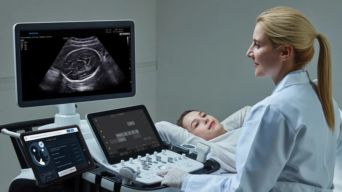
An ultrasound scan is performed using a hand-held scanner, or transducer, connected to a computer. High frequency sound waves are sent into the body. As the sound waves bounce around, the echoes are converted to electrical impulses that show a picture on a screen.

During most ultrasound scans you will be asked to lie on your back or side. Gel will be put on your skin where the scan will take place. The sonographer will move the transducer on the gel. The sonographer may need to press, but this usually does not cause any discomfort.

A woman undergoing an ultrasound scan
Ultrasounds during pregnancy are important for checking the baby’s development, checking for multiple pregnancies and picking up abnormailities

If it is recommended that you have a transvaginal ultrasound scan you will be asked to empty your bladder and undress from the waist down, with a gown or sheet to cover you. The transducer is slightly larger than a tampon. It will be covered in a protective sleeve or condom and lubricated with gel, inserted into the vagina and gently moved around.

Women can request a female sonographer to perform this type of ultrasound. There are certain situations where a transvaginal ultrasound will not be offered, for example in children, or where women may decide not to have one, in which case an external pelvic ultrasound will be performed instead.

A transrectal ultrasound requires that you have an enema beforehand. A narrow transducer, coated in gel, is inserted into the rectum from where it can take images of the prostate and surrounding tissues. This does not normally hurt.

An ultrasound scan usually takes 20 – 60 minutes. It is an outpatient procedure (you will not be admitted to hospital), performed by a specially trained doctor, radiologist or sonographer. There are no after effects and you’ll be able to go about your normal activities afterwards.

Are there any risks with ultrasound?

Because it does not involve radiation, ultrasound is very safe. Because it’s considered very safe it can be used routinely in pregnancy. Generally, ultrasound does not involve any injections.

The sound waves produced during ultrasound are beyond the threshold of human hearing, so you won’t hear them.

Happy
Happy
0 %
Sad
Sad
0 %
Excited
Excited
0 %
Sleepy
Sleepy
0 %
Angry
Angry
0 %
Surprise
Surprise
0 %
Dr. Ahmed Hafez

Average Rating

5 Star
0%
4 Star
0%
3 Star
0%
2 Star
0%
1 Star
0%

Leave a Reply

Your email address will not be published. Required fields are marked *