Clinical values of ECG
By the end of this course you will learn how to detect many diseases by ECG which is important, easy & inexpensive clinical test as following :
- Heart rate : Normal, bradycardia or tachycardia
- How to detect Heart axis and measure α angle
- Hypertrophy of both aria & ventricles on both sides right and left
- Myocardial Infarction, type, localization & stage
- Arrhythmia types, localization & stages
- Heart blocks, types & stages
- unusual syndromes of arrhythmia
Features of Time voltage chart
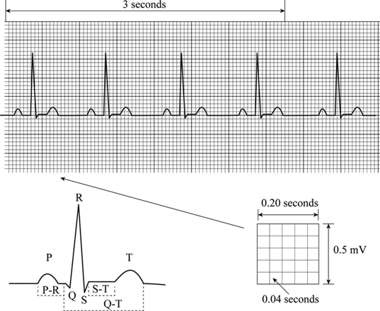
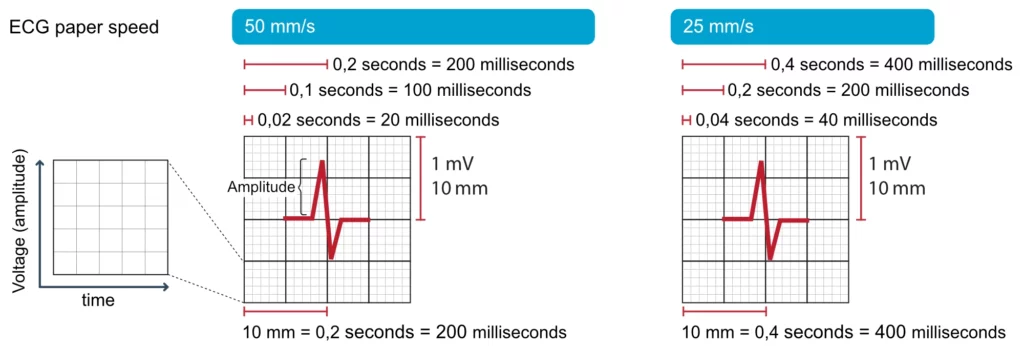
The P-QRS-T sequence is usually recorded on special ECG graph paper that is divided into grid-like boxes.
• Duration is measured on horizontal axis
• 1 small square = 0.04 sec.
• 1bigsquare = 0.2 sec.
• 5 big square = 1 sec.
• 300 big square = 1 min.
• 1500 small square= 1 min.
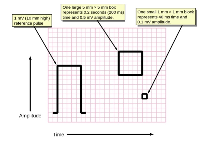
Voltage of any wave is measured on vertical axis in mm.
• 1 small square = 0.1 mv.
• 2 big squares = 1 mv {standard caliberation)
Definition
The electrocardiogram is a special graph that represents the electrical activity of the heart from one instant to the next in the form of time voltage chart, by mean of conductive electrodes
Important values :
» Normal HR : 60 – 90 beats per minute.
» Bradycardia : HR < 60 beats/min.
» Tachycardia : HR > 90 beats/min.
HR : Heart Rate
MI : Myocardial Infarction
SA node (SAN) : Sino-Atrial Node
AV node (AVN) : Atrioventricular node
Rt : Right
Lt : Left
mv : Milli volt
aVR : Augmented Voltage on Right arm
aVL : Augmented Voltage on Left arm
aVF : Augmented Voltage on Foot
RA : Right Atrium
LA : Left Atrium
RV : Right Ventricle
LV : Left Ventricle
SVC : Superior Vena Cava
Features of ECG leads
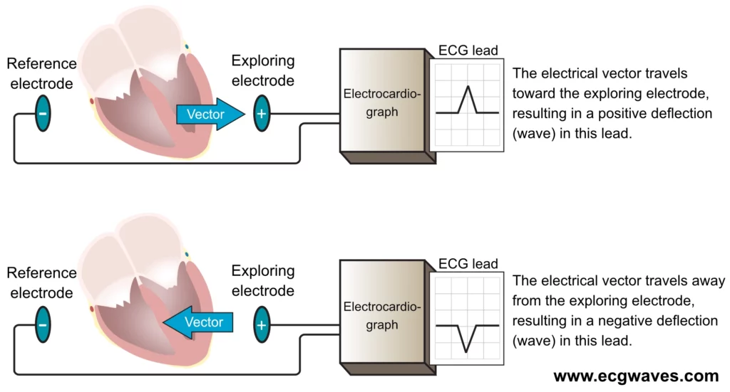
direction of its head at +ve electrode and its tail at negative electrode, it records the summation of voltage.
12 leads
6 limb leads ( coronal plane ) and 6 chest leads ( horizontal )
6 Limb leads
3 Bipolar ( Standard ) leads and 3 Unipolar ( Augmented voltage ) leads
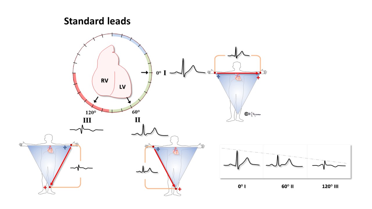
Bipolar leads ( Standard leads )
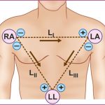
Bipolar recordings use standard limb lead configurations depicted in the figure. By convention, lead I has the positive electrode on the left arm, and the negative electrode on the right arm, and therefore measures the potential difference between the two arms. In this and the other two limb leads, an electrode on the right leg serves as a reference electrode for recording. In the lead II configuration, the positive electrode is on the left leg and the negative electrode is on the right arm. Lead III has a positive electrode on the left leg and a negative electrode on the left arm. These three bipolar leads roughly form an equilateral triangle (with the heart at the center) that is called Einthoven’s triangle in honor of Willem Einthoven, who developed the electrocardiogram in the early 1900s. Whether the limb leads are attached to the end of the limb (wrists and ankles) or at the origin of the limb (shoulder or upper thigh) makes little difference in the recording because the limbs act like a long wire conductor originating from a point on the trunk of the body.
» Lead I : Rt arm → Lt arm
» Lead II : Rt arm → Lt leg ; Lead II = Lead I + Lead Ill
» Lead Ill : Lt arm → Lt leg

![]()
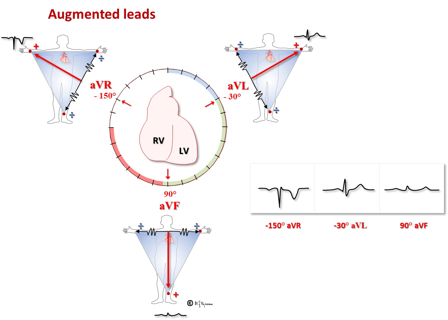
Unipolar leads ( Augmented voltage leads )
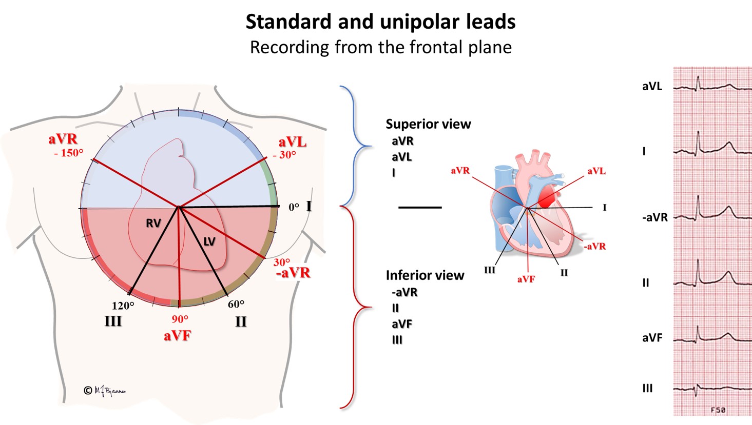
Besides the three bipolar limb leads, there are three augmented unipolar limb leads. These are termed unipolar leads because there is a single positive electrode that is referenced against a combination of the other limb electrodes. The positive electrodes for these augmented leads are on the left arm (aVL), the right arm (aVR), and the left leg (aVF). These are the same electrode locations are used for leads I, II and III. (The ECG recorder does the actual switching and rearranging of the electrode designations). The three augmented leads are depicted as shown by the axial reference system. Lead aVL is at -30° relative to the lead I axis; aVR is at -150° and aVF is at +90°. It is essential to learn which lead is associated with each axis.
» aVF : (Rt/Lt arm) → Lt leg
» aVL : (Rt arm/Lt leg) → Lt arm
» aVR : (Lt arm/Lt leg) → Rt arm

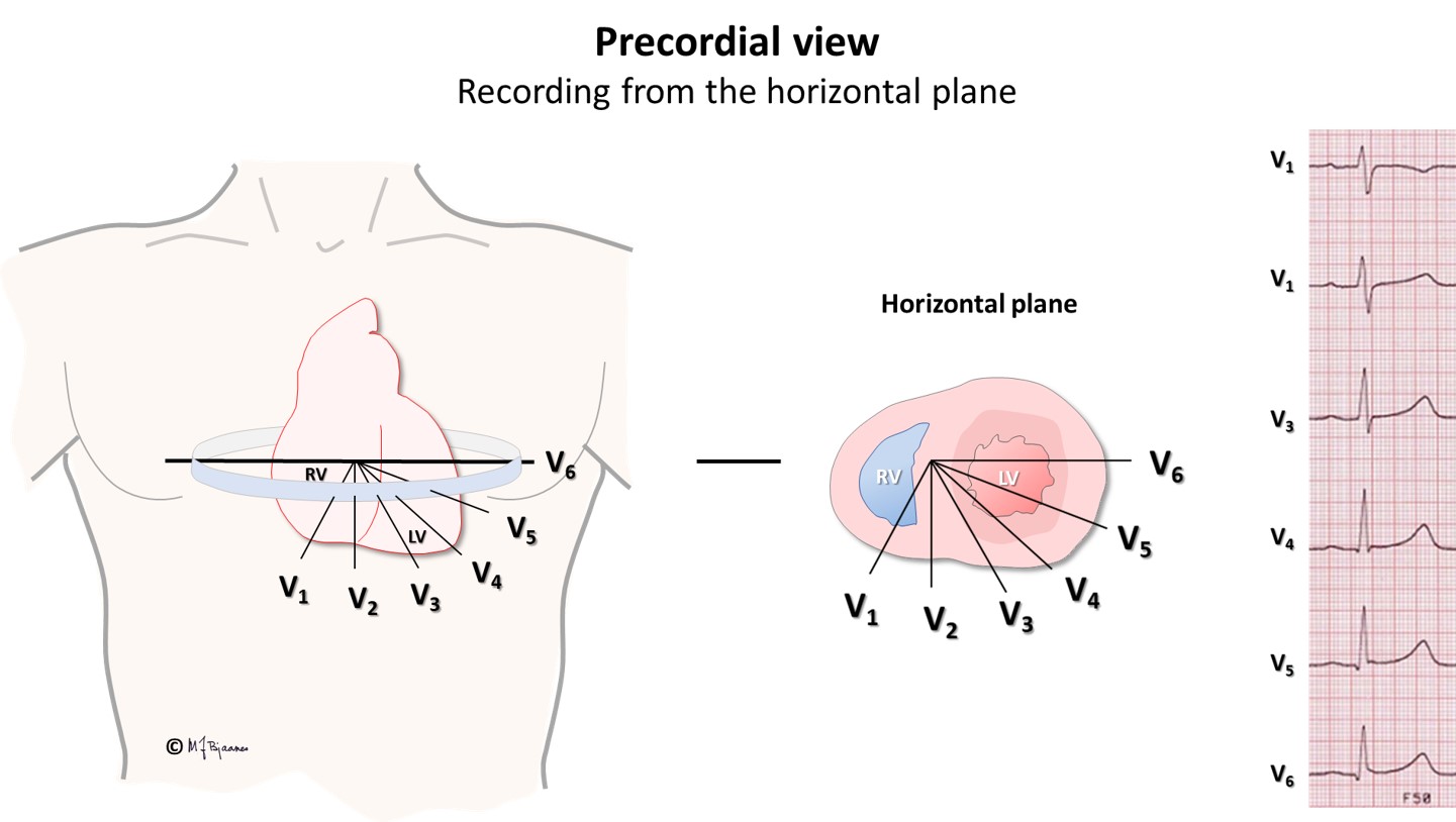
6 Chest leads ( Precordial )
Horizontal leads arranged in a horizontal circle around the heart
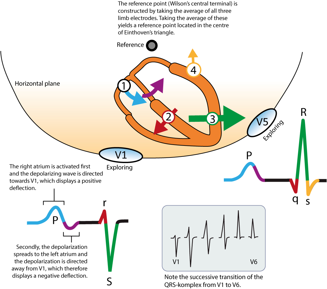
Besides the three standard limb leads and the three augmented limb leads that view the electrical activity of the heart from the frontal plane, there are six precordial, unipolar chest leads. This configuration places six positive electrodes on the surface of the chest over different regions of the heart to record electrical activity in a plane perpendicular to the frontal plane (see figure at right). These six leads are named V1 – V6. The rules of interpretation are the same as for the limb leads. For example, a wave of depolarization traveling toward a particular electrode on the chest surface will elicit a positive deflection.
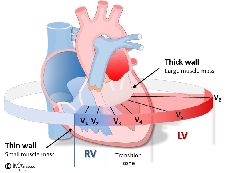
The chest leads overlie the following ventricular regions:
| Leads | Ventricular Region |
| V1-V2 | anteroseptal |
| V3-V4 | anteroapical |
| V5-V6 | anterolateral |
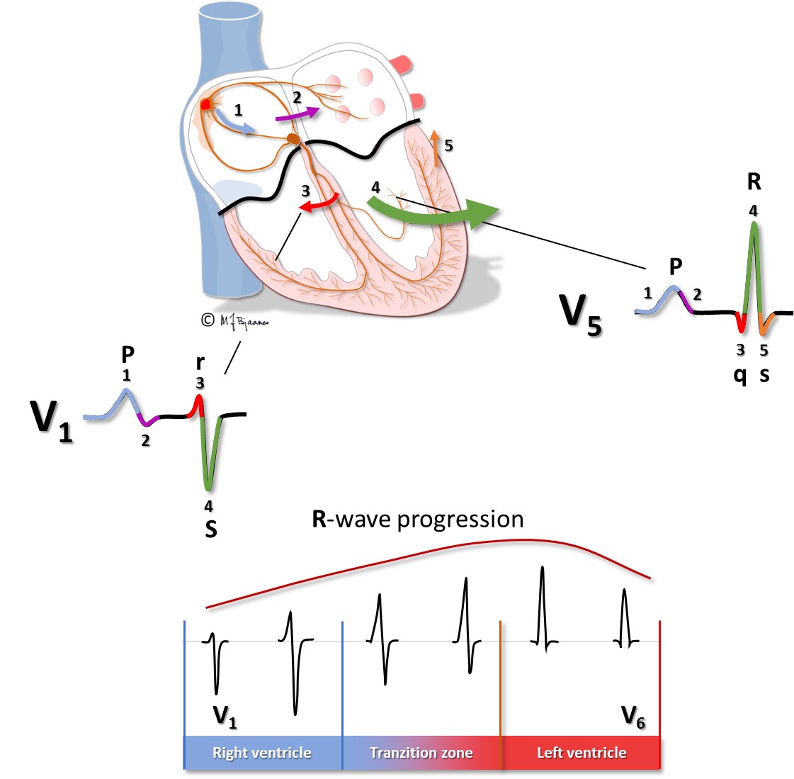
This placement of chest leads produces the following normal ECG tracings:
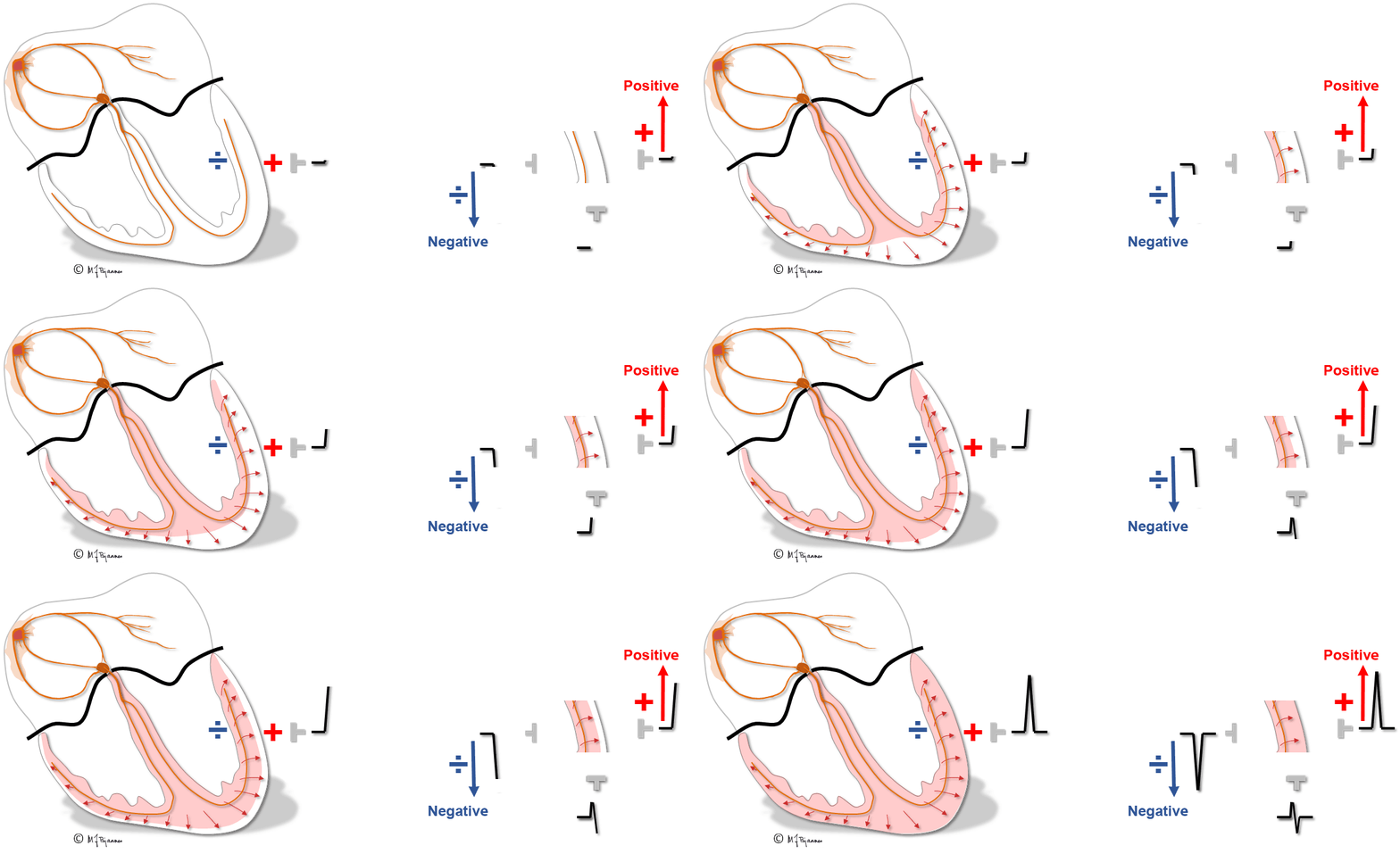
Because initial ventricular depolarization is from left to right across the septum, there is an initial R-wave in V1 followed by an S-wave as the anterior and lateral walls of the left ventricle depolarize. Leads V5and V6 have a large net positive QRS because these leads overlie the anterolateral wall of the left ventricle, which has a large muscle mass undergoing depolarization. Tracings from leads V5and V6 are almost opposite in polarity from V1because they are viewing opposite sides of the heart. Leads V2-V4 are intermediate owing to their electrode placement.
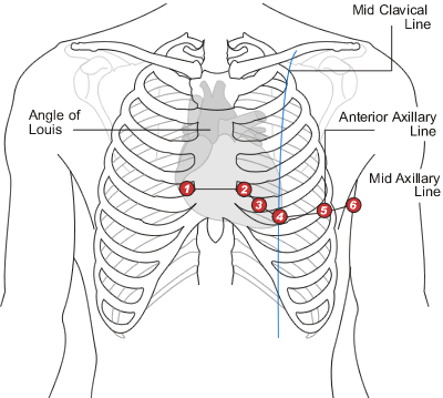
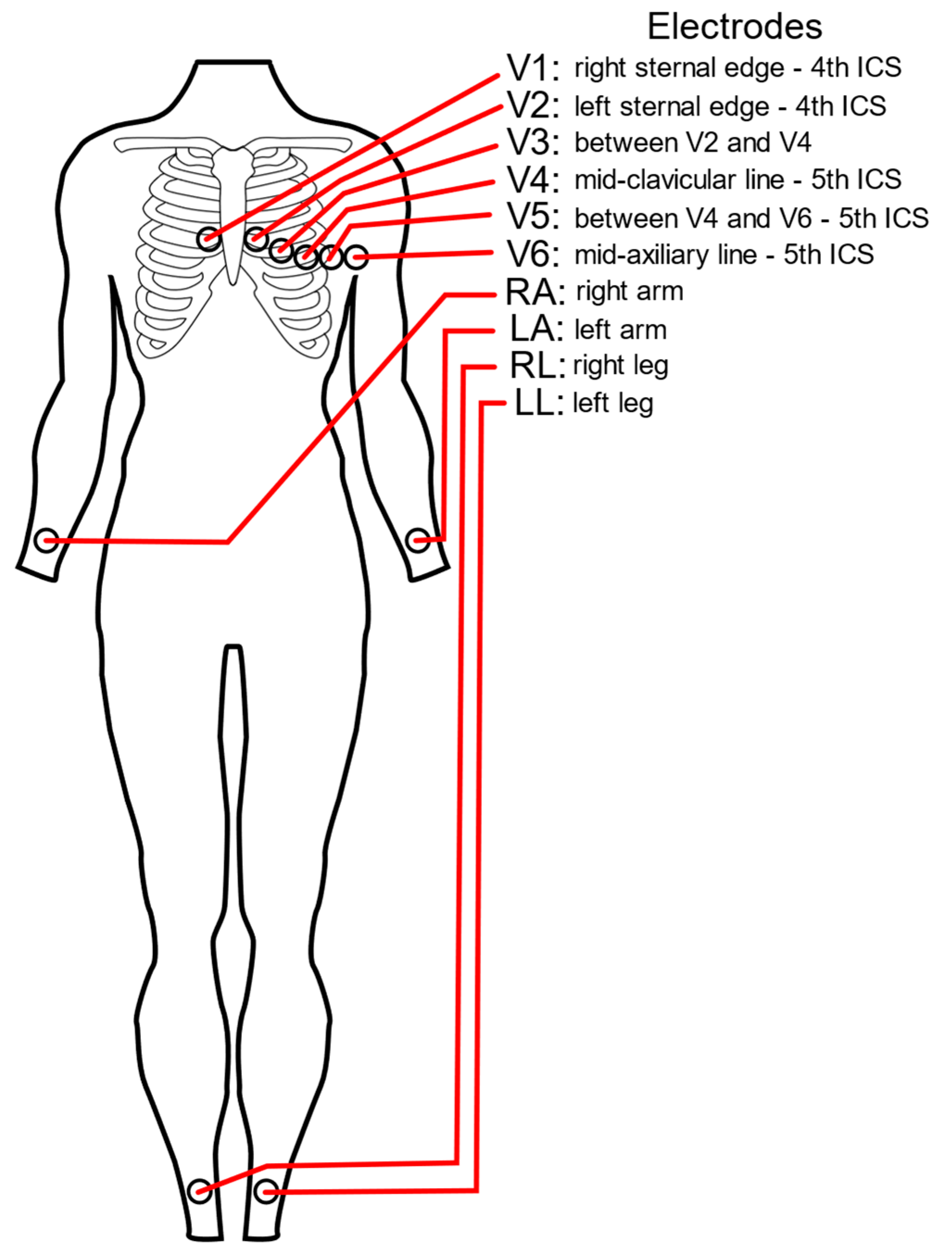
The 6 leads are labelled as “V” leads and numbered V1 to V6. They are positioned in specific positions on the rib cage. To position then accurately it is important to be able to identify the “angle of Louis”, or “sternal angle”.
Conventional placement of ECG chest leads
• Lead V1 : is recorded with the electrode in the fourth intercostal space just to the right of the sternum.
• Lead V2 : in the fourth intercostal space just left to the sternum.
• Lead V3 : in a line midway between leads V2 and V4.
• Lead V4 : in the midclavicular line in t he fifth interspace.
• Lead V5 : in the anterior axillary line at the same level as lead V4.
• Lead V6 : in the midaxillary line at the same level as lead V4.
Features of electrical activity of heart
Cardiac Automaticity
Referes to the capacity of certain cardiac cellsto function as a pace maker by spontaneously generating electrical impulses.
» Example : SA node normally is the 1ry peace maker of the heart because of its inherent automaticity that can suppress and prevent other ectopic foci (2ry pace maker) due to more rapid intrinsic rate of SAN firing.
Depolarization & repolarization of cardiac muscle cells
• When the cell is stimulated it begins to depolarize (stippled area).
• The fully depolarized cell is positively charged on the inside and negatively charged on the outside.
• Repolarization occurs when the stimulated cell returns to the resting state.
– The directions of depolarization and repolarization are represented by arrows.
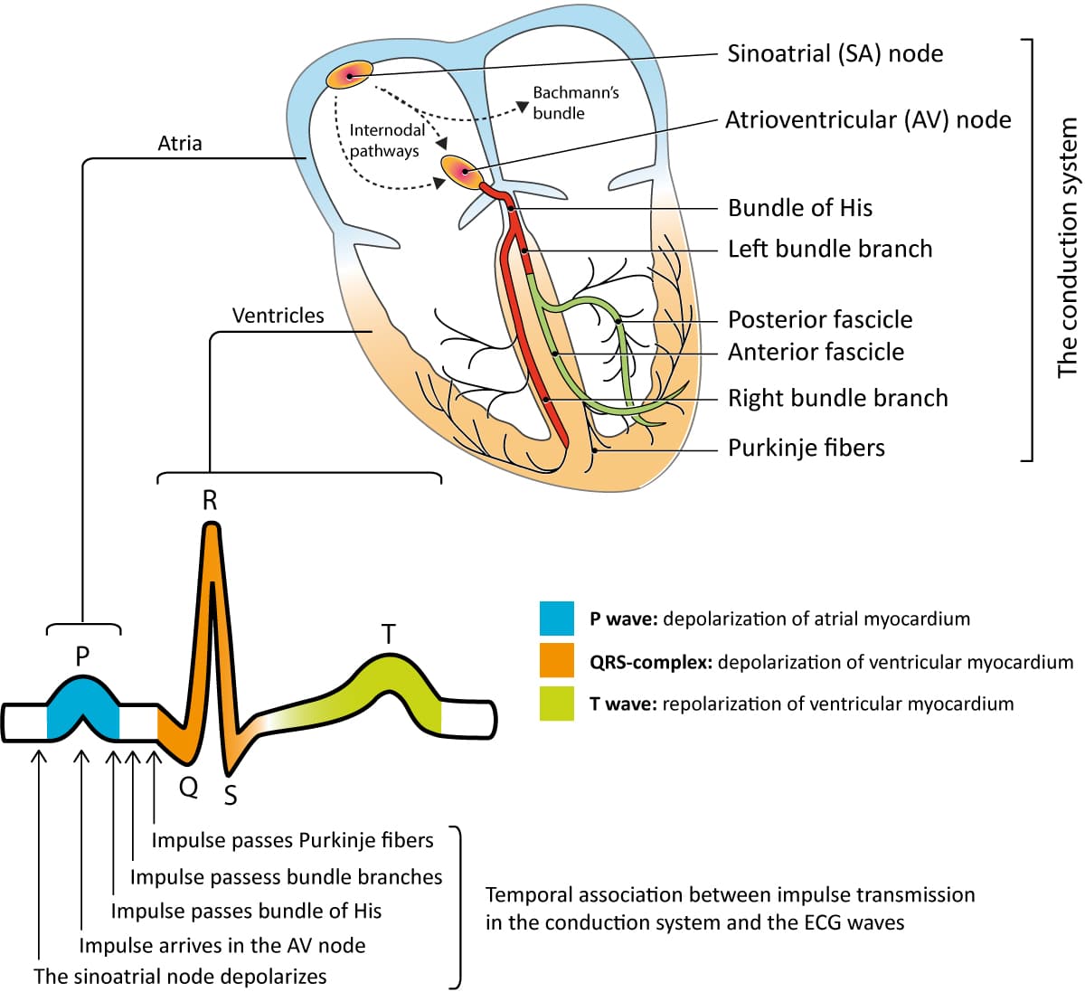
– Depolarization (stimulation) of the atria produces the P wave on the ECG, whereas depolarization of the ventricles produces the QRS complex.
– Repolarization of the ventricles produces the ST-T complex.

Cardiac Conductivity
The speed with which electrical impulses are conducted through different parts of the heart.
» Normally
– Slowest through the AV node due to anatomical labyrinthine structure and electrical depolarization by calcium influx.
– Slow AV nodal conduction allows t he ventricles time to fill with blood before the signal for cardiac cont raction arrives.
– The conduction is fastest through the Purkinje fibers
– The Rapid conduction through the His-Purkinje system ensures synchronous contraction of both ventricles.
NB : pathway of conduction
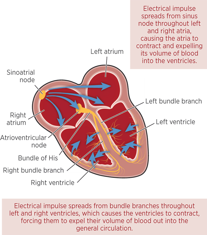
• SAN : initiates the electrical impulse.
Primary pace maker of the heart that is located at right atrium near the opening of SVC.
• Stimulus spreed through Rt atrium then t hrough inter – atrial fibers to Lt atrium.
• Then spread through AV system (AVN & AV bundle)
• Then spread to right & left bundles.
• Then to purkinji fibers (located at subendocardium) to stimulate ventricular muscle cells (from inward to outward or from endocardium to pericardium) .
