Ultrasound

Ultrasound scan of the ovaries

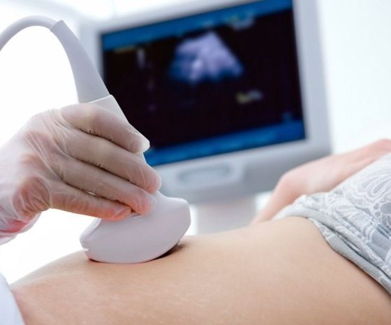
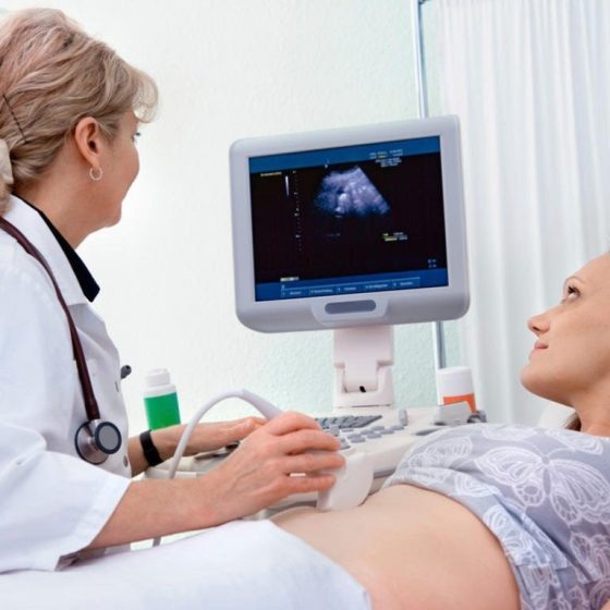
Ultrasound scans use high frequency sound waves to create a picture of a part of the body. It can show the ovaries, womb and surrounding structures. You might have this test to help diagnose ovarian cancer.  To have an ultrasound scan of the ovaries, your doctor or radiographer moves a probe over the lower part of your tummy. You may also have an internal vaginal ultrasound. This is when your doctor gently puts the probe into your vagina. This is also called a transvaginal ultrasound scan. We have separate information about what happens during a transvaginal ultrasound scan.  Why you have it

Ultrasound scan

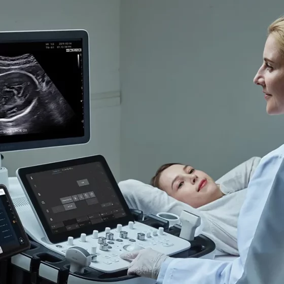
An ultrasound scan is a procedure that uses high frequency sound waves to create a picture of a part of the inside of your body. The ultrasound scanner has a probe that gives off sound waves. The probe looks a bit like a microphone. The sound waves bounce off the organs inside your body, and the probe picks them up. The probe links to a computer that turns the sound waves into a picture on the screen. Ultrasound scans aren’t painful but can cause some discomfort. You might have it at one of the following: your GP surgery local community clinic in your

Neck lymph node ultrasound and biopsy

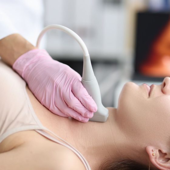
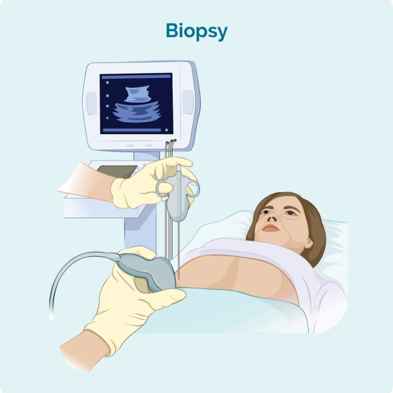
During a neck lymph node biopsy, your doctor uses an ultrasound scanner to help them take a small amount of lymph node tissue using a fine needle. Ultrasound scans use high frequency sound waves to create a picture of a part of the body. The ultrasound scanner has a probe that gives off sound waves. The sound waves bounce off the structures inside your body, and the probe picks them up. The probe links to a computer that turns the sound waves into a picture. You normally have this test as an outpatient procedure in the hospital’s imaging department. Why

Lymph node ultrasound and biopsy – under the arm (axillary) 

Cancer can sometimes spread into the lymph nodes under the arm (axilla). This can happen with different types of cancer, including breast cancer. To check the lymph nodes under your arm for breast cancer cells, you have an ultrasound scan. Ultrasound scans use high frequency sound waves to create a picture of a part of the body. The ultrasound scanner has a microphone that gives off sound waves. The sound waves bounce off the organs inside your body and are picked up by the microphone. The microphone links to a computer and turns the sound waves into a picture. If

Lymph node ultrasound and biopsy – groin

Cancer can sometimes spread into the lymph nodes in the groin. Your groin is the area at the top of your legs, between the hips. This can happen with different types of cancer, such as anal and penile cancer.  To check the lymph nodes in your groin for cancer cells, your doctor checks the groin for swollen (enlarged) lymph nodes. You then might have an ultrasound scan to check the nodes. Ultrasound scans use high frequency sound waves to create a picture of a part of the body. If doctors find any abnormal lymph nodes, they use the ultrasound to

Endoscopic ultrasound for lung cancers

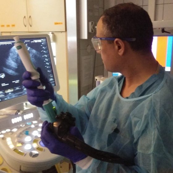
This test combines ultrasound and endoscopy. It helps your doctor to look through the wall of the food pipe at the surrounding tissue. The main airway (windpipe) is close to the food pipe. A doctor or a specialist nurse (endoscopist) does the test. To do the test the endoscopist uses a long flexible tube called an endoscope. They pass the endoscope down your throat into your food pipe. It has a tiny camera and light on the end and an ultrasound probe attached. The ultrasound scan uses high frequency sound waves to create a picture of the inside of your body. The

Breast ultrasound scan 

A breast ultrasound scan is a test that uses high frequency sound waves to create a picture of the inside of the breast.  The ultrasound scanner has a probe that gives off sound waves. The probe looks a bit like a microphone. The sound waves bounce off the tissues in your breast, and the probe picks them up. The probe links to a computer that turns the sound waves into a picture on the screen. A specialist healthcare professional called a sonographer usually does the test.  When you might have a breast ultrasound scan You might have a breast ultrasound: as a first

Ultrasound

What is an ultrasound scan? An ultrasound scan creates a real-time picture of the inside of the body using sound waves. Ultrasound is generally painless and non-invasive. Ultrasound works differently to x-ray in that it does not use radiation. Ultrasound scans are performed by sending out high frequency sound waves aimed at the part of body being examined. The sound waves are emitted by a transducer, which also detects the echoes reflected back. The reflected sound waves (echoes) create a picture which can be recorded on a monitor. What are the types of ultrasound scans? The common types of ultrasound