scan

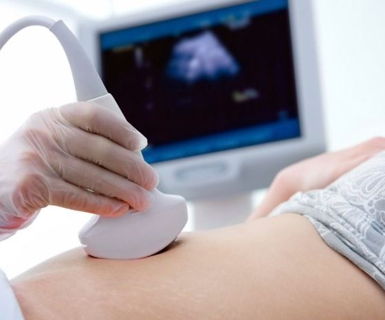
Ultrasound scan of the ovaries

Ultrasound scans use high frequency sound waves to create a picture of a part of the body. It can show the ovaries, womb and surrounding structures. You might have this test to help diagnose ovarian cancer.  To have an ultrasound scan of the ovaries, your doctor or radiographer moves a probe over the lower part of your tummy. You may also have an internal vaginal ultrasound. This is when your doctor gently puts the probe into your vagina. This is also called a transvaginal ultrasound scan. We have separate information about what happens during a transvaginal ultrasound scan.  Why you have it

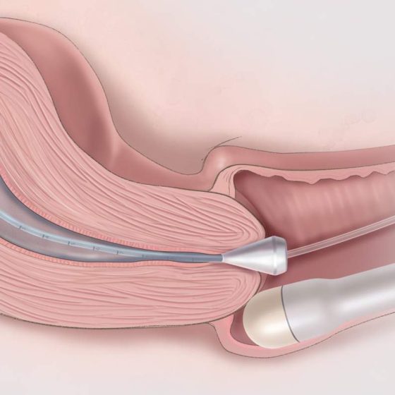
Transvaginal ultrasound scan

An ultrasound scan is a procedure that uses high frequency sound waves to create a picture of a part of the inside of your body. The ultrasound scanner has a probe that gives off sound waves. The probe looks a bit like a microphone. The sound waves bounce off the organs inside your body, and the probe picks them up. The probe links to a computer that turns the sound waves into a picture on the screen. Ultrasound scans aren’t painful but can cause some discomfort. You might have it at one of the following: your GP surgery local community clinic in your

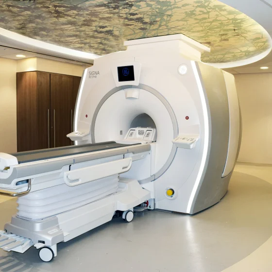
PET-MRI scan

A PET-MRI scan combines a PET scan and an MRI scan into one to give detailed information about your cancer.  PET stands for positron emission tomography. It uses a mildly radioactive liquid (radioactive tracer) to show up areas of your body where cells are more active than normal.  MRI stands for magnetic resonance imaging. It uses magnetism and radio waves to create cross section pictures of the body. It shows up soft tissues very clearly. You usually have a PET-MRI scan in the nuclear medicine or radiology department as an outpatient. These scanners tend to be only in the major

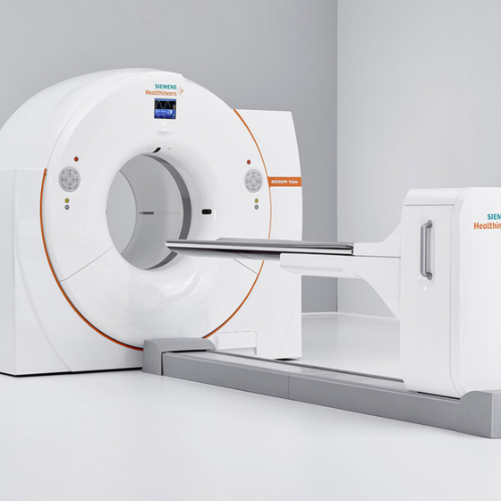
PET-CT scan

A PET-CT scan combines a CT scan and a PET scan. It gives detailed information about your cancer. The CT scan takes a series of x-rays from all around your body and puts them together to create a 3 dimensional (3D) picture. The PET scan uses a mildly radioactive liquid (radioactive tracer) to show up areas of your body where cells are more active than normal.  You usually have a PET-CT scan in the radiology department as an outpatient. A radiographer operates the scanner. It usually takes between 30 and 60 minutes. These scanners tend to be available only in the major cancer hospitals. So you

PET scan

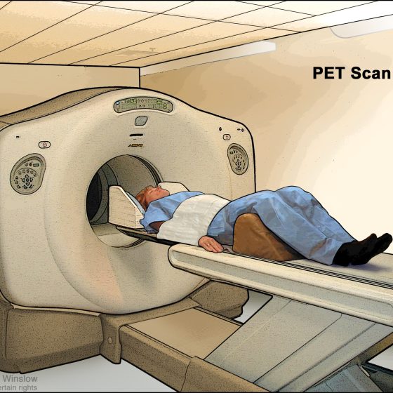
A PET scan is a test that creates 3 dimensional (3D) pictures of the inside of your body. PET stands for positron emission tomography. The PET scan uses a mildly radioactive liquid (radioactive tracer) to show up areas of your body where cells are more active than normal. It’s used to help diagnose some conditions, including cancer. It can also help to find out where and whether cancer has spread.    You usually have a PET scan in the radiology or nuclear medicine department as an outpatient. These scanners tend to be only in the major cancer hospitals. So you might have to

Multiparametric MRI (mpMRI) scan for prostate cancer

MRI stands for magnetic resonance imaging. It is a type of scan that creates pictures using magnetism and radio waves. MRI scans produce pictures from angles all around the body and shows up soft tissues very clearly.  The multiparametric MRI (mpMRI) is a special type of MRI scan that produces a more detailed picture of your prostate gland than a standard MRI scan does. You usually have an mpMRI scan if doctors suspect that you have prostate cancer. Why you might have an mpMRI It is important to know that an mpMRI scan alone cannot diagnose prostate cancer. But it can

MRI scan

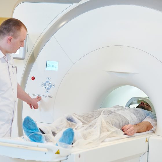
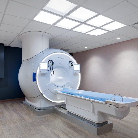
MRI (magnetic resonance imaging) is a type of scan that uses magnetism and radio waves to take pictures of inside the body. The scan takes between 15 and 90 minutes. You might have one to find out whether you have cancer and if you do to measure how big it is and whether it has spread. You might also have one to see how well treatment is working. It is a safe test. If you have an injection of contrast dye it can cause a headache, dizziness or a warm flushed feeling An MRI is a type of scan that creates

Breast ultrasound scan 

A breast ultrasound scan is a test that uses high frequency sound waves to create a picture of the inside of the breast.  The ultrasound scanner has a probe that gives off sound waves. The probe looks a bit like a microphone. The sound waves bounce off the tissues in your breast, and the probe picks them up. The probe links to a computer that turns the sound waves into a picture on the screen. A specialist healthcare professional called a sonographer usually does the test.  When you might have a breast ultrasound scan You might have a breast ultrasound: as a first

Bone scan

A bone scan looks for changes in your bones. Before the test you have a radioactive tracer injection into your bloodstream. You have this through a tube (cannula) into a vein in your hand. It takes 2 to 3 hours for the tracer to go around the body. To have the scan you lie down on a couch while the scanner takes pictures. The scan takes 30 minutes to an hour. It is painless and you can go home after the scan. The body gets rid of the tracer through your urine over the next day. A bone scan shows

Bone density scan (DEXA, DXA)

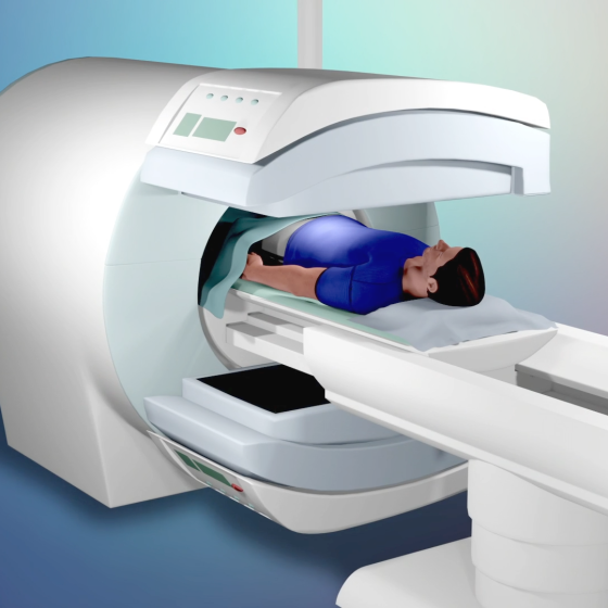
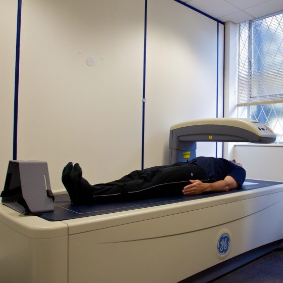
DEXA scan stands for dual energy x-ray absorptiometry. It’s also called a bone densitometry scan. It uses low dose x-rays to take measurements to work out the strength (density) of your bones. You have a DEXA scan in the x-ray (radiology) or nuclear medicine department of the hospital. A radiographer does the scan. It takes about 10 – 20 minutes. You’re not enclosed in a small place as the machine has an arm that hangs over the scanning couch. Why might you have a DEXA scan? You might have this scan if you’re having or have had cancer treatment that