lymph node

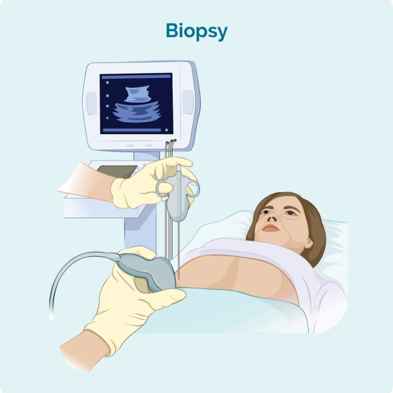
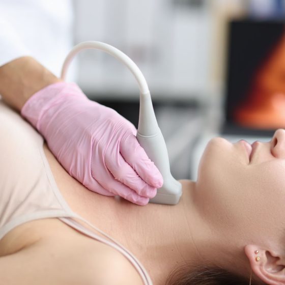
Neck lymph node ultrasound and biopsy

During a neck lymph node biopsy, your doctor uses an ultrasound scanner to help them take a small amount of lymph node tissue using a fine needle. Ultrasound scans use high frequency sound waves to create a picture of a part of the body. The ultrasound scanner has a probe that gives off sound waves. The sound waves bounce off the structures inside your body, and the probe picks them up. The probe links to a computer that turns the sound waves into a picture. You normally have this test as an outpatient procedure in the hospital’s imaging department. Why

Lymph node ultrasound and biopsy – under the arm (axillary) 

Cancer can sometimes spread into the lymph nodes under the arm (axilla). This can happen with different types of cancer, including breast cancer. To check the lymph nodes under your arm for breast cancer cells, you have an ultrasound scan. Ultrasound scans use high frequency sound waves to create a picture of a part of the body. The ultrasound scanner has a microphone that gives off sound waves. The sound waves bounce off the organs inside your body and are picked up by the microphone. The microphone links to a computer and turns the sound waves into a picture. If

Lymph node ultrasound and biopsy – groin

Cancer can sometimes spread into the lymph nodes in the groin. Your groin is the area at the top of your legs, between the hips. This can happen with different types of cancer, such as anal and penile cancer.  To check the lymph nodes in your groin for cancer cells, your doctor checks the groin for swollen (enlarged) lymph nodes. You then might have an ultrasound scan to check the nodes. Ultrasound scans use high frequency sound waves to create a picture of a part of the body. If doctors find any abnormal lymph nodes, they use the ultrasound to