center

Hypovolemic Shock

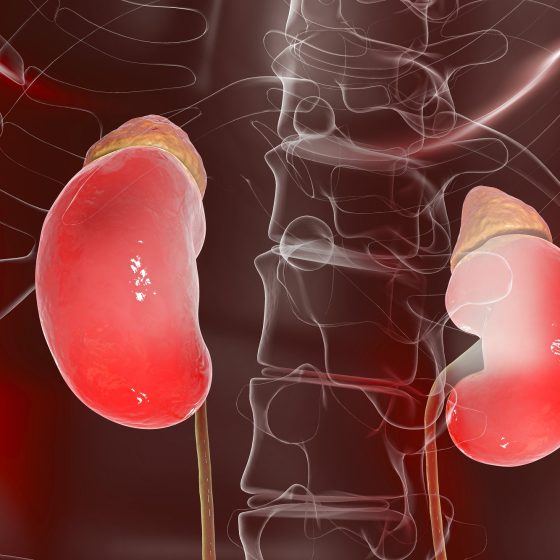
Background Hypovolemic shock refers to a medical or surgical condition in which rapid fluid loss results in multiple organ failure due to inadequate circulating volume and subsequent inadequate perfusion. Endothelium plays a critical role in vascular physiological, pathophysiological, and reparative processes. The functions of the endothelium are highly altered following hypovolemic shock due to ischemia of the endothelial cells and by reperfusion due to resuscitation with fluids. Due to oxygen deprivation, endothelial cell apoptosis is induced following hypovolemic shock.  Most often, hypovolemic shock is secondary to rapid blood loss (hemorrhagic shock). Acute external blood loss secondary to penetrating trauma and

Hypoglycemic coma

Practice Essentials Hypoglycemia is characterized by a reduction in plasma glucose concentration to a level that may induce symptoms or signs such as altered mental status and/or sympathetic nervous system stimulation. This condition typically arises from abnormalities in the mechanisms involved in glucose homeostasis. The most common cause of hypoglycemia in patients with diabetes is injecting a shot of insulin and skipping a meal or overdosing insulin.  The image below depicts a diagnostic algorithm for hypoglycemia. Signs and symptoms of hypoglycemia The glucose level at which an individual becomes symptomatic is highly variable (threshold generally at < 50 mg/dL). Carefully

Metabolic Acidosis Coma

Practice Essentials Metabolic acidosis is a clinical disturbance characterized by an increase in plasma acidity. Metabolic acidosis should be considered a sign of an underlying disease process. Identification of this underlying condition is essential to initiate appropriate therapy.  The initial therapeutic goal for patients with severe acidemia is to raise the systemic pH above 7.1-7.2, a level at which dysrhythmias become less likely and cardiac contractility and responsiveness to catecholamines will be restored. This article discusses the differential diagnosis of metabolic acidosis and presents a scheme for identifying the underlying cause of acidosis by using laboratory tests that are available

Hyperosmolar Hyperglycemic State HHS Coma

Hyperosmolar hyperglycemic state is a life-threatening emergency manifested by marked elevation of blood glucose and hyperosmolarity with little or no ketosis. Although there are multiple precipitating causes, underlying infections are the most common. Other causes include certain medications, nonadherence to therapy, undiagnosed diabetes mellitus, substance abuse, and coexisting disease. In children and adolescents, hyperosmolar hyperglycemic state is often present when type 2 diabetes is diagnosed. Physical findings include profound dehydration and neurologic symptoms ranging from lethargy to coma. Treatment begins with intensive monitoring of the patient and laboratory values, especially glucose, sodium, and potassium levels. Vigorous correction of dehydration is

Cerebral coma

What Is a Coma? A coma is a prolonged state of unconsciousness. During a coma, a person is unresponsive to their environment. The person is alive and looks like they are sleeping. However, unlike in a deep sleep, the person cannot be awakened by any stimulation, including pain. A coma is a prolonged state of unconsciousness. It is caused by an injury to the brain that can be due to increased pressure, bleeding, loss of oxygen, or buildup of toxins. The injury can be temporary and reversible but can also be permanent and lead to death. Coma Causes A coma

Cardiogenic shock

Cardiogenic shock is a medical emergency resulting from inadequate blood flow to the body’s organs due to the dysfunction of the heart. Signs of inadequate blood flow include low urine production (<30 mL/hour), cool arms and legs, and decreased level of consciousness. People may also have a severely low blood pressure and heart rate. Causes of cardiogenic shock include cardiomyopathic, arrhythmic, and mechanical. Cardiogenic shock is most commonly precipitated by a heart attack. Treatment of cardiogenic shock depends on the cause with the initial goals to improve blood flow to the body. If cardiogenic shock is due to a heart

Uremic Encephalopathy

Practice Essentials Uremic encephalopathy is an organic brain disorder. It develops in patients with acute kidney injury or chronic kidney disease, usually when the estimated glomerular filtration rate (eGFR) falls and remains below 15 mL/min.  Manifestations of this syndrome vary from mild symptoms (eg, lassitude, fatigue) to severe signs (eg, seizures, coma). Severity and progression depend on the rate of decline in kidney function; thus, symptoms are usually worse in patients with acute kidney injury. Prompt identification of uremia as the cause of encephalopathy is essential because symptoms are readily reversible following initiation of dialysis. Pathophysiology Uremic encephalopathy has a

Pneumothorax coma

Practice Essentials Pneumothorax is defined as the presence of air or gas in the pleural cavity (ie, the potential space between the visceral and parietal pleura of the lung), which can impair oxygenation and/or ventilation. The clinical results are dependent on the degree of collapse of the lung on the affected side. If the pneumothorax is significant, it can cause a shift of the mediastinum and compromise hemodynamic stability. Air can enter the intrapleural space through a communication from the chest wall (ie, trauma) or through the lung parenchyma across the visceral pleura. See the image below. Signs and symptoms

Kaposi’s sarcoma 

Kaposi’s sarcoma (KS) is a rare type of soft tissue sarcoma. A soft tissue sarcoma is a type of cancer. What is Kaposi’s sarcoma? Kaposi’s sarcoma (KS) develops from cells called endothelial cells, which line the blood and lymph vessels. It is most commonly found on the skin, but it can also affect the inside of the body, such as the lymph nodes, lungs, bowel, liver and spleen. KS growths are also called lesions or tumours.  KS develops in a different way from other types of cancer. Most cancers begin in one place in the body and may then spread to

Treatment for Large bowel and rectal neuroendocrine tumours (NETs)

The treatment you have for a large bowel or rectal neuroendocrine tumour (NET) depends on a number of factors. This includes where the cancer started, its size and whether it has spread (the stage). Surgery is the main treatment for large bowel and rectal NETs and is usually the only treatment that can cure it. Which treatment do I need? A team of doctors and other professionals discuss the best treatment and care for you. They are called a multidisciplinary team (MDT).  The treatment you have depends on: where the large bowel or rectal NET started and its size whether

Linitis plastica of the stomach

Gastric linitis plastica is the medical term for a rare type of stomach (gastric) cancer. It can thicken your stomach wall and affect digestion. Treatment for linitis plastica is like treatment for other types of stomach cancers. What is linitis plastica? Linitis plastica is a type of adenocarcinoma. Adenocarcinoma is the most common form of stomach cancer. Linitis plastica spreads to the muscles of the stomach wall and makes it thicker and more rigid.   This means that the stomach can’t hold as much and doesn’t stretch or move as it should when you’re digesting food. This stiff walled stomach is sometimes

Malignant peripheral nerve sheath tumor (MPNST)

Malignant peripheral nerve sheath tumours (MPNSTs) is rare. It is a type of peripheral nerve sheath tumour that is cancerous (malignant). Most peripheral nerve sheath tumours are not cancerous (benign). A tumour is a lump or growth in the body. One type of non cancerous peripheral nerve sheath tumour is called a schwannoma. What is a malignant peripheral nerve sheath tumour? Malignant peripheral nerve sheath tumours are a type of peripheral nerve sheath tumour. These cancers begin in the layer (nerve sheath) that cover the peripheral nerves. Peripheral nerves send messages between the brain and spinal cord and the rest

Mediastinal germ cell tumours

The mediastinum  is the area in the centre of the chest, between the lungs. It is pronounced media-sty-num. Mediastinal germ cell tumours are a type of tumour that grow in the mediastinum. They develop in a type of cell called germ cells. There are different types of mediastinal germ cell tumours. Some mediastinal germ cell tumours are non cancerous (benign). Your treatment depends on what type of mediastinal germ cell tumour you have. Treatment options include surgery and chemotherapy. What are mediastinal germ cell tumours? Germ cell tumours develop in germ cells. These are the cells in the body that develop into

Treatment for phaeochromocytomas

The main treatment for phaeochromocytoma is surgery. Other treatments include internal radiotherapy, external radiotherapy and chemotherapy. Your treatment depends on different factors including: the size of the tumour whether it has spread your general health and fitness A team of doctors and other professionals discuss the best treatment and care for you. They are called a multidisciplinary team (MDT). The team usually includes a: specialist surgeon doctor specialising in hormone disorders (endocrinologist) cancer doctor (oncologist) doctor specialising in the diseases of tissues or cells (pathologist) doctor specialised in reading scans (radiologist) clinical nurse specialist (CNS) You may need to travel

Treatment for pseudomyxoma peritonei

The main treatments for pseudomyxoma peritonei (PMP) are surgery and chemotherapy. Your treatment depends on the size of the cancer and your general health. You might not start treatment straight away. Your doctor closely monitors your cancer in case you need treatment in the future. This is called watch and wait.  If you need treatment you might have: surgery combined with chemotherapy into the tummy (abdomen) surgery to remove as much cancer as possible (debulking surgery) chemotherapy Watch and wait Your doctor might decide to closely monitor your cancer if it’s small and slow growing and you don’t currently need

Treating retinoblastoma

Retinoblastoma is rare and so your child will go to a specialist centre for treatment. There are different treatments such as freezing and laser therapy, chemotherapy, radiotherapy and surgery. The treatment your child has depends on several things including the size of the cancer and if it is in one or both eyes. Treatment centres There are 2 specialist retinoblastoma centres in England and they are: The Royal London Hospital Birmingham Children’s Hospital These 2 centres serve all parts of the UK including Wales, Scotland and Northern Ireland. They have teams of specialists who know a lot about retinoblastoma and

Treatment for small bowel cancer

Your treatment will depend on what type of small bowel cancer you have. This page is about the treatment you might have if you have small bowel adenocarcinoma. This is the most common type of small bowel cancer.  Treatment for other types of small bowel cancer, such as neuroendocrine tumours and lymphoma will be different. Deciding which treatment you need Your doctor will talk to you about the treatment they suggest. They will explain its benefits and the possible side effects. Your treatment will depend on: where your cancer is how big it is and whether it has spread (the stage) the type

Treatment for small bowel neuroendocrine tumours

The treatment you have depends on a number of things. This includes where the cancer started, its size and whether it has spread (the stage). Surgery is the main treatment for small bowel neuroendocrine tumours. But surgery isn’t always possible. Some small bowel neuroendocrine tumours might have already started to spread when you are diagnosed. Or you might not be well enough to have it. You continue to have treatment to help your symptoms if surgery isn’t an option. Deciding which treatment you need A team of doctors and other professionals discuss the best treatment and care for you. They

Treating tongue cancer

The main treatments for tongue cancer are surgery, radiotherapy and chemotherapy, either combined or on their own. Your treatment depends on how big the cancer is and whether it has spread (the stage). It also depends on which part of the tongue is affected. Treating the oral tongue The front two thirds of the tongue (oral tongue) are treated like a mouth cancer. Early stage Early cancer means your cancer is smaller than 4cm and is contained within the tongue. The most common treatment is surgery to remove the affected area. You might also have radiotherapy after surgery to try

Recovery package for cancer

The recovery package is a set of interventions that your healthcare team uses. An intervention means that you and your team find ways to help you with your care needs and worries as they come up. It helps to improve your experience of living with cancer. The recovery package aims to support people affected by cancer to: feel more in control improve their quality of life Your medical team at the hospital and GP work together to make sure you receive the recovery package. The recovery package is available throughout England. In Northern Ireland, Scotland and Wales, there are similar

How do I mentally prepare for cancer treatment?

You can help yourself to get ready for cancer treatment by looking after your mental wellbeing. Focussing on your mental health is one part of prehabilitation. This is a programme of advice and support that can help to prepare you for treatment. The other parts are eating a varied diet and being physically active. Focusing on these three areas, in whatever time you have before treatment starts, can help you: feel more in control of your health cope better during treatment recover better And by making it part of life with cancer, it can help you to live well in

How do I physically prepare for cancer treatment ?

You can help yourself to get ready for cancer treatment by being physically active. Being more active and exercising is one part of prehabilitation. This is a programme of advice and support that can help to prepare you for treatment. The other parts are eating a varied diet and looking after your mental wellbeing. Focusing on these three areas, in whatever time you have before treatment starts can: help you feel more in control of your health cope better during treatment recover better And by making it part of life with cancer, it can help you to live well in the

What is prehabilitation ?

Prehabilitation (prehab) means getting ready for cancer treatment in whatever time you have before it starts. It is a programme of support and advice that some NHS hospitals are using. It covers three particular parts of your health: what you’re eating and your weight physical activity or exercise mental wellbeing Stopping smoking and cutting down on alcohol can also help. It helps while you’re having treatment, with your recovery, as well as improving your overall health. How does prehabilitation help? At diagnosis, your medical team should screen you for any problems in the three areas above. Research shows that if you

Signs and symptoms of cancer

Spotting cancer at an early stage saves lives, so tell your doctor if you notice anything that isn’t normal for you. You don’t need to try and remember all the signs and symptoms of cancer – listen to your body and talk to your doctor if you notice anything that isn’t normal for you. Signs and symptoms are more often caused by something less serious than cancer – but if it is cancer, spotting it early can make a real difference. This page covers some of the key signs and symptoms of cancer, which could help you spot cancer early. We

X-rays

An x-ray is a test that uses small amounts (doses) of radiation to take pictures of the inside of your body. They are a good way to look at bones and can show changes caused by cancer or other medical conditions. X-rays can also show changes in other organs, such as the lungs. You usually have x-rays in the imaging department of the hospital, taken by a radiographer. But in an emergency they are sometimes done on the ward.  Types There are different types of tests using x-rays, including: chest x-rays to show fluid, signs of infection, an enlarged heart or tumours in the chest

Vulval biopsy 

A vulval biopsy means removing a sample of tissue from the vulva. This is the best way to find out whether or not you have vulval cancer or another vulval condition. Preparing for your vulval biopsy You usually have this test as a day patient using a local anaesthetic. Rarely, you may have it under a general anaesthetic. This is when you are asleep and don’t feel anything. Your doctor or nurse will ask you to sign a consent form once they have given you information about the test. Eating and drinking You usually have a vulval biopsy under local anaesthetic,

Video of your throat and larynx (transnasal oesophagoscopy)

In this test the doctor uses a long, flexible telescope through your nose. This is to make a video of the inside of your: nose throat voice box (larynx) upper part of the food pipe (oesophagus) The test is called a transnasal oesophagoscopy. Why do you have a transnasal oesophagoscopy? This test helps your doctor to view your voice box (larynx) and food pipe (oesophagus). You might have it instead of having an endoscopy. This might happen if you aren’t well enough to have a general anaesthetic. Endoscopy instruments are bigger. You usually need a general anaesthetic for an endoscopy. How to prepare for

Vaginal biopsy

Your doctor may want you to have a biopsy. This means removing a sample of tissue from the vagina. You might have this done during a colposcopy or as a separate test. The doctor sends the sample to the laboratory where a pathologist  examines it.  You might have:  an excision biopsy a punch biopsy Why do you have this test? You have a vaginal biopsy to find out if you have pre cancerous changes (VAIN) or cancer of the vagina. If you have cancer, looking at the cells under a microscope will show which type of vaginal cancer it is. Your doctor may

Ultrasound scan of the ovaries

Ultrasound scans use high frequency sound waves to create a picture of a part of the body. It can show the ovaries, womb and surrounding structures. You might have this test to help diagnose ovarian cancer.  To have an ultrasound scan of the ovaries, your doctor or radiographer moves a probe over the lower part of your tummy. You may also have an internal vaginal ultrasound. This is when your doctor gently puts the probe into your vagina. This is also called a transvaginal ultrasound scan. We have separate information about what happens during a transvaginal ultrasound scan.  Why you have it

Ultrasound scan

An ultrasound scan is a procedure that uses high frequency sound waves to create a picture of a part of the inside of your body. The ultrasound scanner has a probe that gives off sound waves. The probe looks a bit like a microphone. The sound waves bounce off the organs inside your body, and the probe picks them up. The probe links to a computer that turns the sound waves into a picture on the screen. Ultrasound scans aren’t painful but can cause some discomfort. You might have it at one of the following: your GP surgery local community clinic in your

Transvaginal ultrasound scan

An ultrasound scan is a procedure that uses high frequency sound waves to create a picture of a part of the inside of your body. The ultrasound scanner has a probe that gives off sound waves. The probe looks a bit like a microphone. The sound waves bounce off the organs inside your body, and the probe picks them up. The probe links to a computer that turns the sound waves into a picture on the screen. Ultrasound scans aren’t painful but can cause some discomfort. You might have it at one of the following: your GP surgery local community clinic in your

Transrectal ultrasound scan (TRUS) and biopsy for prostate cancer

A transrectal ultrasound scan (TRUS) is an examination of the prostate gland using ultrasound. Your doctor might take samples of tissue from the prostate during this test. This is a TRUS guided biopsy. It can help to diagnose prostate cancer. You usually have an MRI scan before your TRUS guided biopsy. The MRI scan helps your doctor decide whether you need a biopsy and where to take the biopsies from. What is a TRUS? Ultrasound scans use high frequency sound waves to create a picture of a part of the body. A prostate ultrasound scan can show changes in your

Transperineal biopsy for prostate cancer

A transperineal biopsy is a needle biopsy to look for cancer cells in the prostate. This helps to diagnose prostate cancer. Your doctor puts a needle into the prostate through the skin behind the testicles (perineum). They take a number of samples, which are sent to the laboratory. In the laboratory, a specialist doctor looks at the samples under the microscope. Preparing for your transperineal biopsy You usually have this test in the outpatient department under local anaesthetic. Sometimes, you may have it in the operating theatre under a general anaesthetic. This is when you are asleep and don’t feel anything. Your doctor

Tissue typing

Tissue typing is a set of tests that are done with your blood and saliva. You might have it if your doctors think that you need a stem cell or bone marrow transplant  from someone else (donor transplant). It is to show how closely a possible stem cell or bone marrow donor’s tissue matches your own. You might also hear this test called other names such as: HLA tissue typing HLA typing Histocompatibility testing HLA crossmatching You usually have one or more of the following tests: a blood test a swab taken from the inside of your cheek (buccal swab)

Thoracoscopy and pleural biopsy

Thoracoscopy is a test to look at the space between your lung and chest wall. This is the pleural cavity. You might have a thoracoscopy to check for signs of cancer such as mesothelioma. It is a small operation. The doctor uses a flexible tube with a light and video camera attached. This is called a thoracoscope. They use it to take samples (biopsies) from the tissues that cover your lung (pleura). Why you might have it You have a thoracoscopy to look inside your chest to see possible areas of cancer. Doctors use a thoracoscopy to help diagnose cancers

Surgical biopsy for lung cancer

During a surgical biopsy for lung cancer, your surgeon takes a sample of the lung tissue by making small cuts in your chest. The surgeon carries out your biopsy in one of the following ways: keyhole surgery , also called video assisted thoracoscopy surgery (VATS) open surgery, through a small cut in the chest wall (small thoracotomy) You have this test under general anaesthetic . So, you will be asleep and won’t feel anything. Why do I need a surgical biopsy? A surgical biopsy of the lung can help diagnose lung cancer. You might have this test if: a CT

Percutaneous transhepatic cholangiography (PTC)

A percutaneous transhepatic cholangiography (per-kew-tay-nee-us trans-hep-attic col-an-jee-og-raf-ee) is a way of looking at your bile ducts using x-rays. This test is also called PTC.  Your doctor puts a long thin needle through the skin and into your liver and bile ducts. They take x-rays of the pancreas, gallbladder and bile ducts. They can also take samples (biopsies) of any abnormal looking areas. You usually have a PTC if you can’t have an ERCP for any reason or if you have had an ERCP but it didn’t work. For example, if doctors weren’t able to take samples of tissue or drain the bile ducts. Why

Penile dynamic sentinel lymph node biopsy

A dynamic sentinel lymph node biopsy (DSLNB) is a test to find out if there is cancer in the lymph nodes near your cancer. Not everyone with penile cancer needs a sentinel lymph node biopsy. You might have this test if there is no obvious sign that cancer cells have spread to the lymph nodes after: you have been examined by your specialist you have had an ultrasound scan It means that your doctor can avoid removing all the lymph nodes in the groin if this is not necessary. You may have to travel to a specialist hospital for this test. This is

Pelvic examination under anaesthetic

This is an internal examination under general anaesthetic . An examination under general anaesthetic is also called an EUA. The examination includes checking your: cervix, vagina and vulva womb bladder anus and rectum (back passage) Your doctor (gynaecological oncologist) can take samples of tissue (biopsies) during the test if necessary. Why you might have a pelvic examination under anaesthetic You might have this examination to check for signs of cancer. It can help your doctor diagnose a number of cancers such as: Preparing for a pelvic examination under anaesthetic Your doctor checks that you are fit and well enough for a general anaesthetic. Let

Pelvic examination

A pelvic examination is when a doctor or a specialist nurse examines your pelvis. Your pelvis is the area between your hip bones. It contains the pelvic organs, including the: bladder lower part of the bowel In women, it also contains the female reproductive system . This includes the: ovaries fallopian tubes womb cervix vagina You may also hear your doctor calling this test an internal or vaginal examination. During the pelvic examination, your doctor looks and feels for anything that doesn’t feel normal. They might refer you for more tests or treatment if they find anything abnormal. Your doctor

PET-MRI scan

A PET-MRI scan combines a PET scan and an MRI scan into one to give detailed information about your cancer.  PET stands for positron emission tomography. It uses a mildly radioactive liquid (radioactive tracer) to show up areas of your body where cells are more active than normal.  MRI stands for magnetic resonance imaging. It uses magnetism and radio waves to create cross section pictures of the body. It shows up soft tissues very clearly. You usually have a PET-MRI scan in the nuclear medicine or radiology department as an outpatient. These scanners tend to be only in the major

PET scan

A PET scan is a test that creates 3 dimensional (3D) pictures of the inside of your body. PET stands for positron emission tomography. The PET scan uses a mildly radioactive liquid (radioactive tracer) to show up areas of your body where cells are more active than normal. It’s used to help diagnose some conditions, including cancer. It can also help to find out where and whether cancer has spread.    You usually have a PET scan in the radiology or nuclear medicine department as an outpatient. These scanners tend to be only in the major cancer hospitals. So you might have to

Neuroendoscopy to take a sample of tissue

Neuroendoscopy is a way of taking samples of tissue from a brain tumour. Doctors take samples of tissue (biopsy) to look at it under a microscope. This helps your doctor decide the best treatment for you. You may also have a neuroendoscopy to: remove fluid from the fluid filled spaces of the brain (ventricles) remove all or part of the brain tumour You usually have a neuroendoscopy under general anaesthetic. This means that you will be asleep and won’t feel anything. Having a neuroendoscopy sounds like a frightening procedure, but it is quite safe. Doctors who carry out these procedures

Multiparametric MRI (mpMRI) scan for prostate cancer

MRI stands for magnetic resonance imaging. It is a type of scan that creates pictures using magnetism and radio waves. MRI scans produce pictures from angles all around the body and shows up soft tissues very clearly.  The multiparametric MRI (mpMRI) is a special type of MRI scan that produces a more detailed picture of your prostate gland than a standard MRI scan does. You usually have an mpMRI scan if doctors suspect that you have prostate cancer. Why you might have an mpMRI It is important to know that an mpMRI scan alone cannot diagnose prostate cancer. But it can

Mediastinoscopy 

Mediastinoscopy is a test that examines the mediastinum. This is the centre of your chest and area between your lungs. It contains: the heart the main blood vessels lymph nodes (glands) the food pipe (oesophagus) The mediastinoscopy takes between 45 to 60 minutes. You have a general anaesthetic to have this test.  Why do I need a mediastinoscopy? You might have this test to see if cancer cells have spread into the lymph nodes around the windpipe.  Preparing for your mediastinoscopy You see a doctor before the mediastinoscopy. They’ll ask some general questions about your health. You will also need some other tests before

Mammogram

A mammogram is an x-ray of your breasts. X-rays use high energy rays to take pictures of the inside of your body. You might have mammograms: as part of the breast cancer screening programme to check for cancer if you have symptoms that could be due to breast cancer. This is called a diagnostic mammogram The health professionals who take mammograms are called mammographers. The mammogram itself usually only takes a few minutes, but the appointment may last about 30 minutes. Mammograms for breast screening Mammograms for breast screening can help to find breast cancer early when they are too

MRI scan

MRI (magnetic resonance imaging) is a type of scan that uses magnetism and radio waves to take pictures of inside the body. The scan takes between 15 and 90 minutes. You might have one to find out whether you have cancer and if you do to measure how big it is and whether it has spread. You might also have one to see how well treatment is working. It is a safe test. If you have an injection of contrast dye it can cause a headache, dizziness or a warm flushed feeling An MRI is a type of scan that creates

Radioactive scans for neuroendocrine tumours (NETs)

Radioactive scans can help your doctors diagnose some types of neuroendocrine tumours. You have an injection of a low dose radioactive substance, which can show up on a scan. Radioactive scans include: octreotide scans (or Octreoscans) – these are also called somatostatin receptor scintigraphy scans tektroyd scans gallium dotatate PET (positron emission tomography) scans You might also have an mIBG scan, but this is less common now. mIBG stands for metaiodobenzylguanidine. What are radioactive scans and why do I need one? Octreotide, dotatate and mIBG are substances that some neuroendocrine cells take up (absorb). Doctors can attach a radioactive substance

Lymph node ultrasound and biopsy – under the arm (axillary) 

Cancer can sometimes spread into the lymph nodes under the arm (axilla). This can happen with different types of cancer, including breast cancer. To check the lymph nodes under your arm for breast cancer cells, you have an ultrasound scan. Ultrasound scans use high frequency sound waves to create a picture of a part of the body. The ultrasound scanner has a microphone that gives off sound waves. The sound waves bounce off the organs inside your body and are picked up by the microphone. The microphone links to a computer and turns the sound waves into a picture. If

Lymph node ultrasound and biopsy – groin

Cancer can sometimes spread into the lymph nodes in the groin. Your groin is the area at the top of your legs, between the hips. This can happen with different types of cancer, such as anal and penile cancer.  To check the lymph nodes in your groin for cancer cells, your doctor checks the groin for swollen (enlarged) lymph nodes. You then might have an ultrasound scan to check the nodes. Ultrasound scans use high frequency sound waves to create a picture of a part of the body. If doctors find any abnormal lymph nodes, they use the ultrasound to

Lumbar puncture

A lumbar puncture is a test to check the fluid that circulates around the brain and spinal cord. This is called the cerebrospinal fluid or CSF. Why do I need a lumbar puncture? A lumbar puncture can check for cancer cells or for infection in the CSF. Doctors remove some of the CSF to test for cancer cells. They use a needle to take a sample of the CSF from your lower back. You normally have this test in the outpatient department under local anaesthetic. This means you are awake but the area is numb.  What Is a Lumbar puncture? A

Liver biopsy 

A biopsy means removing a sample of cells or tissue and looking at it under a microscope. You may have a liver biopsy to help diagnose liver cancer. But not everyone needs to have a liver biopsy. Doctors can often diagnose liver cancer just by looking at the scans. You usually won’t have a liver biopsy if your doctor thinks: they can remove the cancer you may be able to have a liver transplant This is because there is a small risk that a liver biopsy could spread the cancer along the path of the biopsy needle. It is important to

Laparoscopy

Laparoscopy is a small operation to look inside your tummy (abdomen) and the area between your hips (pelvis). Laparoscopy is also known as keyhole surgery or minimally invasive surgery. It can help diagnose certain types of cancer. It is also a very common procedure used to treat and diagnose many other non cancerous conditions. A laparoscopy involves your surgeon making several small cuts (incisions) instead of a large incision across your abdomen. The surgeon places hollow tubes inside the incisions. These are called ports. Your surgeon puts a thin tube with a light and a camera (laparoscope) through one of

LLETZ – Large Loop Excision of the Transformation Zone

LLETZ stands for large loop excision of the transformation zone. It’s also known as loop electrosurgical excision (LEEP) or loop diathermy. Your colposcopist uses a thin wire loop to remove the transformation zone of the cervix. The wire has an electrical current running through it, which cuts the tissue and seals the wound at the same time. The transformation zone is the area around the opening of the cervix. LLETZ is an outpatient treatment and takes up to 15 minutes. You usually have it under local anaesthetic. Why you might have LLETZ LLETZ is a treatment for abnormal cervical cells picked

Kidney biopsy

A kidney biopsy is a procedure to take a small piece of tissue from the kidney. A specialist doctor (pathologist) looks at it under a microscope and can tell if you have kidney cancer and which type it is. Knowing this helps your doctor decide on the best treatment for you.  Why you might have a kidney biopsy Your doctor will ask you to have a kidney biopsy to: find out if you have kidney cancer if other tests haven’t been clear help them decide if a small kidney cancer can be watched before being treated (active surveillance) tell them

IVU (Intravenous urogram) or IVP (Intravenous pyelogram)

An intravenous urogram (IVU) is a test that looks at the whole of your urinary system. It’s sometimes called an intravenous pyelogram (IVP). It looks at the: kidneys bladder tubes that connect the kidney with the bladder (ureters) The male urinary system The female urinary system The test uses a colourless dye, also called contrast medium. This shows up the soft tissues of the urinary system on a normal x-ray. Why do I need an IVU test? An IVU test can show if cancer is growing in any part of your urinary system. The cancer will show up as a

Gastroscopy for stomach cancers

A gastroscopy is a test that looks at the inside of your food pipe (oesophagus), stomach and the first part of your small intestine (small bowel). A doctor or specialist nurse (endoscopist) does the test. They use a long flexible tube which has a tiny camera and light at the end. This tube is called a gastroscope or endoscope.  You may also hear this test called an endoscopy or oesophago gastric duodenoscopy (OGD). Why do I need a gastroscopy? You might have a gastroscopy to find out the cause of symptoms such as: abnormal bleeding indigestion or heartburn that doesn’t go away with

Fluorescein angiogram of the eye

Your eye specialist might do a fluorescein angiogram (FA) to look at the blood vessels in your eye. You may also have a photograph taken of the surface of the inside of the eye (the fundus). Doctors call this colour fundus photography. What is it? A fluorescein angiogram looks at the blood vessels and the lining at the back of your eye (the retina). This test can pick up changes in the condition of your eyes. It can help your specialist make a diagnosis of eye cancer or tell them how well treatment is working. What happens You usually have the test in the

Flexible sigmoidoscopy

Flexible sigmoidoscopy is a test to look inside the lower part of your large bowel. It is also called bowel scope or flexi sig. The flexible sigmoidoscopy is a thin flexible tube called a colonoscope. This has a small light and camera at one end. The endoscopist (specially trained doctor or nurse) puts the tube into your back passage (anus). They then gently move it up into the lower part of your bowel. They can see the pictures of the inside of your bowel on a TV screen. You usually have this test in the endoscopy unit at the hospital. It may

FIT (Faecal Immunochemical Test)

FIT (Faecal Immunochemical Test) is a test that looks for blood in a sample of your poo. It looks for tiny traces of blood that you might not be able to see and which could be a sign of cancer. Traces of blood in your poo can be caused by other medical conditions and doesn’t necessarily mean you have cancer. But if it is cancer, finding it at an early stage means treatment is more likely to work. This information is about using the FIT test for people who have symptoms that could be caused by bowel cancer. We have separate

FISH (fluorescence in situ hybridisation)

FISH stands for fluorescence in situ hybridisation. It is a test that looks for gene changes in cells. Genes are made of DNA. They control everything the cell does, including when it grows and reproduces. FISH tests look for specific genes or parts of genes.  Changes in genes can make the cancer cell: produce particular proteins make far more of the protein than normal stop making a particular protein If a gene change occurs, the cell may produce too much of a protein or not enough. This can make the cancer cells grow and reproduce more than normal. Cancer treatment There are now

Eye examination

The specialist eye doctor (ophthalmologist) will examine your eyes using a few different instruments. They look at the different structures of your eye to check for changes or abnormalities. What happens before the eye examination? You usually have drops into your eyes before the examination. These enlarge the pupils to make it easier for the eye specialist to look in your eyes. The eye drops might sting a bit when they first go in. They can cause blurred vision, and your eyesight may be blurry for a few hours, so you shouldn’t drive yourself home. It might be helpful to take

Examination of anus and rectum (back passage)

This examination involves your doctor feeling inside the anus and rectum using their finger. This is also called a digital rectal exam (DRE). You might have a DRE if you have symptoms that could be due to bowel or anal cancer.  Why you might have an examination of the anus and rectum You might have this to help work out the cause of your symptoms. Preparing for the examination It’s normal to feel anxious about this test but it usually only takes a few minutes. It might be uncomfortable but you shouldn’t feel any pain. Tell your doctor if you

Endoscopic ultrasound for lung cancers

This test combines ultrasound and endoscopy. It helps your doctor to look through the wall of the food pipe at the surrounding tissue. The main airway (windpipe) is close to the food pipe. A doctor or a specialist nurse (endoscopist) does the test. To do the test the endoscopist uses a long flexible tube called an endoscope. They pass the endoscope down your throat into your food pipe. It has a tiny camera and light on the end and an ultrasound probe attached. The ultrasound scan uses high frequency sound waves to create a picture of the inside of your body. The

Endoscopic ultrasound (EUS)

This test combines an ultrasound and endoscopy. It can look at parts of your upper gastrointestinal tract. This includes the: food pipe (oesophagus) stomach pancreas bile ducts gall bladder first part of your small bowel (duodenum) lymph nodes  An endoscopy is a test to look inside your body. Your doctor uses a long flexible tube (endoscope) with a tiny camera and light on the end. The endoscope also has an ultrasound probe at its tip.  An ultrasound scan uses high frequency sound waves to create a picture of the inside of your body. The preparations for this test are the

Echo (echocardiogram)

An echo is an ultrasound scan of the heart. This scan is very similar to the scans women have during pregnancy. It uses high frequency sound waves to create a picture of your heart. Doctors can look at the structure of your heart and how well it is pumping, and the nearby blood vessels.  The ultrasound scanner has a microphone that gives off sound waves. The sound waves bounce off the heart and the microphone picks them up. The microphone links to a computer that turns the sound waves into a picture on the screen. This microphone device is called the probe.

ERCP (endoscopic retrograde cholangiopancreatography)

ERCP test ERCP stands for endoscopic retrograde cholangio pancreatography. It is a test to help diagnose conditions of the liver, bile ducts, pancreas or gallbladder. What is an ERCP? Your doctor uses a long flexible tube with a small camera and light at the end, called an endoscope. It’s also sometimes called a duodenoscope. They pass this tube through your mouth, throat, stomach and into the first part of your small bowel (duodenum). Your doctor can look down the endoscope or see pictures on an X-ray screen of the pancreas, gallbladder and bile ducts.   They can take samples (biopsies) of any abnormal looking areas. 

Draining fluid from your chest or tummy

Cancer cells can inflame the lung lining (pleura) or abdominal lining (peritoneum). This can cause fluid to build up. The fluid might contain cancer cells. Fluid around the lungs is called a pleural effusion. It can make it difficult to breathe. Draining fluid from your chest is called thoracocentesis or pleural aspiration. Fluid in the tummy (abdomen) is called peritoneal ascites. It can make the abdomen feel swollen, tight and uncomfortable. Draining fluid from your abdomen is called an abdoparacentesis or peritoneal aspiration.  Why you might have it The doctors might drain fluid from around your lungs or abdomen to: see if the fluid contains

Looking at your mole or skin (dermoscopy)

You will see a skin specialist (dermatologist). They will ask you questions about your mole or abnormal area of skin, such as how long you have had it and what changes you have noticed. They will look closely at the abnormal area, and will check the rest of your skin for any changes. They usually use a dermatoscope to do this.  What is a dermatoscope? A dermatoscope is a handheld instrument, a bit like a magnifying glass. It can make things bigger (magnify) by up to 10 times. Your specialist puts some oil or gel onto your skin. They then hold the dermatoscope

Cystoscopy to check for cancer

A cystoscopy is a test to look at the inside of your bladder and tube that carries urine from your bladder out of your body (urethra). It uses a thin tube called a cystoscope.  There are different types of cystoscopies: flexible cystoscopy rigid cystoscopy narrow band imaging (NBI), blue light cystoscopy or photodynamic diagnosis (PDD) Your doctor will discuss with you what type of cystoscopy you’re having. Flexible cystoscopy The flexible cystoscope has optic fibres inside it, a light and camera attached to it. Because it’s flexible it can bend around the tubes as it passes through your urethra. This is generally done under local

Colposcopy

A colposcopy is a test to look at the cervix in detail. A colposcope is a large magnifying glass that a doctor or specialist nurse (a colposcopist) uses to look closely at the skin-like covering of the cervix. By looking through it, the colposcopist can see changes that may be too small to see with the naked eye. They can take samples (biopsies) of any abnormal areas. You usually have a colposcopy in the hospital outpatient clinic. Why you might have a colposcopy You have a colposcopy if you’ve had an abnormal result after a cervical screening test. You may also have a colposcopy if you have symptoms

Cholangiography

Cholangiography means looking at the structure of the bile ducts and gallbladder. It can help to find the size of a cancer and whether it has spread. What is cholangiography? Cholangiography means putting a dye called a contrast medium into the bile ducts and gallbladder to show them up clearly on x-ray. Preparing for your test You might have a blood test 2 days beforehand to check how well your blood clots. Tell your doctor if you’re having medicine that changes how your blood clots. This includes: aspirin clopidogrel arthritis medicines warfarin or heparin apixaban, rivaroxaban, dabigatran, edoxaban or betrixaban Your doctor will

CT urogram

A CT urogram is a test using a CT scan and special dye (contrast medium) to look at the urinary system. The contrast medium helps show up the urinary system more clearly.  You have a CT scan of your: kidney bladder tubes that connect the kidneys to your bladder (ureters) A CT scan uses x-rays to take detailed pictures of your body from different angles. A computer then puts them together to make a 3 dimensional (3D) image. CT stands for computed (axial) tomography. It can see details like the soft tissues and blood vessels.  You usually have a CT urogram in the x-ray (radiology) department as

CT scan

A CT scan is a test that uses x-rays and a computer to create detailed pictures of the inside of your body. It takes pictures from different angles. The computer puts them together to make a 3 dimensional (3D) image.   CT (or CAT) stands for computed (axial) tomography. You usually have a CT scan in the x-ray (radiology) department as an outpatient . A radiographer operates the scanner. The whole appointment can take up to an hour and a half depending on which part of your body they are scanning.  You might have a CT scan combined with another test such as

Bronchoscopy

A bronchoscopy is a test to look at the inside of the breathing tubes (airways) in your lungs. You might have this test to allow your doctor to: look for the cause of your symptoms help your doctor see any areas that look abnormal on an x-ray or scans take samples of cells These samples might be a tissue sample called a biopsy. Or your doctor might take some cells by using a small brush or using a liquid to collect them. To have the test your doctor puts a narrow, flexible tube called a bronchoscope down your windpipe (trachea)

Breast biopsy

A breast biopsy means having a sample of breast tissue taken to look at under a microscope. It is the only way to find out whether you have breast cancer or other breast conditions. Types of breast biopsy There are different types of breast biopsy. The type of biopsy you have depends on a number of factors such as: how big the abnormal area is where it is Before having a biopsy, you have scans such as a mammogram and breast ultrasound scan . Your doctor looks at the results of the scans and decides on the best type of

Brain angiogram

An angiogram is an x-ray that can show blood vessels. A specialist doctor called a neuroradiologist injects a dye (contrast medium) and then takes x-ray images of your brain. They look at your brain on the x-ray screen to see: which blood vessels supply the tumour if the tumour is near any major blood vessel This test is also called cerebral angiography.  Why you might have it You might have a brain angiogram if: your tumour is growing very deep inside your brain you have a type of brain tumour called meningioma Preparing for your brain angiogram You usually go into hospital on the morning

Bone biopsy

A bone biopsy is where a surgeon takes a small sample of bone and sends it to the laboratory for a specialist doctor to look at under a microscope. They can find out whether an abnormal area of bone is cancer or not. You have this test in the interventional radiology department or operating theatre with the help of an x-ray. The x-ray shows the surgeon where exactly to place the biopsy needle. It is a minor procedure. You have it under local or general anaesthetic. Why you might have a bone biopsy You have this test if your doctor

Biopsy for pancreatic cancer 

A biopsy means taking a sample of tissue and sending it to the laboratory so it can be looked at under a microscope. There are different ways of taking biopsies to check for pancreatic cancer. But not everyone with a likely diagnosis of pancreatic cancer will have a biopsy. Why do I need a biopsy? The most sure way of diagnosing pancreatic cancer is by taking a biopsy and looking at it under a microscope. Your doctor takes a biopsy by putting a needle into the area of suspected cancer. Doctors don’t usually take biopsies from the pancreas if they think

Biopsy

A biopsy means taking a sample of tissue so that it can be looked at under a microscope. This is the only way to be certain if an abnormal area is cancer or not. You may also have a biopsy to find out more about the cancer. This includes what type of cancer it is and how fast it is growing. You can have a biopsy from almost anywhere in your body. This includes your skin, organs and other structures. Your doctor takes a sample of the abnormal area and sends the sample to the laboratory. This is where a doctor called

Barium x-ray

A barium x-ray is a test to look at the outline of any part of your digestive system .  Barium is a white liquid that shows up clearly on an x-ray. Once it is inside the body, it coats the inside of the oesophagus , stomach or bowel. This then shows the outline of the organs on the x-ray. If there is a tumour, it shows up as an irregular outline extending out from the wall of the affected part of the body. Barium liquid does not do you any harm and passes through your digestive system so you don’t absorb

Anal examination (anoscopy) and biopsy

A specialist doctor examines your anus with a thin tube called an anoscope. Your doctor can take a sample of tissue (a biopsy) from any abnormal areas. Women might have an internal examination of the vagina as well. This is because it is close to the anal canal. Why you might have an anal examination and biopsy You might have an anal examination and biopsy to help diagnose anal cancer. Or to find out more about the size and position of anal cancer (the stage). Preparing for the anal examination and biopsy It’s normal to feel anxious about this test but it usually

Types of cancer

The main types of cancer Our bodies are made up of billions of cells. The cells are so small that we can only see them under a microscope.  Cells group together to make up the tissues and organs of our bodies. They are very similar. But vary in some ways because body organs do very different things. For example, nerves and muscles do different things, so the cells have different structures. There are more than 200 types of cancer and we can classify cancers according to where they start in the body, such as breast cancer or lung cancer.   We can also group

Why some cancers come back

Why cancer might come back Cancer might come back some time after the first treatment. This idea can be frightening. There are different reasons for why cancer might come back. These reasons are: the original treatment didn’t get rid of all the cancer cells and those left behind grew into a new tumour some cancer cells have spread elsewhere in the body and started growing there to form a tumour After surgery Cancer can come back after surgery because: some cancer cells were left behind during the operation some cancer cells had already broken away from the primary cancer but were too small to see. These are called micrometastases.

Genes, DNA and cancer

DNA and genes Inside almost every cell in your body is a structure called the nucleus. This is the control centre of the cell. Inside the nucleus are 23 pairs of chromosomes. These are long strings of DNA.  DNA stands for deoxyribonucleic acid (pronounced dee-oxy-rye-bow-nu-clay-ik acid). Each string of DNA looks like a twisted ladder. Scientists call this a double helix. You have more than 2 metres of DNA inside every cell. But is very tightly coiled up so it all fits. DNA is like a code containing all the instructions that tell a cell what to do. It is made up of genes. Humans have around

Sex hormones, heart problems and diabetes in women

Some cancer treatments lower the amount of sex hormones in the body. The main female sex hormones are oestrogen and progesterone.  Low levels of these hormones may increase your risk of heart problems and diabetes. Hormone therapy and the risk of heart problems There is some evidence to suggest that aromatase inhibitors may increase the risk of heart problems. Aromatase inhibitors are a type of hormone therapy used to treat breast cancer. The group of aromatase inhibitors includes: anastrozole (Arimidex) exemestane (Aromasin) letrozole (Femara)   Research into hormone therapies and heart problems Evidence shows that before the natural menopause, oestrogen

Sex hormones and weight changes in women

Some cancer treatments lower the amount of sex hormones in the body. Low levels of sex hormones can sometimes make you put on weight. What are the sex hormones?  Hormones are natural substances made by the glands of our hormone (endocrine) system. They are carried around our body in our bloodstream. The main female sex hormones are oestrogen and progesterone. The main male sex hormone is testosterone. Sex hormones and weight changes After the menopause the way that fat is distributed around the body changes. Extra weight tends to build up around the waist, rather than on the hips and

Mood changes and sex hormones in women

Some cancer treatments lower the levels of sex hormones in the body. Low levels of sex hormones can sometimes cause mood changes. Hormones and mood changes Hormones are natural substances made by the glands of our hormone (endocrine) system. They are carried around our body in our bloodstream. The main male sex hormone is testosterone. The main female sex hormones are oestrogen and progesterone. There is evidence that a fall in oestrogen levels can cause mood changes. This could be due to the cancer treatment, the menopause , or both.  The causes of mood changes can be difficult to assess

Thinking and memory changes in women

Some cancer treatments lower the amount of sex hormones in the body. Low levels of sex hormones can cause problems with concentration, thinking, and memory. What are the sex hormones? Hormones are natural substances made by the glands of our hormone system. They are carried around our body in our bloodstream. They act as messengers to carry signals between one part of the body and another. The main female sex hormones are oestrogen and progesterone.  Hormone therapy to treat breast cancer can work in one of two ways: lowering the levels of oestrogen blocking the effect of oestrogen on cancer cells 

Hot flushes and sweats in women

Cancer or cancer treatment can lower the levels of sex hormones in the body. This can lead to hot flushes and sweats. The main female sex hormones are oestrogen and progesterone. Hot flushes are one of the most common symptoms women have when they go through the menopause. But hot flushes can also happen because of treatment for cancer. Women having a natural menopause may find their hot flushes start before the menopause. They usually become less frequent and less severe during the 5 years after their last period. But for many, the hot flushes can go on longer.  

Bone problems and osteoporosis in prostate cancer

Hormone therapy for prostate cancer can lower the levels of sex hormones (testosterone) in the blood. This can cause different side effects including problems with your bones. How does hormone therapy for prostate cancer affect the bones? Hormone therapy lowers the levels of testosterone. This can cause thinning and weakening of the bones. How can I reduce the risk of bone problems? Make sure your diet has enough calcium and vitamin D. This helps to keep your bones healthy.  Can I take medicines to help with bone loss? Your doctor may suggest you take medicines called bisphosphonates to help with bone

Sex hormones, heart disease and diabetes 

Hormone therapy for prostate cancer reduces the level of testosterone in the body. Low levels of testosterone can increase your risk of heart problems and diabetes. Does hormone therapy for prostate cancer affect the heart? Yes, research suggests that hormone therapy increases the risk of heart problems. Why does hormone therapy cause heart problems? Researchers think it’s because it increases the amount of fat in the body. Can hormone therapy cause diabetes? Yes, hormone therapy can increase the risk of diabetes. Some cancer treatments can lower the levels of sex hormones in the body. The sex hormones are oestrogen and

Breast swelling (gynaecomastia)

Some hormone therapies for prostate cancer can cause swelling and tenderness in the breasts. This is gynaecomastia (pronounced guy-nee-co-mass-tee-ah). What is gynaecomastia? Gynaecomastia is swelling or enlargement of the breast in men. What causes breast swelling in men with prostate cancer? Hormone therapy is a common treatment for prostate cancer. This can change the balance of hormones in your body and cause breast tissue to grow. What is the treatment for gynaecomastia? You might have radiotherapy, a drug called tamoxifen or surgery to remove the breast tissue. Talk to your doctor about what is best for you. Why does hormone

Less interest in sex (libido)

Prostate cancer and its treatments can cause a loss of interest in sex. This is also called a low libido or sex drive. Your sex drive might be lower because of a number of reasons. These include: tiredness anxiety about having prostate cancer  loss of confidence and self esteem  It may also be a side effect of hormone therapy.  How hormone therapy causes a loss of libido Hormone therapy blocks or lowers the amount of testosterone in the body. This can cause less interest in sex.  Different hormone therapy drugs can cause different side effects. So your doctor might suggest changing

Infertility after prostate cancer treatment 

You might not be able to have children after prostate cancer treatment. This can be difficult to cope with. Understanding why your fertility can be affected and what can help may help you to cope with these changes. How prostate cancer surgery affects your fertility A common treatment for prostate cancer is surgery to remove the prostate gland. This is a radical prostatectomy.  After a radical prostatectomy, you will not be able to have children by sexual intercourse. If you want to have children, your doctor might suggest collecting sperm before surgery. The sperm is frozen and stored until you

Sex and erection problems after treatment for prostate cancer

Prostate cancer treatments can affect your sex life. Some treatments can damage the nerves that you need to get an erection. Other treatments can affect the levels of hormones needed to get and maintain an erection.   Even if you can’t get a full erection, you can still have a sex life. There are several treatments that may help you but not everyone chooses to have treatment. Treatments might include: drug treatments such as tablets and creams vacuum pumps implants Sex therapy and counselling can also help. Speak to your doctor or specialist nurse if this is something you think might help.  

Surgery to remove your testicles (orchidectomy)

You might have an operation to remove your testicles (orchidectomy). The testicles produce testosterone, which can help prostate cancer grow. So removing the testicles can help to control the growth of prostate cancer. After the removal of the testicles, the level of testosterone in the blood falls quickly.   Having an orchidectomy doesn’t cure prostate cancer. But it can control the cancer and reduce your symptoms. Removing your testicles is a type of hormone therapy for prostate cancer. It is not a common treatment. You’re more likely to have injections or tablets to reduce the level of testosterone in your blood. Some

Problems with sleeping

Hormone therapy for prostate cancer can change the levels of sex hormones in your body. This can cause different side effects including difficulty sleeping (insomnia).  What is insomnia? Insomnia is when you have difficulty falling asleep or staying asleep at night. Does hormone therapy for prostate cancer cause insomnia? Hormone therapy can cause side effects such as hot flushes and anxiety. This can make it difficult to sleep. What are the best treatments for insomnia?   Relaxation techniques such as meditation can help. Your doctor might also suggest medicines if you’re finding it very difficult to sleep. Difficulty sleeping and

Hot flushes in men 

Hormone therapy for prostate cancer can change the levels of sex hormones in your body. This can cause hot flushes.  This page is about hot flushes in men with prostate cancer. But some of the tips on how to cope with hot flushes are also applicable to men who have hot flushes for other reasons.  What causes hot flushes in men? Some prostate cancer treatments lower the amount of sex hormones in the body. These lower levels of hormones can cause hot flushes. What do they feel like? They usually start as a feeling of warmth in your neck and

How does cancer affect the lymphatic system?

What the lymphatic system is The lymphatic system is a system of thin tubes and lymph nodes that run throughout the body. These tubes are called lymph vessels or lymphatic vessels. The lymph system is an important part of our immune system. It plays a role in: fighting bacteria and other infections destroying old or abnormal cells, such as cancer cells The lymphatic system The diagram shows the lymph vessels, lymph nodes and the other organs that make up the lymphatic system. How it works The lymphatic system is similar to the blood circulation system. The lymph vessels branch through all parts of