cancer research

ERCP (endoscopic retrograde cholangiopancreatography)

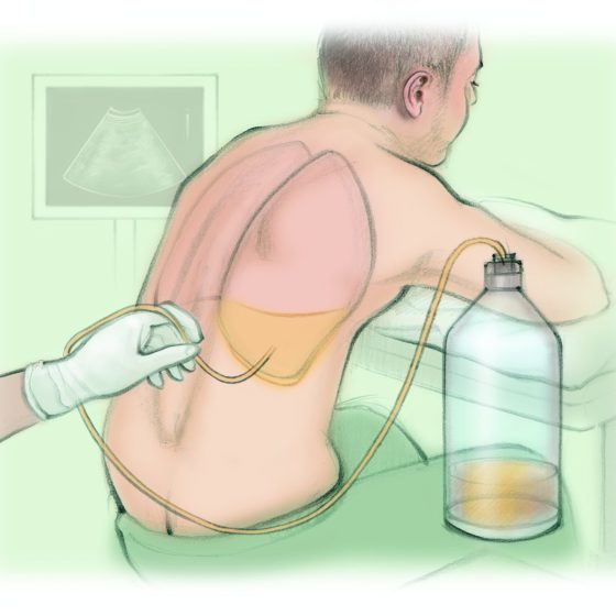
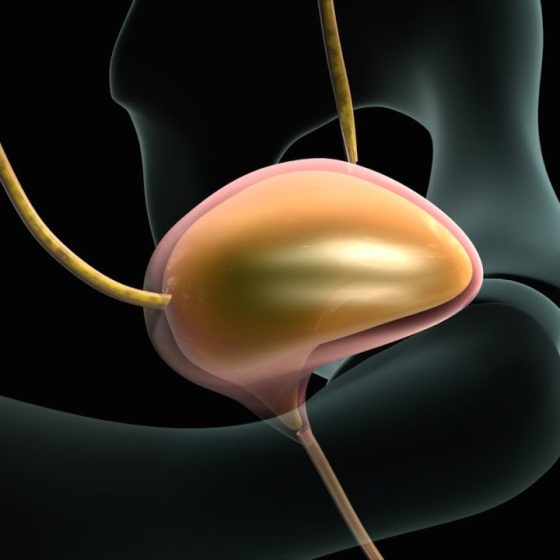
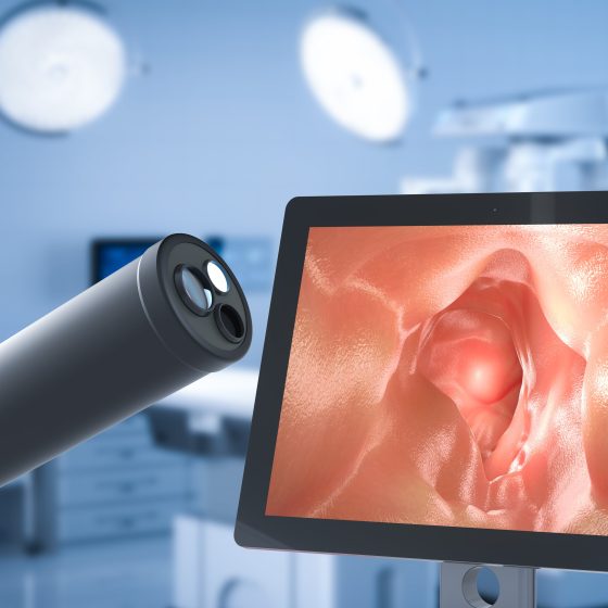
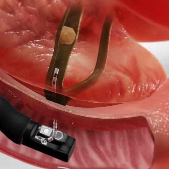
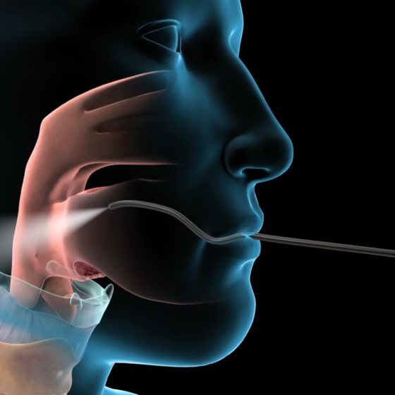
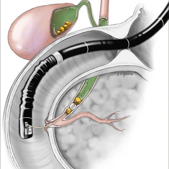
ERCP test ERCP stands for endoscopic retrograde cholangio pancreatography. It is a test to help diagnose conditions of the liver, bile ducts, pancreas or gallbladder. What is an ERCP? Your doctor uses a long flexible tube with a small camera and light at the end, called an endoscope. It’s also sometimes called a duodenoscope. They pass this tube through your mouth, throat, stomach and into the first part of your small bowel (duodenum). Your doctor can look down the endoscope or see pictures on an X-ray screen of the pancreas, gallbladder and bile ducts.   They can take samples (biopsies) of any abnormal looking areas. 

Draining fluid from your chest or tummy

Cancer cells can inflame the lung lining (pleura) or abdominal lining (peritoneum). This can cause fluid to build up. The fluid might contain cancer cells. Fluid around the lungs is called a pleural effusion. It can make it difficult to breathe. Draining fluid from your chest is called thoracocentesis or pleural aspiration. Fluid in the tummy (abdomen) is called peritoneal ascites. It can make the abdomen feel swollen, tight and uncomfortable. Draining fluid from your abdomen is called an abdoparacentesis or peritoneal aspiration.  Why you might have it The doctors might drain fluid from around your lungs or abdomen to: see if the fluid contains

Cystoscopy to check for cancer

A cystoscopy is a test to look at the inside of your bladder and tube that carries urine from your bladder out of your body (urethra). It uses a thin tube called a cystoscope.  There are different types of cystoscopies: flexible cystoscopy rigid cystoscopy narrow band imaging (NBI), blue light cystoscopy or photodynamic diagnosis (PDD) Your doctor will discuss with you what type of cystoscopy you’re having. Flexible cystoscopy The flexible cystoscope has optic fibres inside it, a light and camera attached to it. Because it’s flexible it can bend around the tubes as it passes through your urethra. This is generally done under local

Colonoscopy

A colonoscopy looks at the whole of the inside of the large bowel. A doctor or nurse (endoscopist) uses a flexible tube called a colonoscope. The tube has a small light and camera at one end. The endoscopist puts the tube into your back passage and passes it along the bowel. They can see pictures of the inside of your bowel on a TV monitor.  Why might you have a colonoscopy You may need a colonoscopy: to help find the cause of bowel symptoms to look for early signs of bowel cancer as part of the national bowel cancer screening programme

Cholangiography

Cholangiography means looking at the structure of the bile ducts and gallbladder. It can help to find the size of a cancer and whether it has spread. What is cholangiography? Cholangiography means putting a dye called a contrast medium into the bile ducts and gallbladder to show them up clearly on x-ray. Preparing for your test You might have a blood test 2 days beforehand to check how well your blood clots. Tell your doctor if you’re having medicine that changes how your blood clots. This includes: aspirin clopidogrel arthritis medicines warfarin or heparin apixaban, rivaroxaban, dabigatran, edoxaban or betrixaban Your doctor will

Cervical biopsy (cone biopsy)

A cone biopsy is a small operation to remove a cone shaped piece of tissue from your cervix. You usually have it under general anaesthetic, which means you are asleep. The operation takes about 20 to 30 minutes. You will probably stay in hospital overnight.  Why you might have a cone biopsy You might have a cone biopsy if you have symptoms that could be caused by cervical cancer. It’s also a treatment for abnormal cervical cells picked up through cervical screening. The abnormal cells might be on the outer surface of the cervix (ectocervix) or the inner part of the cervix (endocervix).

Capsule endoscopy

A capsule endoscopy looks at the inside of your bowel. This is also called a video capsule endoscopy or PillCam.  You swallow a capsule that contains a small disposable camera. The capsule is the size of a vitamin pill. The camera takes thousands of pictures as it travels along your gut. The camera sends the pictures wirelessly to a data recorder that you wear on your waist. The test is complete once the capsule has passed through your bowel and out into the toilet. A doctor or specialist nurse looks at the pictures from the data recorder to help find out the

CT scan

A CT scan is a test that uses x-rays and a computer to create detailed pictures of the inside of your body. It takes pictures from different angles. The computer puts them together to make a 3 dimensional (3D) image.   CT (or CAT) stands for computed (axial) tomography. You usually have a CT scan in the x-ray (radiology) department as an outpatient . A radiographer operates the scanner. The whole appointment can take up to an hour and a half depending on which part of your body they are scanning.  You might have a CT scan combined with another test such as

Bronchoscopy

A bronchoscopy is a test to look at the inside of the breathing tubes (airways) in your lungs. You might have this test to allow your doctor to: look for the cause of your symptoms help your doctor see any areas that look abnormal on an x-ray or scans take samples of cells These samples might be a tissue sample called a biopsy. Or your doctor might take some cells by using a small brush or using a liquid to collect them. To have the test your doctor puts a narrow, flexible tube called a bronchoscope down your windpipe (trachea)

Breast ultrasound scan 

A breast ultrasound scan is a test that uses high frequency sound waves to create a picture of the inside of the breast.  The ultrasound scanner has a probe that gives off sound waves. The probe looks a bit like a microphone. The sound waves bounce off the tissues in your breast, and the probe picks them up. The probe links to a computer that turns the sound waves into a picture on the screen. A specialist healthcare professional called a sonographer usually does the test.  When you might have a breast ultrasound scan You might have a breast ultrasound: as a first

Breast biopsy

A breast biopsy means having a sample of breast tissue taken to look at under a microscope. It is the only way to find out whether you have breast cancer or other breast conditions. Types of breast biopsy There are different types of breast biopsy. The type of biopsy you have depends on a number of factors such as: how big the abnormal area is where it is Before having a biopsy, you have scans such as a mammogram and breast ultrasound scan . Your doctor looks at the results of the scans and decides on the best type of

Bone scan

A bone scan looks for changes in your bones. Before the test you have a radioactive tracer injection into your bloodstream. You have this through a tube (cannula) into a vein in your hand. It takes 2 to 3 hours for the tracer to go around the body. To have the scan you lie down on a couch while the scanner takes pictures. The scan takes 30 minutes to an hour. It is painless and you can go home after the scan. The body gets rid of the tracer through your urine over the next day. A bone scan shows

Bone marrow test

What is a bone marrow test? A bone marrow test is a way of testing cells from your bone marrow. Bone marrow is the spongy tissue inside your bones that makes blood cells. Why do I need it? To find out whether there are any cancer cells in the bone marrow. You might also have one to check whether treatment is working. How do you have it and how long does it take? You have a local anaesthetic injection to numb a small area on your hip. The doctor puts a needle into your hip to suck out some bone

Bone density scan (DEXA, DXA)

DEXA scan stands for dual energy x-ray absorptiometry. It’s also called a bone densitometry scan. It uses low dose x-rays to take measurements to work out the strength (density) of your bones. You have a DEXA scan in the x-ray (radiology) or nuclear medicine department of the hospital. A radiographer does the scan. It takes about 10 – 20 minutes. You’re not enclosed in a small place as the machine has an arm that hangs over the scanning couch. Why might you have a DEXA scan? You might have this scan if you’re having or have had cancer treatment that

Biopsy for pancreatic cancer 

A biopsy means taking a sample of tissue and sending it to the laboratory so it can be looked at under a microscope. There are different ways of taking biopsies to check for pancreatic cancer. But not everyone with a likely diagnosis of pancreatic cancer will have a biopsy. Why do I need a biopsy? The most sure way of diagnosing pancreatic cancer is by taking a biopsy and looking at it under a microscope. Your doctor takes a biopsy by putting a needle into the area of suspected cancer. Doctors don’t usually take biopsies from the pancreas if they think

Biopsy

A biopsy means taking a sample of tissue so that it can be looked at under a microscope. This is the only way to be certain if an abnormal area is cancer or not. You may also have a biopsy to find out more about the cancer. This includes what type of cancer it is and how fast it is growing. You can have a biopsy from almost anywhere in your body. This includes your skin, organs and other structures. Your doctor takes a sample of the abnormal area and sends the sample to the laboratory. This is where a doctor called