cancer center

Echo (echocardiogram)

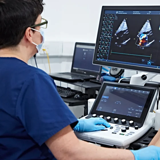
An echo is an ultrasound scan of the heart. This scan is very similar to the scans women have during pregnancy. It uses high frequency sound waves to create a picture of your heart. Doctors can look at the structure of your heart and how well it is pumping, and the nearby blood vessels.  The ultrasound scanner has a microphone that gives off sound waves. The sound waves bounce off the heart and the microphone picks them up. The microphone links to a computer that turns the sound waves into a picture on the screen. This microphone device is called the probe.

ERCP (endoscopic retrograde cholangiopancreatography)

ERCP test ERCP stands for endoscopic retrograde cholangio pancreatography. It is a test to help diagnose conditions of the liver, bile ducts, pancreas or gallbladder. What is an ERCP? Your doctor uses a long flexible tube with a small camera and light at the end, called an endoscope. It’s also sometimes called a duodenoscope. They pass this tube through your mouth, throat, stomach and into the first part of your small bowel (duodenum). Your doctor can look down the endoscope or see pictures on an X-ray screen of the pancreas, gallbladder and bile ducts.   They can take samples (biopsies) of any abnormal looking areas. 

Draining fluid from your chest or tummy

Cancer cells can inflame the lung lining (pleura) or abdominal lining (peritoneum). This can cause fluid to build up. The fluid might contain cancer cells. Fluid around the lungs is called a pleural effusion. It can make it difficult to breathe. Draining fluid from your chest is called thoracocentesis or pleural aspiration. Fluid in the tummy (abdomen) is called peritoneal ascites. It can make the abdomen feel swollen, tight and uncomfortable. Draining fluid from your abdomen is called an abdoparacentesis or peritoneal aspiration.  Why you might have it The doctors might drain fluid from around your lungs or abdomen to: see if the fluid contains

Looking at your mole or skin (dermoscopy)

You will see a skin specialist (dermatologist). They will ask you questions about your mole or abnormal area of skin, such as how long you have had it and what changes you have noticed. They will look closely at the abnormal area, and will check the rest of your skin for any changes. They usually use a dermatoscope to do this.  What is a dermatoscope? A dermatoscope is a handheld instrument, a bit like a magnifying glass. It can make things bigger (magnify) by up to 10 times. Your specialist puts some oil or gel onto your skin. They then hold the dermatoscope

Cystoscopy to check for cancer

A cystoscopy is a test to look at the inside of your bladder and tube that carries urine from your bladder out of your body (urethra). It uses a thin tube called a cystoscope.  There are different types of cystoscopies: flexible cystoscopy rigid cystoscopy narrow band imaging (NBI), blue light cystoscopy or photodynamic diagnosis (PDD) Your doctor will discuss with you what type of cystoscopy you’re having. Flexible cystoscopy The flexible cystoscope has optic fibres inside it, a light and camera attached to it. Because it’s flexible it can bend around the tubes as it passes through your urethra. This is generally done under local

Colposcopy

A colposcopy is a test to look at the cervix in detail. A colposcope is a large magnifying glass that a doctor or specialist nurse (a colposcopist) uses to look closely at the skin-like covering of the cervix. By looking through it, the colposcopist can see changes that may be too small to see with the naked eye. They can take samples (biopsies) of any abnormal areas. You usually have a colposcopy in the hospital outpatient clinic. Why you might have a colposcopy You have a colposcopy if you’ve had an abnormal result after a cervical screening test. You may also have a colposcopy if you have symptoms

Cholangiography

Cholangiography means looking at the structure of the bile ducts and gallbladder. It can help to find the size of a cancer and whether it has spread. What is cholangiography? Cholangiography means putting a dye called a contrast medium into the bile ducts and gallbladder to show them up clearly on x-ray. Preparing for your test You might have a blood test 2 days beforehand to check how well your blood clots. Tell your doctor if you’re having medicine that changes how your blood clots. This includes: aspirin clopidogrel arthritis medicines warfarin or heparin apixaban, rivaroxaban, dabigatran, edoxaban or betrixaban Your doctor will

Capsule endoscopy

A capsule endoscopy looks at the inside of your bowel. This is also called a video capsule endoscopy or PillCam.  You swallow a capsule that contains a small disposable camera. The capsule is the size of a vitamin pill. The camera takes thousands of pictures as it travels along your gut. The camera sends the pictures wirelessly to a data recorder that you wear on your waist. The test is complete once the capsule has passed through your bowel and out into the toilet. A doctor or specialist nurse looks at the pictures from the data recorder to help find out the

CT urogram

A CT urogram is a test using a CT scan and special dye (contrast medium) to look at the urinary system. The contrast medium helps show up the urinary system more clearly.  You have a CT scan of your: kidney bladder tubes that connect the kidneys to your bladder (ureters) A CT scan uses x-rays to take detailed pictures of your body from different angles. A computer then puts them together to make a 3 dimensional (3D) image. CT stands for computed (axial) tomography. It can see details like the soft tissues and blood vessels.  You usually have a CT urogram in the x-ray (radiology) department as

CT scan

A CT scan is a test that uses x-rays and a computer to create detailed pictures of the inside of your body. It takes pictures from different angles. The computer puts them together to make a 3 dimensional (3D) image.   CT (or CAT) stands for computed (axial) tomography. You usually have a CT scan in the x-ray (radiology) department as an outpatient . A radiographer operates the scanner. The whole appointment can take up to an hour and a half depending on which part of your body they are scanning.  You might have a CT scan combined with another test such as

CT colonography

CT (computed tomography) colonography is a test that uses CT scans to check the large bowel (colon) and back passage (rectum). It’s also called a virtual colonoscopy. You have this test as an outpatient in the CT scanning (or radiology) department at the hospital. A radiographer or specialist doctor (radiologist) carries out the test. It usually takes around 30 minutes but you should expect to be in the department for about an hour or so. Why do I need a CT colonography? You usually have this test to help find the cause of your symptoms. Before your test You need

Bronchoscopy

A bronchoscopy is a test to look at the inside of the breathing tubes (airways) in your lungs. You might have this test to allow your doctor to: look for the cause of your symptoms help your doctor see any areas that look abnormal on an x-ray or scans take samples of cells These samples might be a tissue sample called a biopsy. Or your doctor might take some cells by using a small brush or using a liquid to collect them. To have the test your doctor puts a narrow, flexible tube called a bronchoscope down your windpipe (trachea)

Breast biopsy

A breast biopsy means having a sample of breast tissue taken to look at under a microscope. It is the only way to find out whether you have breast cancer or other breast conditions. Types of breast biopsy There are different types of breast biopsy. The type of biopsy you have depends on a number of factors such as: how big the abnormal area is where it is Before having a biopsy, you have scans such as a mammogram and breast ultrasound scan . Your doctor looks at the results of the scans and decides on the best type of

Brain angiogram

An angiogram is an x-ray that can show blood vessels. A specialist doctor called a neuroradiologist injects a dye (contrast medium) and then takes x-ray images of your brain. They look at your brain on the x-ray screen to see: which blood vessels supply the tumour if the tumour is near any major blood vessel This test is also called cerebral angiography.  Why you might have it You might have a brain angiogram if: your tumour is growing very deep inside your brain you have a type of brain tumour called meningioma Preparing for your brain angiogram You usually go into hospital on the morning

Bone scan

A bone scan looks for changes in your bones. Before the test you have a radioactive tracer injection into your bloodstream. You have this through a tube (cannula) into a vein in your hand. It takes 2 to 3 hours for the tracer to go around the body. To have the scan you lie down on a couch while the scanner takes pictures. The scan takes 30 minutes to an hour. It is painless and you can go home after the scan. The body gets rid of the tracer through your urine over the next day. A bone scan shows

Biopsy for pancreatic cancer 

A biopsy means taking a sample of tissue and sending it to the laboratory so it can be looked at under a microscope. There are different ways of taking biopsies to check for pancreatic cancer. But not everyone with a likely diagnosis of pancreatic cancer will have a biopsy. Why do I need a biopsy? The most sure way of diagnosing pancreatic cancer is by taking a biopsy and looking at it under a microscope. Your doctor takes a biopsy by putting a needle into the area of suspected cancer. Doctors don’t usually take biopsies from the pancreas if they think

Biopsy

A biopsy means taking a sample of tissue so that it can be looked at under a microscope. This is the only way to be certain if an abnormal area is cancer or not. You may also have a biopsy to find out more about the cancer. This includes what type of cancer it is and how fast it is growing. You can have a biopsy from almost anywhere in your body. This includes your skin, organs and other structures. Your doctor takes a sample of the abnormal area and sends the sample to the laboratory. This is where a doctor called

Anal examination (anoscopy) and biopsy

A specialist doctor examines your anus with a thin tube called an anoscope. Your doctor can take a sample of tissue (a biopsy) from any abnormal areas. Women might have an internal examination of the vagina as well. This is because it is close to the anal canal. Why you might have an anal examination and biopsy You might have an anal examination and biopsy to help diagnose anal cancer. Or to find out more about the size and position of anal cancer (the stage). Preparing for the anal examination and biopsy It’s normal to feel anxious about this test but it usually