Transient Tachypnoea of the New-born (TTN)
This is a condition which is the most common cause of respiratory distress in babies.
– In the fetus, the lungs are filled with fluid, however this normally gets squeezed out during vaginal birth and the remainder gets absorbed shortly after birth into the bloodstream.
– The problem arises when there is a delay in the reabsorption of lung fluid, which “drowns” lungs
– A risk factor is C-section delivery (as fluid is not squeezed out of the lungs)
Symptoms:
– Respiratory distress (tachypnoea, tachycardia, breathlessness)
Diagnosis:
– Diagnosis of exclusion once other more serious conditions have been ruled out
– CXR –> shows hyperinflated lungs with fluid in individual lung lobes
Management:
– Give oxygen to maintain O2 saturations
– Usually, it is a self-resolving condition as the fluid is absorbed into blood within days
Meconium Aspiration Syndrome
This is a condition which is caused by the foetus aspirating its meconium (first stool that the foetus passes)
– The meconium is a lung irritant and can lead to mechanical obstruction and chemical pneumonitis
Symptoms:
– Respiratory distress (tachypnoea, breathlessness, coughing) and hypoxia
– Risk factor for developing persistent pulmonary hypertension of the Newborn
– May also develop pneumonia and sepsis
Diagnosis:
– Can be diagnosed if you see baby with respiratory distress in infant born in dark meconium stained amnionic fluid
Management:
– Respiratory support (with mechanical ventilation) with anti-inflammatories/pulmonary dilators if pulmonary hypertension
Persistent Pulmonary Hypertension of the New-born (PPHN)
This is a condition which causes pulmonary hypertension in neonates.
– It is caused by failure of the pulmonary circulation to undergo the normal transition after birth
– There is sustained high pulmonary vascular resistance after birth which leads to pulmonary hypertension
– This can cause right-to-left shunting across persistent foetal shunts (PFO, PDA) and thus hypoxia
Causes:
– Primary –> idiopathic
– Secondary –> after birth asphyxia, meconium aspiration, sepsis
Symptoms:
– Gives cyanosis soon after birth
– Can lead to chronic lung disease leading to respiratory distress
– Increased incidence of hospital acquired infections
Test:
– Echocardiogram shows high pulmonary resistance
Management:
– Oxygen therapy (with mechanical ventilation) and pulmonary vasodilators to reduce resistance
Jaundice of the new-born
This refers to yellowing of the skin which occurs in 50% of all new-born infants
– It occurs as there is marked physiological release of Haemoglobin from RBC breakdown because of the high haemoglobin concentration at birth
– In addition, the RBC lifespan of neonates is shorter than that of adults (70d v 120d)
– Also, hepatic bilirubin metabolism is less efficient in the first few days of life
– It is also more common in breastfed-babies
Jaundice in the first 24 hours
The problem with neonatal jaundice is that is can lead to a condition known as Kernicterus:
– Unconjugated bilirubin is fat soluble so it can pass through the BBB
– It is neurotoxic, damaging the basal ganglia causing encephalopathy
Symptoms:
– Muscle hypertonia or hypotonia with spasm (torticollis)
– Opisthotonos –> hyperextension of back with extreme arching
– Inability to move the eyes up and down –> “sun setting” sign
– Seizures
– Lethargy and poor feeding

Diagnosis:
– Measure serum bilirubin level (blood test or transcutaneous bilirubinometer)
– The level is then plotted on a chart to see whether need to treat
Management:
Need for treatment and treatment options are determined using charts of the bilirubin level
– If below treatment line –> encourage feeds, avoid dehydration, and give support for breastfeeding
– If above treatment line –> 1st line is phototherapy (makes bilirubin water soluble to stop it crossing BBB)
–> 2nd line is exchange transfusion (removing baby’s blood and replace with donor)
Sudden infant death syndrome (SIDS)
This is the sudden unexplained death of a child <1-year-old, which occurs due to an unknown reason.
– It usually occurs during sleep, between the hours of midnight and 9am and is known as “cot-death”
– Whilst the exact cause is unknown, there are many risk factors which contribute to the syndrome
– In addition, certain factors like sharing the room with the baby and breastfeeding are known to be protective
Risk factors:
These are additive and together can give a much higher risk of death
– Infant –> Low birth weight, prematurity, male sex
– Parents –> Parental smoking, multiple births, maternal drug use, young mother <21 years old
– Environment –> Putting the baby to sleep prone (instead of on back)
– Bed sharing, infant pillow use
– Hyperthermia (over-wrapping) or head covering)
Diagnosis:
– It is a diagnosis of exclusion.
– Can only be diagnosed if the death remains unexplained after an autopsy, finding out the medical history of patient and family and investigation the death scene.
Management:
– After death, families are offering emotional support and grief counselling
Congenital Diaphragmatic Hernia
This is a condition where the abdominal organs herniate a foramen in the diaphragm
– Most cases occur on the left-side and lead to compression of the lungs
– It occurs due to a failure of the pleuroperitoneal folds which form the diaphragm to seal, which leaves an opening through which the organs can pass through
– This allows the abdominal organs such as the bowel to pass through into the thorax

Symptoms:
– Pulmonary hypoplasia –> causes respiratory distress (SOB, cyanosis)
– Poor air entry into the left chest
– Raised blood pressure
– Apex beat and heart sounds displaced to right
Diagnosis:
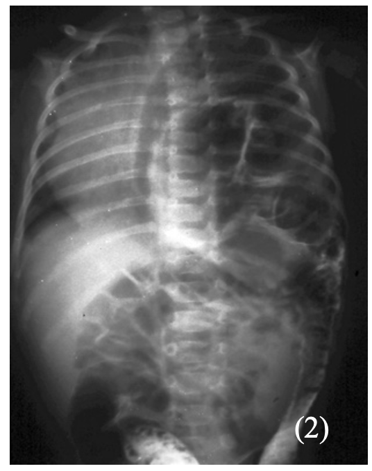
– X-ray chest and abdomen shows loops of bowel in the chest:
Management:
– NG tube (suction of bowel contents) and intubation to secure airway
– Surgical correction is required
Hypoglycaemia
This refers to a low glucose level, generally considered as <2.6mM in the bloodstream
– A transient drop in blood glucose is commonly seen after birth as part of the adaptation to postnatal life
– Even term babies often get hypoglycaemic in the first 24 hours without any complications
– However, severe or prolonged hypoglycaemia may result in long-term neurological damage
Risk factors:
– Prematurity, infection, diabetic mothers, IUGR and macrosomia
Symptoms:
– Can be asymptomatic
– Can cause irritability, lethargy, apnoea, grunting, sweating, seizures
Management:
– If mild –> promote breast or bottle feeding
– If severe –> IV 10% dextrose
Haemorrhagic Disease of the new-born
This is a condition which causes bleeding in the neonate due to vitamin K deficiency.
– Vitamin K is needed as it is a cofactor for the carboxylation and maturation of various clotting factors
– It can lead to a spectrum of disease from minor bruising to large haemorrhages which can be seen up to 8 weeks after birth
Risk factors:
– Liver disease
– Maternal antiepileptic therapy –> these impair synthesis of vitamin K-dependent clotting factors
– Breast fed babies –> the breast milk is a poor source of Vitamin K
Symptoms:
These can occur up to 8 weeks after birth
– Bruising
– Haematemesis and melaena
– Potential intracranial haemorrhage
Management:
– IM Vitamin K and fresh frozen plasma if severe bleeding
Prevention:
– Vitamin K is offered all infants immediately after birth, usually as single IM injection
– Mothers on antiepileptics should receive oral prophylaxis from 36 weeks’ gestation

