The female reproductive system lies posterior to the bladder and anterior to the rectum.
– It includes the uterus, ovaries, cervix, fallopian tubes and the vagina.
Ovaries
The ovaries are the main female reproductive organs, as they produce egg cells and sex hormones oestrogen and progesterone.
– They lie lateral to the uterus close to the pelvic wall
– The right lies close to caecum/appendix and left near sigmoid
Blood supply:
– Ovarian artery (direct from abdominal aorta)
Venous drainage:
– Ovarian vein –> IVC on right/renal vein on left
Nerve supply:
– Sympathetics from T10 –> pain referred to umbilicus.

Uterus
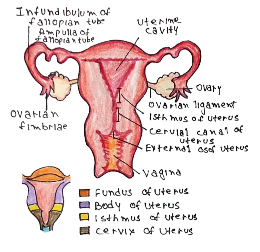
The uterus (womb) is the organ where offspring are conceived and held during gestation, which can be divided into 3 parts:
i) Fundus (at the top)
ii) Body
iii) Cervix
The lining of the uterus is also divided into 3 main layers:
i) Inner Endometrium –> made of outer columnar epithelium with glands and inner connective tissue
ii) Middle Myometrium –> smooth muscle layer which contracts during delivery
iii) Outer serosa/adventitia
Blood supply:
Dual Supply from uterine artery (from internal iliac) + ovarian artery.
Venous drainage:
Uterine Vein (in the broad ligament) –> internal iliac vein
Nerve supply:
– Sympathetic from T11-L2 give contraction and vasoconstriction
– Parasympathetic from S2-S4 (pelvic splanchnic) gives vasodilation
The uterus is held in place by ligaments which attach it to pelvic structures.
Round ligament | Anchors the uterus anteriorly |
Broad ligament | Supports the uterus laterally |
Cardinal ligament | Supports the uterus laterally |
Uterosacral ligaments | Anchors the uterus to the sacrum |
Cervix
This is the lowest part of the uterus composed of fibroelastic connective tissue.
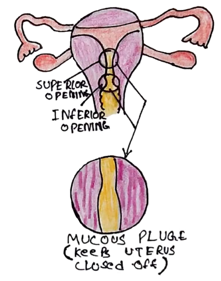
It has two openings:
– Internal os connects the cervix to the uterus
– External os connects the cervix to the vagina
The cervix produces mucus sealing off the uterus from the environment
– This mucus varies in pH and thickness throughout the uterine cycle (under the influence of oestrogen/progesterone) to either facilitate or inhibit the passage of sperm into the uterus
– The cervix also allows broken down endometrium to pass out the uterus to the vagina during menstruation.

The cervix is divided into 3 parts:
i) Endocervix:
The innermost part of cervix above the uterine external os (opening of uterus). Lined by simple columnar epithelium with mucous producing cells.
ii) Ectocervix:
Outer part of cervix distal to the uterine external os, lined by squamous epithelium
iii) Cervical transformation zone:
The junction between the endo/ectocervix
– Here there is a border between the inner columnar epithelium and distal squamous epithelium
Fallopian Tubes
These tubes connect the ovaries with the uterus
– They are responsible for transporting ova from ovary to uterus, but also sperm in the opposite direction
– Divided into 4 parts: (Starting at ovary) Fimbriae –> Infundibulum –> Ampulla –> Isthmus
– The isthmus is the narrowest part, whereas the ampulla is the site where fertilization usually occurs
– The tubes are lined with cilia which are needed to transport cells to uterus and secretes nutrients
Blood supply:
– Ovarian artery (lateral 2/3) and uterine artery
Nerve supply:
– Same as uterus
Vagina
This is an elastic muscular canal that connects the uterus to the outside world.
– It is lined by non-keratinized stratified squamous epithelium
– It is supported by ligaments of the pelvic floor, which attach to the perineal body between the urogenital and anal triangles –> if damaged then risk of rectum, bladder, uterine prolapse.
– The hymen is a membrane the partially covers the vaginal opening
Vulva
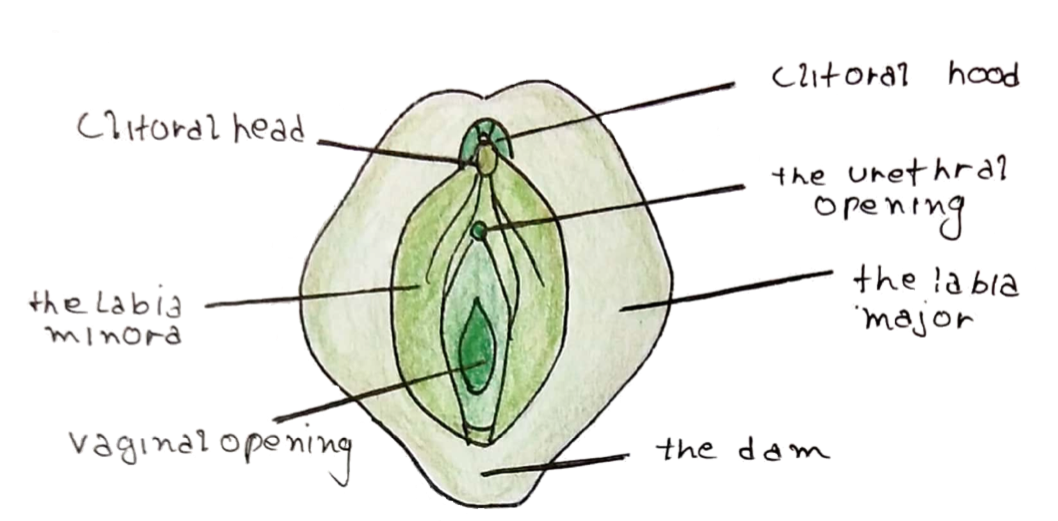
This is the outermost part of the female genitalia.
– Includes the opening of the vagina (vestibule), and the vaginal lips and clitoris (main erogenous zone of the female).
– Lined by Bartholin’s glands, which secrete alkaline fluid for lubrication during sex.

There are 2 sets of skin folds on the vulva:
– Labia majora – contains sweat gland, sebaceous glands and pubic hair.
– Labia minora- hairless but contains sebaceous glands
Oestrogen has an important role in the keratinization and proliferation of the vulvar epithelium. It maintains thickness and prevents atrophy (this is seen in menopause).
Anatomy of the Breast
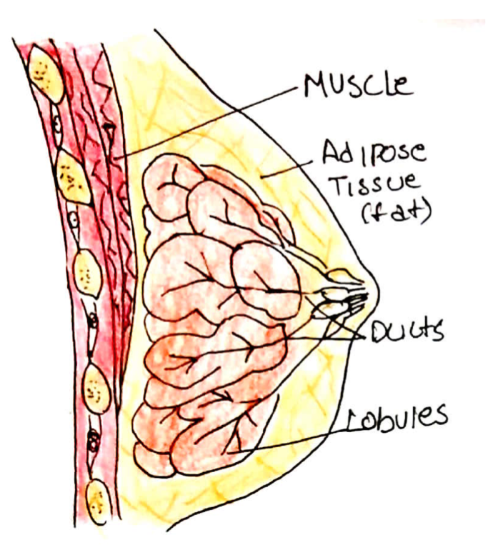
The breast is a sexual organ which is made of fat, connective tissue and glands
– Glands are arranged in 15-20 lobules which secrete milk into lactiferous ducts
– Milk drains from smaller ducts to the major ducts and to the lactiferous sinus (reservoir behind the nipple) and ultimately onto the nipple
– The breast tissue is enclosed in anterior and posterior leave of body fascia, which lies on pectoralis major
– It is attached to the chest wall by loose connective tissue to the pectoral fascia.
– Bands of connective tissue called Coopers’ Ligaments run from the fascia to dermis of the breast to maintain shape
– The breast can give bilateral nipple discharge naturally

Blood supply:
– Axillary, Intercostal + Internal Thoracic Arteries
Lymph drainage:
– Axillary nodes –> apical nodes in apex of axilla
The breast is hormone sensitive and development is mainly driven by oestrogen and progesterone:
– Oestrogen –> this increases cell proliferation and causes hypertrophy of the milk duct system
– Progesterone –> this increases cell differentiation and proliferation of the lobules.

