The upper limb mainly comprises of three main joints – the shoulder, elbow and wrist.
Shoulder
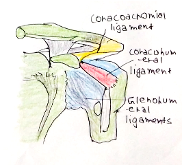
This is a synovial ball-and-socket joint. The surface area of the humeral head is 3x that of the glenoid fossa.
The capsule is strengthened by coracohumeral ligaments and 3 anterior glenohumeral ligaments.
Muscles:
These can be described by the action they cause on the joint. The shoulder is stabalised by the rotator cuff muscles which attach onto greater tuberosity of humerus, expect teres minor (lesser tuberosity)
i) Abduction:
– Supraspinatus –> provides the initial 15 degrees of abduction, by suprascapular nerve
– Deltoid –> From acromion onto deltoid tuberosity of humerus, main abductor, axillary nerve
ii) Adduction:
– Coracobrachialis –> From coracoid process to humerus

iii) Flexion:
– Deltoid, coracobrachialis + Pectoralis Major
iv) Extension:
– Triceps Brachii + Latissimus Dorsi
v) Lateral Rotation:
– Infraspinatus + Teres minor
vi) Medial Rotation:
Subscapularis, teres major + latissimus dorsi and pectoralis major
Arm
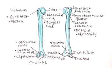
Bones: The main bone which forms the arm is the humerus.
– The neck separates the head from the greater and lesser tuberosities, which give attachments for the rotator cuff muscles.
– The spiral groove houses the radial nerve and profunda brachii artery
– The epicondyles are attachment points for muscles of the forearm.

Muscles: Categorized into anterior and posterior compartments:
i) Anterior:
– Coracobrachialis –> From coracoid process onto humerus, adducts shoulder
– Brachialis –> From humerus onto ulna, main flexor of the elbow
– Biceps brachii –> short head from coracoid process + long head from shoulder joint
–> inserts into radial tuberosity, powerful supinator and flexor of elbow
All three muscles supplied by musculocutaneous nerve, off lateral cord of brachial plexus.
ii) Posterior:
Triceps Brachii –> Formed of three heads: long, lateral and medial (deepest), radial nerve
–> Inserts onto olecranon of ulna, main extensor of the elbow joint
Elbow
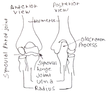
At this joint, the trochlea of the humerus articulates with the trochlear notch of the ulna and capitulum with head of radius.
– Anteriorly, the coronoid fossa accepts coronoid process of the ulna in flexion.
– Posteriorly the olecranon fossa accepts the olecranon during extension.
– Stabilized by medial and lateral collateral ligaments and the anular ligament around radial neck
The radial nerve, winds around the radial neck before dividing into superficial and posterior interosseous branch.
– In the elbow, the three key structures (medial to lateral) are median nerve, Brachial artery, biceps tendon.

Forearm
Bones:
The forearm is composed of the radius and the ulna
– Radius – proximal head articulates with capitulum of humerus + radial fossa of ulna.
—> Lateral aspect makes the styloid process, and posteriorly there is Lister’s tubercle on radial side.
– Ulna – articulates with trochlea of humerus. Has a small distal head and small styloid process.
Muscles: The intrinsic muscles of the forearm are divided into the anterior and posterior compartments.
i) Superficial anterior:
These arise from common flexor origin on medial epicondyle of humerus.
– Pronator teres (pronates arm), Flexor carpi radialis, palmaris longus + flexor carpi ulnaris (ulnar nerve)
– All supplied by median nerve (except FCU), and work to flex wrist and cause radial/ulnar deviation.
ii) Deep anterior:
These include flexor digitorum superficialis/profundus + flexor pollicis longus + pronator quadratus
– All supplied by anterior interosseous branch of median nerve and flex wrist/fingers/thumb
iii) Superficial posterior:
These all arise from the common extensor origin on lateral epicondyle (radial nerve)
– Brachioradialis (Flexor) + extensor carpi radialis longus/brevis, extensor digiti communis, extensor digiti minimi + extensor carpi ulnaris
– These mainly extend the wrist and also the fingers + cause radial/ulnar deviation
iv) Deep posterior:
Main 3 are abductor pollicis longus, extensor pollicis brevis + extensor pollicis longus
– These form the boundaries of the anatomical snuffbox causing thumb extension + abductor
– Supinator is a supinator of the forearm, whilst extensor digiti minimi extends little finger
– All supplied by posterior interosseous branch on radial nerve.
Wrist
This is formed by the distal surface of the radius articulating with scaphoid and lunate.
– Distal head of ulna is separated from triquetral bone by a fibrocartilaginous disc, which separates wrist from distal radio ulnar joint.
– Capsule strengthened by medal and lateral collateral ligaments from radius/ulna to carpal bones
– Movements include, flexion/extension + ad/abduction and circumduction.

Hand
Bones:
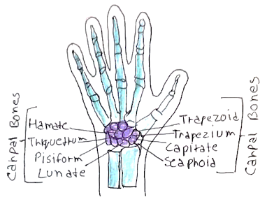
The hand is formed of two rows of eight carpal bones.
– The proximal row (radial to ulna) = Scaphoid, lunate, Triquetral, Pisiform
– The distal row (radial to ulna) = Trapezium, Trapezium, Capitate + Hamate
The next row is made up of metacarpals, which have a base, shaft and head
– Minimal movement occurs between carpals and metacarpals
– The shafts are connected by transverse metacarpal ligaments.
The most distal bones are the phalanges, comprised of a base, shaft and head, and are stabalised by radial and ulnar collateral ligaments.
Muscles:
The intrinsic muscles of the hand lie in three groups:
i) Thenar eminence:
Abductor pollicis brevis, flexor pollicis brevis and opponens pollicis
Action –> Control fine movements of the thumb
Nerve supply –> Recurrent branch of the median nerve.
ii) Hypothenar eminence:
Abductor digiti minimi, flexor digiti minimi and opponens digiti minimi
Action –> Control fine movements of the little finger
Nerve Supply –> Ulnar nerve
iii) Deep muscles:
Adductor pollicis, interossei and lumbricals.
Action –> Palmar interossei adduct, dorsal interossei abduct (PAD-DAB). Lumbricals flex metacarpophalangeal joints + extent interphalangeal joints.
Nerve Supply –> Ulnar nerve, except for lateral two lumbricals (median nerve).
Blood supply:
– Ulnar artery gives rise to the superficial palmar arch
– Radial artery gives rise to deep palmar arch, with rich anastomoses between the two
Skin:
– The skin of the hand is attached to the palmar aponeurosis, which is the deep fascia of the palm.
– This receives the tendon of palmaris longus and covers the flexor sheaths of the fingers
– It can become tense leading to Dupuytren’s contracture giving fixed flexion deformities of the fingers.

