The lower limb comprises of three main joints – the hip, knee and ankle.
Hip
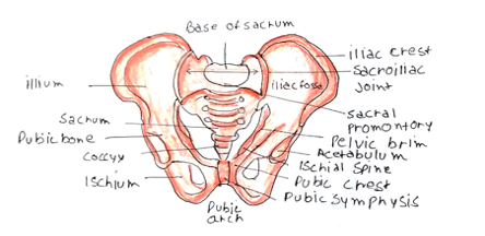
This is formed of the head of the femur interacting with the acetabulum
– The articular surface of the acetabulum is a horseshoe – finished by transverse ligament and fibrocartilage labrum.
– Fovea of femoral head connects to acetabulum by ligamentum teres.
The capsule is attached around acetabulum to the femoral neck, above trochanters.
– It is strengthened by 3 ligaments –ischiofemoral, pubofemoral and iliofemoral (this is the strongest Y-shaped which goes from AIIS to intertrochanteric line preventing hyperextension)
Muscles:
These are categorized by their actions on the hip joint:
Flexion – Psoas major + Iliacus (attaches to lesser trochanter) + Sartorius (from ASIS to medial tibial condyle)
Extension – Gluteus maximus and hamstrings
Adduction – done medial compartment of thigh, supplied by obturator nerve
Abduction – gluteus medius and minimus insert onto greater trochanter, supplied by superior gluteal nerve
Medial rotation – Anterior fibres of gluteus medius and minimus
Lateral rotation – gluteus maximus + lateral rotators (piriformis, obturators) which attach to greater trochanter
Blood supply:
Femoral head gets blood from retinacular fibres of capsule from superficial femoral artery
– Ligamentum teres in children has a branch of obturator artery, but this is negligible in adults.
Thigh
Bones:
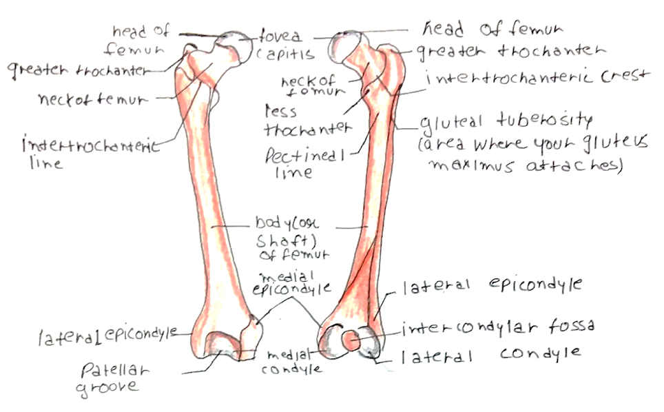
This is made up of the femur, which has an anatomical head with a central depression (fovea), attached to the ligamentum teres.
– Neck forms an angle of 125º with shaft (angle of inclination) and angle of 12º with femoral condyles (angle of anteversion)
– Proximal end has greater and lesser trochanters which demarcate neck and shaft.
– Distal end has medial and lateral femoral condyles, which are separated posteriorly by the intercondylar notch.
Muscles:
These are divided into anterior, medial and posterior compartments:
i) Anterior:
Quadriceps femoris (Rectus femoris, vastus lateralis/medialis/intermedius)
– Arise from femur and hip bone and have a common tendon via patella onto tibial tuberosity
– They all extend the knee and rectus femoris also flexes hip
– The anterior compartment is supplied by the femoral nerve
ii) Medial:
3 adductors (adductor magnus, brevis, longus) arise from ischiopubic ramus
– They insert down the femoral shaft onto the linea aspera
– Gracilis and Pectineus run down medial thigh and also adduct thigh
– Obturator externus is a lateral rotator of the hip
– These are all supplied by the obturator nerve
iii) Posterior:
This compartment extends the hip and flexes the knee
– Semimembranosus and semitendinosus arise from ischial tuberosity –> insert onto medial tibial condyle
– Biceps femoris has two heads from ischial tuberosity + femur –> inserts onto lateral head of fibula
– They are all supplied by the tibial nerve, except short head of biceps (common fibular nerve)
Soft tissue:
– Fascia lata is thick fascia which divides muscles into compartments.
– It forms the iliotibial tract laterally which receives tensor fascia latae + gluteus maximus
– As this fascia is so tight a fracture of the femur can lead to compartment syndrome
Knee
Femoral condyles articulate with tibial condyles and patella articulates with the anterior surface of femur.
– Patella sits in trochlea groove, and is at risk of dislocating laterally, especially in teenage girls
– Capsule is deficient anteriorly and is instead completed by quadriceps tendon.
– The medial tibial condyle has an attachment area for 3 important muscles (sartorius, gracilis and semitendinosus). This is known as the pes anserinus (“Goose’s foot”)
Ligaments:
Bony contours do not give stability and so you need support from the ligaments.
Collateral:
Medial and lateral collaterals extend from femur to tibia medially and head of fibular laterally
– Medial collateral injured by valgus strain, whereas lateral is hurt by varus strain
Cruciate:
ACL connects anterior part of intercondylar eminence to lateral femoral condyle
–> Prevents forward displacement of tibia on femur
PCL connects posterior part of intercondylar eminence to medial femoral condyle
–> Prevents backward displacement of tibia on femur, important when going downhill
A completely extended knee is “locked” – this means you can stay upright without muscular effort.
– Hence flexion must first be initiated by popliteus to rotate femur on tibia, “unlocking” the knee.
Behind the knee joint is a diamond-shaped space called the popliteal fossa, bounded by diverging hamstrings and distally heads of gastrocnemius. It contains:
– Popliteal artery + vein
– Lymph nodes
– Tibial nerve and common fibular nerve
– Sural nerve
Leg
Bones:
The leg is formed by the tibia medially and the fibula laterally. The medial margins of these bones forms interosseous membrane, which makes inferior tibiofibular joint fibrous.
– Tibia – Tibial plateau articulates with femur, extends distally to articulate with talus and has medial malleolus
– Fibula- Head articulates with tibia and extends laterally to articulate with tales and has lateral malleolus.
Muscles: These can be divided into anterior, lateral and posterior compartments of the leg.
i) Anterior:
Tibialis anterior + Extensor hallucis longus, extensor digitorum longus + Fibularis tertius
– These muscles are dorsiflexors of the ankle (+toes/hallux) and cause inversion.
– These are supplied by the deep branch of the common fibular nerve
ii) Lateral:
Fibularis longus (inserts into first metatarsal) + fibularis brevis (fifth metatarsal)
– These cause eversion of the foot at subtalar joint + plantarflexion
– Supplied by the superficial branch of the common fibular nerve
iii) Posterior:
– Superficial muscles: Gastrocnemius, Plantaris and soleus
–> These insert into common calcaneal (Achilles) tendon to plantarflex ankle + flex knee
– Deep muscles: Popliteus + flexor hallucis longus, flexor digitorum longus + tibialis posterior
–> Tendons pass under medial malleolus flexor retinaculum (tarsal tunnel) to cause dorsiflexion of the ankle and toes
– All the posterior compartment is supplied by branches of the tibial nerve.
Ankle
This is a synovial hinge joint, with a deep mortise made of the distal tibia + fibula which houses body of talus
– The terminal ends of the tibia and fibula form the medial and lateral malleoli, which prevent inversion and eversion at the ankle joint.
– The only movements possible are plantarflexion and dorsiflexion.
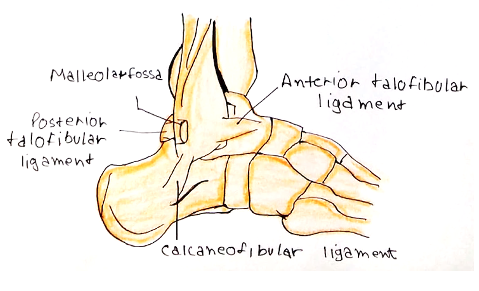
Medially it is stabalised by medial (deltoid) ligament from tibia to calcaneus + navicular
– Laterally stabalised by lateral ligament which has 3 bands: anterior/posterior talofibular ligament and calcaneofibular ligament.
– Twisted ankles commonly tear anterior talofibular lateral ligament.
Foot
Bones:
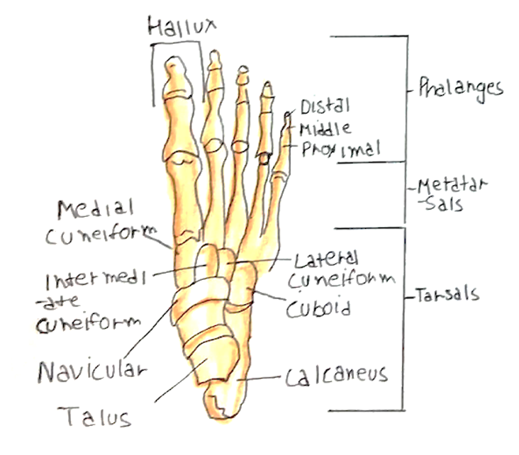
The foot is formed by 7 tarsal bones, 5 metatarsals and 5 phalanges.
– The tarsal bones are calcaneus, talus, navicular, cuboid + 3 cuneiforms
– Inversion and eversion of the foot occur at the subtalar joint, which is composed of the talocalcaneal + talocalcaneonavicular joint, but function as one joint
– This is important as the malleoli do not allow inversion/eversion at the ankle.
Neurovascular Status of lower Limb
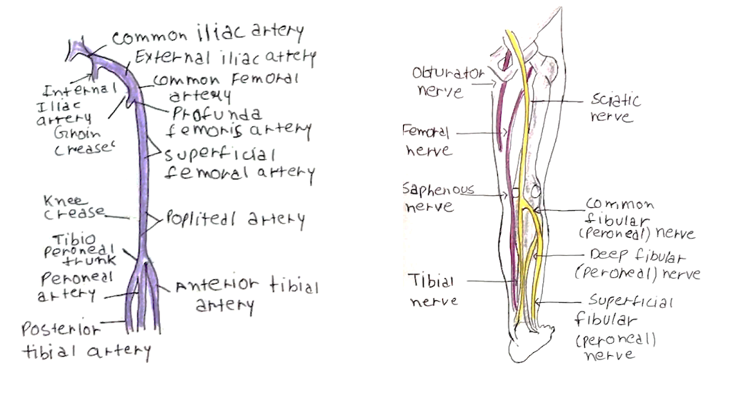
The main neurovascular structures enter the lower limb through the femoral triangle:
– Bounded superiorly by inguinal ligament, Laterally medial border of Sartorius + medially by adductor longus.
– Lateral to medial: Femoral nerve –> Femoral Artery –> Femoral vein –> Femoral canal (space with lymphatics)
Arteries:
After the femoral artery has passed through, it gives profunda femoris and superficial femoral artery.
– Continues as popliteal artery before dividing into 3 branches, anterior + posterior tibial and peroneal artery.
Nerves:
– Femoral nerve (L2-4) is anterior and supplies quadriceps + Sartorius.
– Obturator nerve (L2-4) supplies adductors + medial thigh
– Sciatic nerve (L4-S3) is major nerve posterior which is composed of tibial + common fibular nerve.
–> Tibial branch supplies whole of posterior compartment of leg
–> Superficial branch of the common fibular nerve supplies lateral compartment of leg
–> Deep branch of the common fibular nerve supplies anterior compartment of leg
