A fracture is a medical condition where the continuity of the bone is broken. When describing a fracture, it is essential to describe the type and severity of the fracture. Adequate description includes providing information about a number of different categories:
Open or closed?
Closed fracture = a broken bone with no open wound
Open fracture = a broken bone with a break in the skin
Simple or comminuted?
Simple = a fracture where the bone is broken into two fragments
Comminuted = bone is splintered or crushed into several pieces
Angle of break?
Transverse = this is when the break is perpendicular to the long axis of the bone
Oblique = bone is broken at an angle across the bone
Spiral = a fracture in which the bone has been twisted apart.
Level of displacement?
Displaced = a fracture where the ends no longer retain their normal alignment
Non-displaced fracture = bone fracture where the ends do retain their normal alignment.
How to Describe Displacement
Type of fracture
Hairline = a stress fracture which causes a very thin break – but bones retain the same shape
Compression = bone becomes fractures due to pressure from other bones, often in the vertebrae
Distal Radius Fracture
This is a fracture of the radius which usually occurs due to a fall on an outstretched hand.
It can be divided into two main types of fractures
Colles’ Fracture
This is a fracture which occurs due to a fall on an outstretched hand.
It is an extra-articular fracture of the distal radius (within 2 centimeters of the joint) which involves dorsal angulation with radial tilt.
It causes a dinner fork deformity, due to dorsal angulation of the distal segment.
Smith’s fracture
This is a fracture which occurs with a fall onto flexed wrists.
It is an extra-articular fracture of the distal radius (within 2 cm of joint) which involves volar displacement of the distal segment and is less common than a Colles’ fracture.
These are inherently unstable and usually require surgical fixation.

Key tests
Plain film x-ray of the wrist
Management
If not displaced, immobilisation in cast
If displaced or neurovascular compromise, then operative fixation is often required
Supracondylar Fracture
This is a fracture which occurs at the distal end of the humerus, usually due to a fall on an outstretched hand
It can also be due to a direct blow to the lateral elbow (more common in children)
Symptoms
Pain and swelling
Key tests
Elbow X-ray
Complications
Vascular – acute ischaemia due to disruption of the brachial artery
Neurological – damage to the anterior interosseous nerve, median nerve
Volkmann’s ischaemic contracture
Scaphoid Fracture
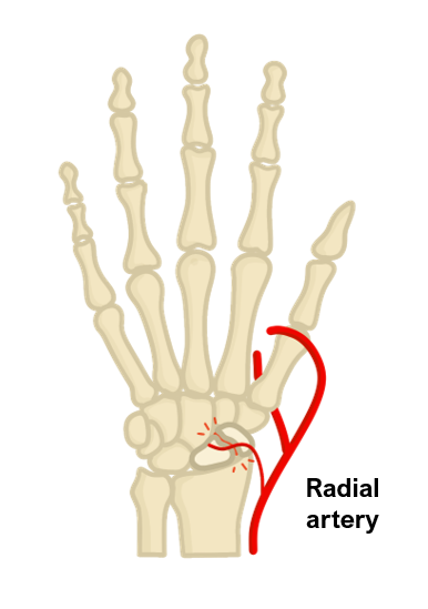
This is a fracture which occurs across the scaphoid, one of the small hand bones.
It usually occurs due to a fall on an outstretched hand.
The blood supply to the scaphoid is from a nutrient branch of the radial artery which runs from distal to proximal.
Therefore, with a proximal pole scaphoid fracture, there is a high risk of avascular necrosis of the proximal segment.
Symptoms
Hand pain and swelling
Tenderness in the anatomical snuffbox
Pain on longitudinal compression of the thumb and on wrist movement

Key tests
Wrist X-ray. If unclear, MRI is gold standard
Management
Usually conservative in a plaster/splint for 6–8 weeks. If there is a delay in union, then it may require operative fixation.
Neck of femur
This is a hip fracture which is often seen in elderly women, usually secondary to a fall.
It can be categorised according to whether the capsule is affected.
Intracapsular fractures
These are divided into subcapital, transcervical and basicervical.
In these types, there is a high risk of damage to blood vessels causing avascular necrosis of the femoral head.
Therefore, in these types, the femoral head often requires replacement with a hemiarthroplasty or total hip replacement.
Extracapsular fractures
These are divided into intertrochanteric or subtrochanteric.
In these types, is less of a risk of avascular necrosis to the femoral head, which means that it does not have to be replaced as often.

Symptoms
Hip/groin pain
Inability to weight bear
Shortening of the leg due to iliopsoas pull with external rotation
Key tests
Hip X-ray shows disruption of Shenton’s line and discontinuity of the cortex
Management
Intracapsular
If non-displaced – consider internal fixation in situ with screws or a plate
If displaced and patient is unfit/elderly – hemiarthroplasty
If displaced and patient is younger/fit – total hip replacement
Extracapsular
If intertrochanteric – dynamic hip screw or intramedullary nail
If subtrochanteric – intramedullary nail

