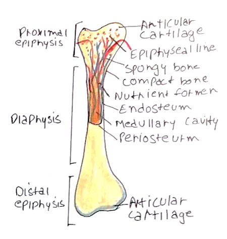
The long bones that make up the body have a distinct structure, and can be subdivided into many parts:
– Bones usually have high levels of compressional and tensile strength, but less torsional strength.

| Term | Definition |
| Diaphysis | The shaft of the bone – This is made up of compact bone which has a dense structure |
| Epiphysis | The end of the bone – This is made up of cancellous/spongy bone. – Made of sheets of trabeculae that branch to form a sponge like network. |
| Epiphyseal line | Also known as the growth plate – Separates the Diaphysis and Epiphysis during development – Ossifies at the end of puberty |
| Metaphysis | The zone adjacent to the GP on the diaphysis side |
| Cortex | The outer layer of the bone – Denser (and therefore whiter on X-rays) |
| Medulla | The inner layer of the bone – Less dense (and therefore darker on X-rays) |
| Sesamoid bones | A bone that lies within a tendon – Some form part of the normal human anatomy (e.g. patella) – Others are normal anatomical variants |
Cellular Components:
Osteoprogenitor – Located in the endosteum and periosteum. These cells differentiate into osteoblasts
Osteoblasts – These are bone producing cells. These differentiate into osteocytes when trapped in forming bone
Osteoclasts – Bone-resorbing cells. Large and multinucleate. They arise from macrophage precursors in the blood
Osteocytes – Quiescent osteoblasts which are located in lacunae
Non-cellular Components (Matrix)
Organic – composed of Collagen fibres (mainly type I) + Amorphous material (e.g. glycosaminoglycans)
– This gives tensile strength
Inorganic – Hydroxyapatite crystals (calcium and phosphorus) + Bicarbonate + Citrate
– This gives compressional strength
Bone Development
There are two types of bone development. Whilst the bones of the head and neck form by intramembranous ossification, the long bones of the body are made by endochondral ossification.
1) Cartilage model
Condensation occurs forming a cartilage model of the bone
2) Primary ossification centre
Chondrocytes in the centre of the diaphysis hypertrophy then die allowing invasion of blood vessels
– Osteoprogenitor cells enter and differentiate into osteoblasts, which secrete bone matrix.
– Osteoid binds calcium to form trabeculae – primary ossification centre
3) Secondary ossification centre
Blood vessels invade epiphysis (around time of birth) forming secondary centres of ossification
4) Epiphyseal growth plate
Cartilage remains at the junction between epiphysis and diaphysis (growth plate)
– Cartilage on epiphyseal side grows and diaphysis side is replaced by bone

