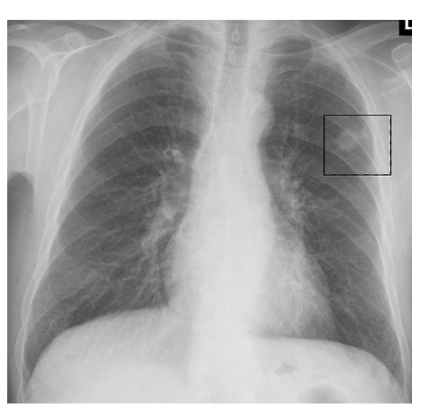
This is a broad term which is most common cancer in the UK, which typically occurs in people of 60 years.
It can be broken down into several types of specific types of cancers, which have different histology.
Metastases are more common, typically arising from breast, prostate, colon, kidney and uterus.
NICE Referral Guidelines
Carcinoma of the bronchus
This type of carcinoma is generally divided into 2 main categories:
Small Cell Carcinoma
This is a tumour which arises from neuro-endocrine cells in the lungs.
It is named “small cell” because the cancerous cells look small when viewed under a
microscope (compared to other lung cancer types).
It has a rapid growth and early metastasis and produces hormones like ADH/ACTH resulting in paraneoplastic syndromes (SIADH and Cushing’s disease respectively)
Non-Small Cell Carcinoma (NSCLC)
This refers to lung cancers which are not composed of “small cells”. 3 types:
The most common is a squamous cell carcinoma (most common in male smokers).
The second most common is adenocarcinoma, a malignant proliferation of epithelial tissue that has glandular characteristics. It is the most common lung tumour in nonsmokers and female smokers.
The other type is large cell carcinoma. This gets its name as the cancer cells are large, with excess cytoplasm, large nuclei and conspicuous nucleoli.

Causes
Smoking
Asbestos
Radiation from radon gas (from uranium decay therefore increased risk for uranium miners and also characteristically present in soil in basements)
Symptoms
Persistent cough and haemoptysis
Weight loss
Slow onset chest pain and dyspnoea
Post obstructive pneumonia
Voice hoarseness (due to Pancoast tumours compressing recurrent laryngeal nerve)
Specific Symptoms
Different tumours also secrete hormones, resulting in paraneoplastic syndromes
Complications
Key tests
Chest X-ray – this may reveal a mass
CT chest with contrast – this is the investigation of choice for diagnosis
Bronchoscopy – this is used to take a biopsy for histological diagnosis
PET-CT/CT chest, abdomen, pelvis – used to detect metastases to stage the cancer

Staging
When assessing a lung cancer, patients are offered PET-CT to stage the cancer. This gives information about the primary tumour (T), if it has spread to regional nodes (N) and metastases (M).
Primary Tumour (T)
The first 2 grades are TX (malignant cells in bronchial secretions) and TIS (carcinoma in situ).
T0 = no evidence of primary tumour
T1 = < 3cm
T2 = >3cm tumour
T3 = involves chest wall, diaphragm, pleura.
T4 = involves mediastinum, heart or other organs
Regional Nodes (N)
This goes from N0 (no nodes) to N4 (contralateral hilum)
Metastasis (M)
M0 = no metastasis
M1 =nodule in other lung or other organ – metastasises to adrenal gland, brain, bone
Management
Non-small cell cancers – surgery (lobectomy) is the definitive treatment. Chemo/radiotherapy and immunotherapy are used for later-stages.
Small cell carcinoma – the mainstay of treatment is chemotherapy
Malignant Mesothelioma
This is a cancer of the pleural cells, which is usually caused by asbestos exposure.
It has a very poor prognosis with mean survival around 8–14 months from diagnosis.
It leads to progressive shortness of breath, chest pain, weight loss and fatigue.
It is also associated with recurrent pleural effusions.
The mainstay of management is palliative chemotherapy.
Symptoms
Progressive difficulty breathing and chest pain.
Weight loss and fatigue.
Recurrent pleural effusions as tumour encases the lung
Management
Palliative chemotherapy usually

