The kidneys lie with their hila at the level L1, with the right kidney being lower
– Kidneys are retroperitoneal organs so access is often obtained from the back
– The kidneys are enclosed in a fibrous capsule and embedded in perinephric fat.
The kidney is composed of an outer region called the cortex and a central medulla.
– The medulla is formed of renal pyramids –> converge on calyces –> inner pelvis
– The pelvis then continues on as the ureter carrying urine to the bladder.

Blood supply
The kidney is perfused by the renal arteries, which directly branch off the descending abdominal aorta at the level of L1.
– The abdominal aorta extends from T12 to L4, dividing into the common iliac arties.
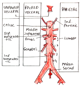
– The aorta gives 3 anterior unpaired visceral branches: the coeliac trunk, superior mesenteric and inferior mesenteric arteries.
– Also gives 3 lateral paired branches: Renal, Middle adrenal and Gonadal arteries
– It finally gives 4 pairs of lumbar arteries and the median sacral artery before dividing into common iliac arteries at L4.
The renal arteries emerge at L1 below the origin of the SMA but above the origin of the inferior mesenteric artery

Venous drainage
The kidneys are drained by the renal veins which drain into the inferior vena cava.
– The IVC is made by the union of the common iliac veins to the right of L5.
– It then passes through the diaphragm at the level of T8.
The IVC is on the right of the midline, so the right renal vein is shorter than the left.
– The left renal vein crosses the aorta below the origin of the SMA to reach the IVC
– On the right, adrenal and gonadal veins drain directly in the IVC whereas on the left, they drain into the left renal vein first.
N.B. Venous draining from the GI tract is to the portal vein, not the IVC.

Urinary Anatomy
After the leaving the kidney, the urine flows via two ureters to the bladder.
– The ureters are 25cm in length and lie anterior to genitofemoral nerves.
– They descend in line with the tips of the transverse processes of L1-5 vertebrae, sacroiliac joints and ischial spines
– They lie anterior to bifurcation of the common iliac arteries on both sides
– The narrowest parts are the PUJ (pelviureteric junction), pelvic brim, and VUJ (vesico-ureteric junction)

Bladder
The bladder is an extraperitoneal organ lying posterior to the pubic symphysis.
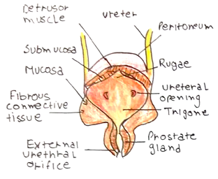
– It has a trigone on the pelvic floor which is formed by the two ureteric orifices and the internal urethral orifice.
– The ureters enter the bladder obliquely making a flap stopping urine backflow.
– Is made of smooth muscle called the detrusor.
– Supplied by parasympathetic nerves S2-4 for micturition. These cause contraction of the muscle to cause urination.
– M3 receptor –> Phospholipase C –> IP3 + DAG –> Contraction

At bladder opening there is the internal sphincter made of smooth muscle that relaxes when the bladder is full.
– In males, there is sympathetic supply to the bladder neck closing it preventing retrograde ejaculation.
The mucosa of the renal pelvis, ureter and the bladder is lined by specialised transitional epithelium. This can withstand the toxicity of the urine and accommodate a high degree of stretching.

