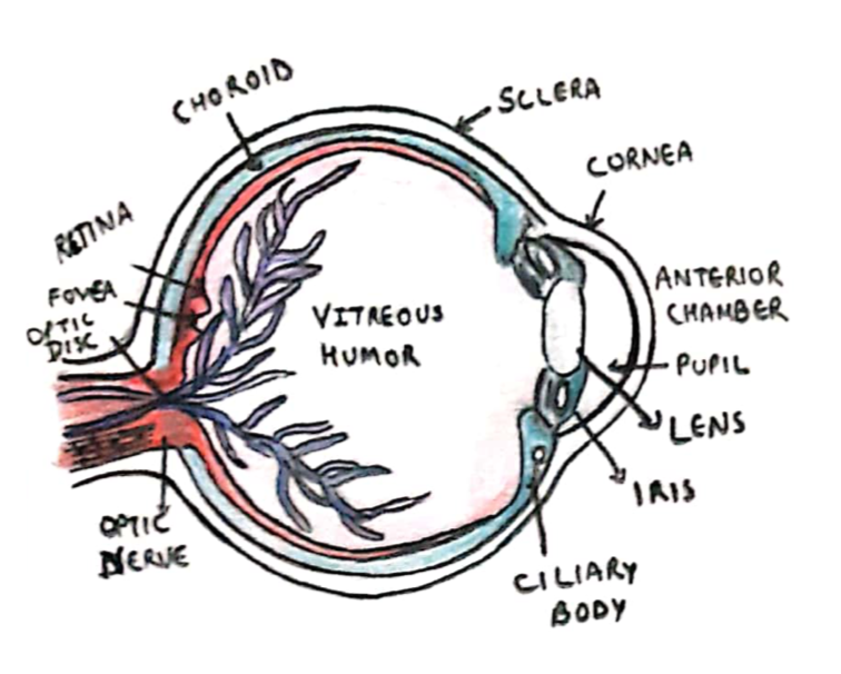
The eye is supplied by the optic nerve (cranial nerve II) and it is divided in anterior and posterior chambers:
– Anterior chamber is the space that lies between the cornea and iris
– Posterior chamber is the space from the iris back to the lens.

Cornea
This is the clear bulging surface in front of the eye which is the main refractive surface of the eye.
– It gets nutrition from the aqueous humor and from tears.
Iris
This contains the sphincter muscle to constrict and dilate pupil
– Parasympathetic stimulation constricts whereas sympathetic dilates
Lens
This is a transparent body enclosed in an elastic capsule, made of proteins and water
– The lens can change shape via the ciliary muscles, in order to focus the image onto the retina.
– Ciliary muscles contract, suspensory ligaments relax, and lens becomes more curved (looking closer)
– Ciliary muscles relax, suspensory ligaments pulled tight, lens flattens (looking further)
Uvea
This is the middle, vascular layer of the eye, lying under the sclera layer and above the nervous layer.
– The three components of the uvea are the iris, ciliary body and choroid
Aqueous Humour
The anterior and posterior chambers of the eye are filled with aqueous humour.
– Secreted by the ciliary epithelium, it has a similar composition to plasma, but a lower protein concentration.
– It maintains the intraocular pressure which gives the eyeball its shape + provides nutrition for avascular ocular tissues (cornea, lens and trabecular meshwork)
The flow of aqueous humour is initially from the posterior chamber through the pupil to anterior chamber
– Then it exits through trabecular meshwork into Schlemm’s canal at the joining point of cornea and sclera
– Can also flow out of the eye via uveoscleral drainage (10%)
– Fluid is normally 15mmHg above atmospheric pressure –> this pressure becomes raised in glaucoma.
Optic Disk
This is the point where the optic nerve meets the retina, giving a small blind spot in each eye
– Is a direct extension of the brain, and has CSF so gets inflamed in raised ICP (papilledema)
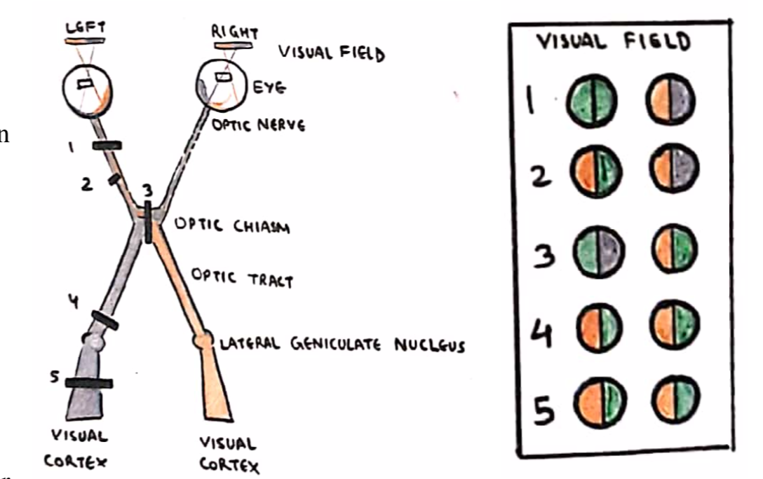
– From disc, visual info flows via optic tract to optic chiasm –> optic radiation –> primary visual cortex.
Visual Tract Lesions
1. Complete monocular blindness
2. Unilateral nasal hemianopia
– Usually due to intracranial optic nerve compression
3. Bitemporal hemianopia
– This occurs due to pressure on the optic chiasm
– If upper quadrants worse:
–> Inferior compression e.g. pituitary tumour
– If lower quadrants worse:
–> Superior compression e.g. craniopharyngioma

4/5. Contralateral homonymous hemianopia
Usually occurs due to a stroke of the posterior circulation affecting the optic tract/radiation or occipital cortex.
– If the hemianopia is incongruous (visual field defects in both eyes not same) = lesion of optic tract
– If hemianopia is congruous (visual field defects in eyes equal) = lesion of optic radiation or occipital cortex
– If hemianopia is macula sparing = lesion of the occipital cortex.
It is also possible to get homonymous quadrantinopias due to specific lesions of the optic tract/radiation.
– To distinguish these, remember PITS
Parietal cortex lesion = Inferior Quadrantanopia
Temporal Cortex lesion = Superior Quadrantanopia

