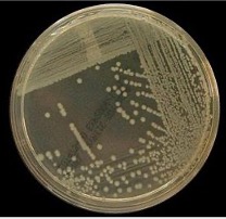
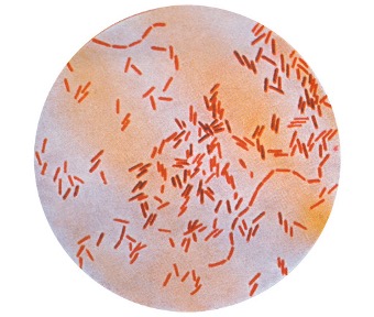
Bacteria are unicellular microorganisms which are composed of a naked DNA chromosome, cytoplasm, cell membrane and cell wall.
They can be divided into Gram Positive and Negative bacteria
i) Gram Positive:
These have a cell wall made of peptidoglycan, take up Gram stain
ii)Gram Negative:
These have a cell wall of peptidoglycan but then an additional outer membrane too, do not take up Gram Stain
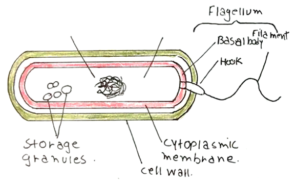
Bacteria can be classified by Gram Stain, shape, enzymes and whether they are aerobic or anaerobic. This will all affect diagnosis and treatment options.
Gram Positive Bacteria
Cocci
These look like round blobs – the most common ones are facultative anaerobes (not obligate).
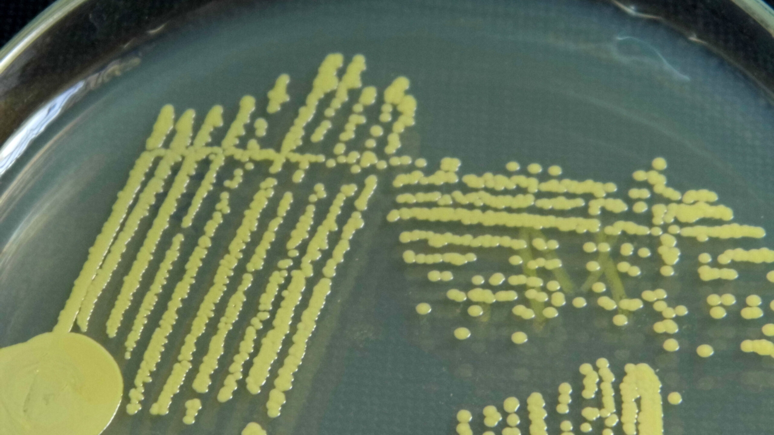
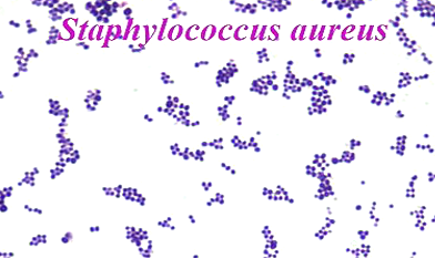
Staphylococcus Aureus
– Catalase positive
– Coagulase positive
– Grows in clusters
– Gives round yellow colonies
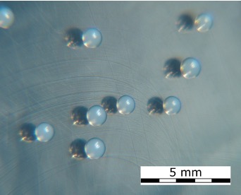
Staphylococcus Epidermidis
– Catalase positive
– Coagulase negative
– Gives small white colonies
Streptococcus Species
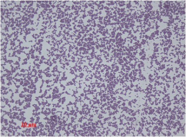
i) S. Pneumoniae (shown in photo)
– Grows in diplococci
ii) S. Pyogenes
– Beta-haemolytic
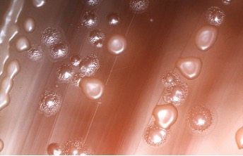
iii) E. Faecalis
– Non-hemolytic
– Colonies milky in the centre
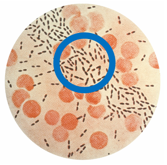
Gram Positive Bacteria
Bacilli
These look like rods. They can be divided according to whether they are aerobes or anaerobes:
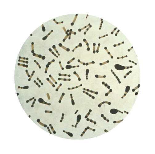
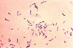
Corynebacterium Diphtheria
– Obligate Aerobe
– Responsible for diphtheria
– Seen on Albert’s stain
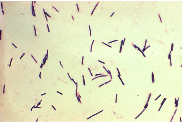
Clostridium Species
– Obligate Anaerobe
– All spore forming and seen by hot malachite green stain
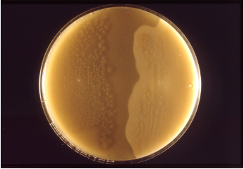
i) C. Perfringens
– Volcano like colonies
– Main cause of gas gangrene
– Secrete a-toxin which is seen using Nagler reaction on egg-white agar
ii) C. Tetani
– Swarm over plate
– Both haemolytic and toxigenic
Gram Negative Bacteria
Cocci
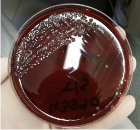
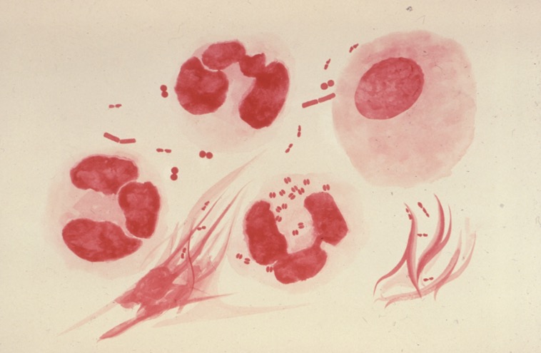
The main Gram-Negative cocci to be aware of is the Neisseria Species, which is responsible for infections like Gonorrhea and Meningitis.
Neisseria Species
– Gram Negative diplococci
– Oxidase positive
– Colonies are round and shiny white
The type of species is distinguished by the site of where the swab was taken from:
e.g. groin -> N. Gonorrhoea
Gram Negative Bacteria
Bacilli
These appear as Gram Negative (pink) rods on culture plates.
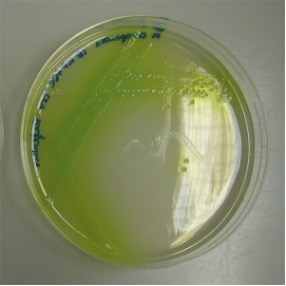
Pseudomonas Aeruginosa
– Obligate Aerobe
– Very large colonies
– Secrete green pigment on agar
– Give acrid smell
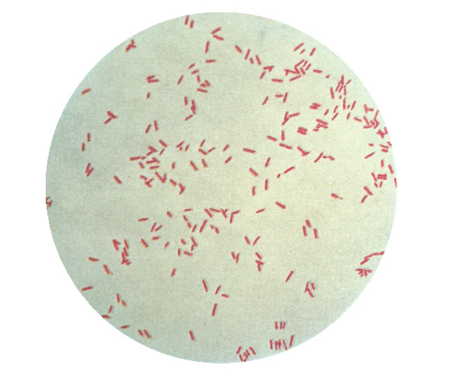
E. Coli
– Facultative anaerobe
– Large colonies with rough margins
– Lactose fermenting
– RED on MacConkey Plate and Yellow on CLED plate
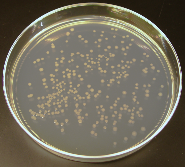
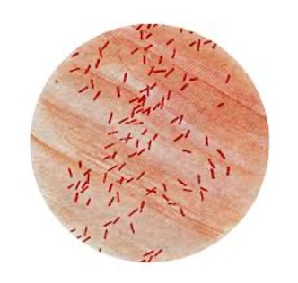
Salmonella
– Facultative anaerobe
– Large colonies with rough margins
– Non-lactose fermenting
– Yellow on MacConkey Plate and Blue on CLED plate
