Lymphomas
This is a neoplastic proliferation of lymphocytes which accumulate in lymph nodes or tissue forming a mass
– They are divided into Hodgkin’s lymphoma (characterized by Reed-Sternberg Cells) and Non-Hodgkin’s
Hodgkin Lymphoma (HL)
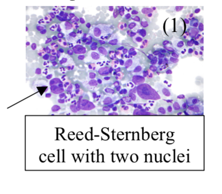
This is a malignant proliferation of Reed-Sternberg (RS) cells. These are large B cells with prominent nucleoli in their multilobed nuclei (owl eyed nuclei)
– The RS cells are CD15 and CD30, and secrete cytokines giving B cell symptoms
– They attract inflammatory cells which make up the tumour bulk
– It usually occurs in a bimodal distribution – seen in 20s and in 60-70-year-old patients

Subtypes of Hodgkin Lymphoma
Risk factors:
– Infection with Epstein Barr Virus, HIV
– Immunosuppression
Symptoms:
– Enlarged, painless superficial lymph node (cervical or mediastinal)
– Lung symptoms –> bronchial/SVC obstruction + cough + hemoptysis + dyspnea
– Systemic –> Weight loss + night sweats
– Pal-Ebstein (1-2-week cyclical) fever
– Lymph nodes hurt with alcohol
Diagnosis:
– Lymph node biopsy is diagnostic
– Raised LDH and eosinophilia
Staging:
1 = singly lymph node
2 = >1 node on same side diaphragm
3 = nodes on both sides of diaphragm
4 = extra nodal site involvement
Management:
Chemoradiotherapy – one regime that is used is ABVD – Adriamycin + Bleomycin + Vinblastine + Dacarbazine
Non-Hodgkin’s Lymphoma
This refer to a group of lymphomas without Reed-Sternberg cells. Overall, these are more common and affects the elderly.
– For low grade lymphomas, there usually indolent but incurable. High grade lymphomas are more curable in early stages but have a worse prognosis
Risk Factors:
EBV infection, immunodeficiency (HIV), autoimmune disease, Sjogren’s syndrome
Types:
i) Follicular lymphoma
This is a proliferation of small B cells that form a follicle-like nodule
– Due to Chr 14 –> 18 translocation activating Bcl2 inhibiting apoptosis + treated with Rituximab
– This is low grade but can transform into diffuse large B cell lymphoma.
ii) Marginal zone lymphoma
This is a proliferation of B cells expanding into the lymph node marginal zone.
– It is associated with chronic inflammatory states like Hashimoto disease + H. Pylori
iii) Diffuse large B-cell lymphoma
This is a proliferation of large B cells growing in sheets
– Most common form of Non-Hodgkin’s lymphoma and very aggressive
iv) Burkitt lymphoma
This is a proliferation of medium B cells, which is most aggressive form.
– Due to c-Myc gene translocation from Chr 8 –> 14 and associated with EBV
– Histology shows “starry sky” appearance of B cells and macrophages with dead cells
– There is an African form, which usually involves the jaw –> risk factor is EBV + Plasmodium Falciparum
– There is also a sporadic form which commonly involves the abdomen, giving ileo-caecal tumours
Symptoms:
– Superficial lymphadenopathy is most common finding (but no alcohol induced pain)
– Systemic B cell symptoms –> fever, night sweats, weight loss (occur later than Hodgins’ s lymphoma)
– Extra-nodal disease (more common) –> gut + bone marrow, lungs
Diagnosis:
Blood tests – FBC shows raised WCC, LDH, inflammatory markers
Biopsy of the lymph node is diagnostic –> then CT scan to assess staging
Staging:
Ann Arbor system –> works same as for Hodgkin’s using stages 1-4
– Also add the letter A (no B cell symptoms) or B (if B cell symptoms are present) e.g. Stage 3B
Management:
– Chemotherapy and radiotherapy
– Surgery can also be used to excise large lymph nodes
Tumour Lysis Syndrome (TLS)
This is a fatal condition which occurs after chemotherapy for high-grade hematological malignancies
– It occurs from the breakdown of the tumour cells and the subsequent release of chemicals from these cells
– This gives rise to electrolyte abnormalities + clinical features
Symptoms:
– AKI –> high urate can lead to acute tubular necrosis, giving azotemia
– Palpitations and arrhythmias –> caused by the high potassium due to AKI
– Electrolyte abnormalities –>Raised Uric Acid, Raised K+, Raised PO43-, Low Ca2+
Management:
Supportive treatment for AKI and hyperkalaemia
– The most important aspect is of prevention. Therefore, during chemotherapy cycles, patients are given prophylactic allopurinol or rasburicase (makes uric acid more easily excreted)
– Therefore it is very important to monitor uric acid levels and U&Es in lymphoma/leukaemia patients who are receiving chemotherapy

