Benign Lesions
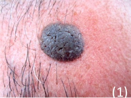
Seborrheic keratosis
This is a benign squamous cell proliferation, common in elderly
– Sudden onset of many lesions suggests GI carcinoma, known as the Leser-Trelat sign
Appearance
– Raised brown/black plaques on extremities/face
– Coin like, “stuck on” appearance that can be very variable
Management
Completely benign but can be removed if suspicion of cancer
Dermatofibroma
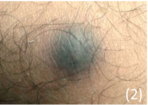
This is an overgrowth of fibrous tissue in the dermis
– Made up of fibroblasts, it is thought to be a reactive process or a neoplasm
– It is completely benign and does not turn into cancer
Appearance
– Single dermal firm nodule found usually on the legs and arms
– Usually <1cm and do not cause symptoms (but can be painful/itchy)
Pre-Malignant Lesions
Actinic Keratoses
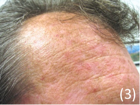
This is a premalignant lesion which is found on sun-exposed skin
– It is caused by abnormal skin proliferation after UV-induced DNA damage
Appearance
– Small crusty scaly lesions usually on sun exposed areas
– Skin coloured with a white/yellow scaly horny surface
Management
– Removed due to skin cancer risk
Keratoacanthoma
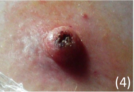
This is a benign tumour (mostly) in sun-exposed skin
– It grows rapidly at a site of minor injury and can be up to 2cm in diameter
Appearance
– Starts as a boil but then becomes dome-shaped with keratin filled crater
– Cannot be clinically distinguished from more severe forms of skin cancer
Management
– Surgical excision as hard to exclude SCC
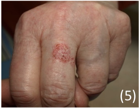
Bowen’s disease (Squamous cell carcinoma in situ)
This is an early superficial stage of squamous cell carcinoma.
– Squamous cells proliferate through epidermis but do not invade dermis.
– Often found in sun-exposed areas such as the legs/hands.
Risk factors:
UV radiation, arsenic, HPV infection and immune suppression
Appearance
– Irregular orange-red scaly patches several centimetres
Management
– Must be removed surgically or 5-flourouracil, but high recurrence rate
Malignant Lesions
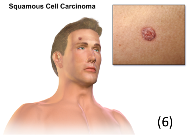
Squamous cell carcinoma (SCC)
This is a proliferation of squamous cells
– Although malignant, it is very slow growing and unlikely to metastasise
Risk factors: UV radiation + Actinic keratoses + Bowen Disease + Smoking
– Immunosuppression –> classically seen after renal transplant or steroid use
– Marjolijn’s ulcer –> this is an aggressive type of SCC which occurs at a site of injury such as an ulcer on the legs
Appearance
– Looks like a scaly, crusty ill defined nodule which may ulcerate
– Can occur on the face, lips, ears and limbs
Management
– Surgical excision with larger margins for larger diameter lesions
– Mohs micrographic surgery used in high-risk patients.
Basal Cell carcinoma (BCC)
This is a proliferation of epidermal keratinocytes
– Most common cutaneous malignancy usually seen in elderly
– Metastasis is very rare as it usually grows slowly and invades locally
Risk factors: UV, sunlight, xeroderma pigmentosum, old age
Appearance – Elevated skin coloured papule with surface telangiectasia
– Commonly scabs and bleeds and then reforms scab
– Pearly appearance, usually seen over the head and neck
Management – 1st line is surgical removal


