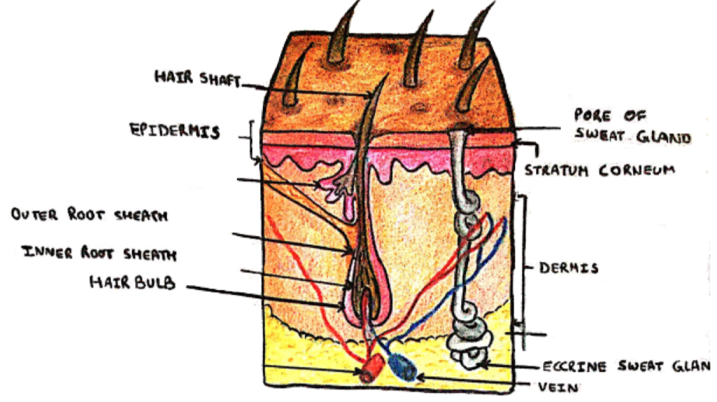
The skin functions as a barrier – made of the dermis and epidermis:
The epidermis can be split into 4 main layers:
1. Stratum corneum – composed of dead keratinocytes
2. Stratum granulosum – has granules in keratinocytes
3. Stratum spinosum – this layer is characterized by desmosomes between keratinocytes
4. Stratum basalis – regenerative stem cell layer
When taking an assessment of the skin, the history is very important.
i) Nature of the problem
ii) Duration of the problem
iii) Treatments so far
iv) Allergies or Sensitivities:
– Including atopy (eczema, asthma, allergy)
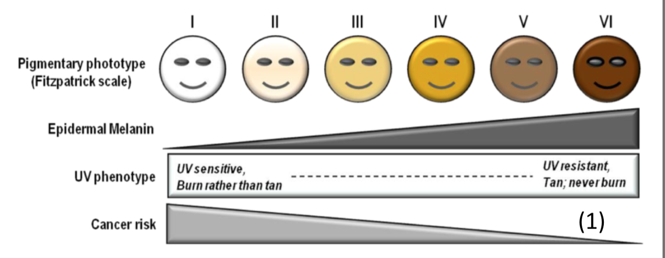
v) Sun exposure:
– Skin response to sun
– Can be classified by the Fitzpatrick skin type scale:
Skin lesions can be classified into many different categories. These depend on the size, texture, colour and whether the lesion is raised or flat.
Macules –> Patches
These are flat, impalpable areas of skin without elevation or depression, but which does have a change in surface colour. They do not have any textural difference from the surrounding skin.
– Macule = <0.5cm
– Patch = >0.5cm
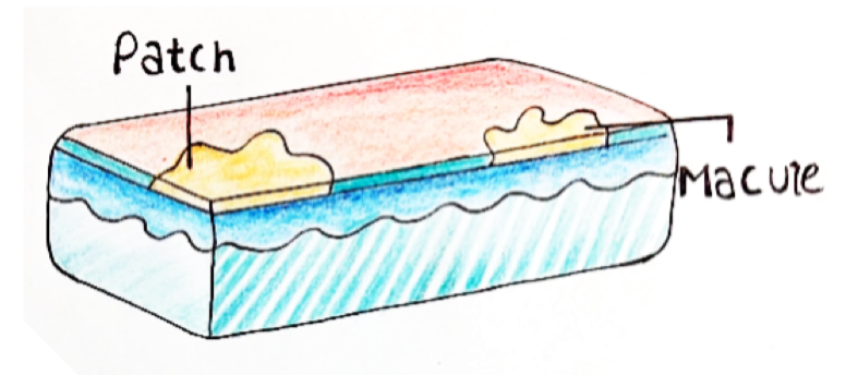
Macules and Patches are described by their colour, in 4 categories:
– Hyperpigmentation –> darker colour than the surrounding skin
– Hypopigmentation –> lighter colour than the surrounding skin
– Erythema –> reddening of the area
– Purpura –> purple spots that do not blanche when pressed
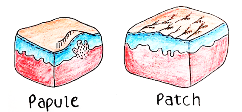
Papules –> Plaques
These are circumscribed, solid elevations of skin with no fluid underneath.
– They are palpable and can be skin-coloured or different coloured
– Papule = <0.5cm
– Plaque = >0.5cm
Cyst
This is a hollow mass that is surrounded by an epithelium-lined wall. It is well demarcated from the adjacent tissue and can contain fluid.
Nodule
This is a solid, circumscribed mass. Similar to a papule but is centred deeper in the dermis – Size =< 0.5cm
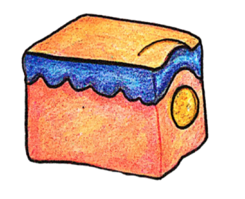
Wheal
A rounded, flat, red plaque that typically disappears in 24 hours, which is part of the inflammatory response
– Characterized by dermal oedema due to histamine release e.g. urticarial rash
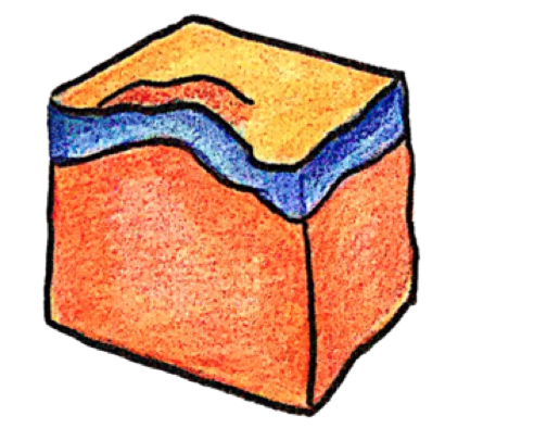
Vesicles –> Bullae
These are blisters full of clear serous fluid. Unlike cysts, they do not have a complete epithelial lining and are therefore less well demarcated. e.g. cold sores
– Vesicles = <0.5cm
– Bullae = >0.5cm

Pustules
These are full of pus and consist of necrotic inflammatory cells.
– They can be sterile or infected

Fissures ➔ Erosions ➔ Ulcers
These are lesions which differ by the depth by which they penetrate the different layers of the epidermis
– Fissure = crack through epidermis to dermis
– Erosion = partial loss of the epidermis
– Ulcers = full thickness loss of epidermis

Scales and Flakes
This is excess shedding of the epidermis.
– e.g. Ichthyosis –> A rare genetic condition making dry, thickened, scaly skin
Crusting
This is when serous fluid or blood leaks under the skin and dries out. It can contain bacterial debris
How to Describe Skin Lesions
When describing skin lesions, we have to refer to 7 things to give a complete description. Remember by the 7S’s:
1) Site
– Site on the body
– Distribution relative to each other
2) Shade
– Pigmented
– Erythematous
– Yellow/orange
– Purpuric
3) Style
– Macule/patch, cyst etc.
4) Size

