free histology & cytology textbooks

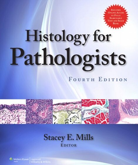
Mills – Histology for Pathologists. 4 ed. 2012. 1362 p.

“This new edition also brings quite a few new authors and their fresh perspectives, sadly necessitated in several instances by the deaths of former world-class senior contributors. Those no longer with us include, Drs. Margaret Billingham, John McNeal, and Sunitha Wickramasinghe. Shortly after submission of his chapter for this edition, my friend Dr. Bernd Scheithauer also left us”–Provided by publisher. Download pdf Back to the same category Note :  All files uploaded here with reserved copyrights for Medicine-21.com & Dr. Ahmed Hafez These materials are for personal use and not for commercial use, so avoid copying or transferring these materials to

Linda – Color Atlas of Fetal and Neonatal Histology. 1 ed. 2011. 414 p.

Color Atlas of Fetal and Neonatal Histology serves as a comprehensive atlas of the developmental histology of the major organs from 15 weeks gestation to the neonatal period. Each section includes text on basic embryologic processes that influence the development of each organ and highlight major histologic features that correspond with certain developmental periods. In addition, there are many color photomicrographs at key developmental stages to assist the reader in identifying appropriate histologic changes at each developmental stage. This book will be of great value to students of embryology, pathology residents and fellows, and attending pathologists who perform fetal autopsies.

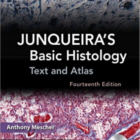
Jinqueira’s Basic Histology. Text and atlas. 14 ed. 2016. 572 p.

Junqueira Histology 14e Addeddate 2017-09-18 10:55:35 Identifier JunqueirasBasicHistologyTextAndAtlas14thEdition Identifier-ark ark:/13960/t8z95fs1v Ocr ABBYY FineReader 11.0 Ppi 600 Scanner Internet Archive HTML5 Uploader 1.6.3 Download pdf Back to the same category Note :  All files uploaded here with reserved copyrights for Medicine-21.com & Dr. Ahmed Hafez These materials are for personal use and not for commercial use, so avoid copying or transferring these materials to other websites or blogs to avoid being subject to local and international legal accountability.
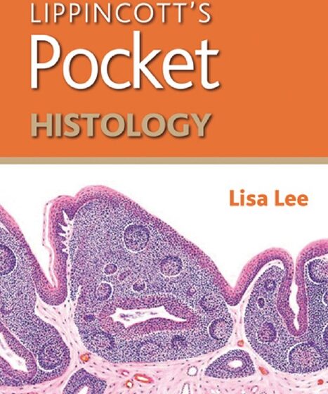
Lippincott’s pocket histology (Lee) 1 ed (2014)

Lippincott’s Pocket Histology is a go-to reference, review and study tool for histology with a strong focus on high-yield topics and presentation. It presents the essential information needed for course and board exam review in a concise, quick-reference format with tables, full-color images, and bullet-point text. The book contains multiple features identifying the clinical significance of concepts, as well as mnemonics to aid in the retention of facts. An index of terms and color-coded systems organization provide easy access to histological facts. This pocket-sized reference intuits how students typically study for exams and provides highly distilled content in one easily

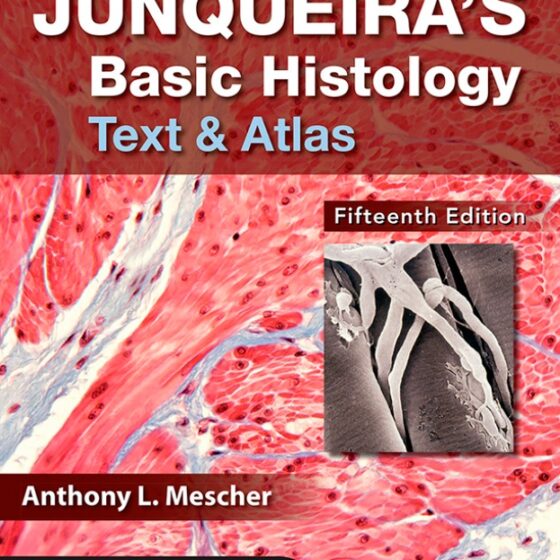
Junqueira’s Basic Histology Text and Atlas (Mescher) 15 ed (2018)

For more than four decades, Junqueira’s Basic Histology has built a global reputation as the most accessible, yet comprehensive overview of human tissue structure and function available. This trusted classic delivers a well-organized and concise presentation of cell biology and histology that integrates the material with that of biochemistry, immunology, endocrinology, and physiology, and provides an excellent foundation for subsequent studies in pathology. Junqueira’s is written specifically for students of medicine and other health-related professions, as well as for advanced undergraduate courses in tissue biology – and there is nothing else like it. Electron and light micrographs comprise a definitive

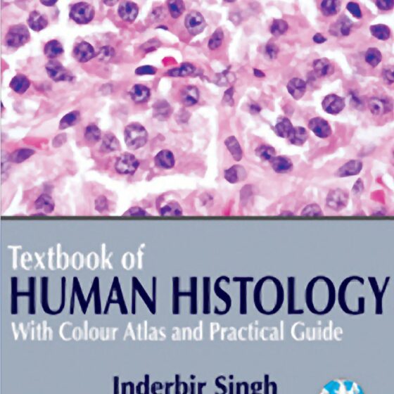
Textbook of Human Histology. With Colour Atlas & Practical Guide (Singh) 6 ed (2011)

This new edition of a well-established textbook has been enriched by the addition of a large number of new photomicrographs of human tissues. The text has also been fully updated and the layout redesigned. High-quality slides are arranged in groups based on similarity of appearanceto aid accurate identification. For medical and biomedical science students, students of anatomy, human physiology and dentistry. This new edition of a well-established textbook has been enriched by the addition of a large number of new photomicrographs of human tissues. The text has also been fully updated and the layout redesigned. High-quality slides are arranged in

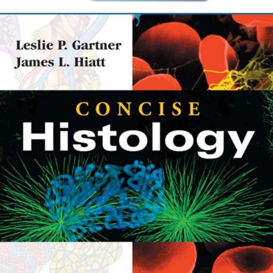
Concise Histology (Gartner) 1 ed (2011)

Description CONCISE HISTOLOGY, by Leslie P. Gartner, PhD and James L. Hiatt, PhD, thoroughly reviews all the histology knowledge required for the USMLE Step 1 in an easy-access outline format. Designed for students who need to learn a large amount of material in a limited time, it presents key information in a readable, concise manner, accompanied by full-color illustrations that clarify complex concepts. Online access via Student Consult includes an online testing centre providing students with class style tests using electron and photomicrographs, cross referenced to the corresponding sections of the textbook. Key Features Efficiently absorb each topic through a

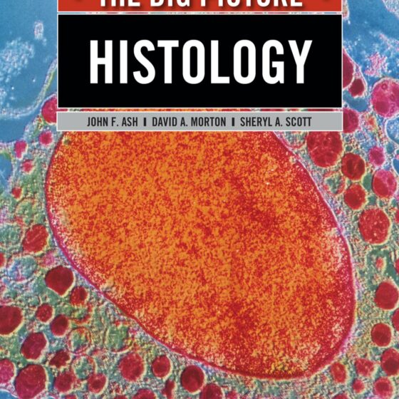
Histology. The Big PicTure (Ash) 1 ed (2013)

Publisher’s Note: Products purchased from Third Party sellers are not guaranteed by the publisher for quality, authenticity, or access to any online entitlements included with the product. Get the BIG PICTURE of Histology – and zero-in on what you really need to know to ace the course and board exams! A Doody’s Core Title for 2020!     400 FULL-COLOR ILLUSTRATIONS Medical Histology: The Big Picture is a different kind of study tool. With an emphasis on what you “need to know” versus “what’s nice to know”, and featuring more than 400 full-color illustrations and micrographs, it offers a focused,

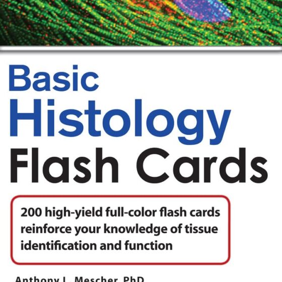
Lange Basic Histology Flash Cards (Mescher) 1 ed (2011)

Flashcards derived from one of the best histology texts ever published Based on Junquira’s Basic Histology 5 STAR DOODY’S REVIEW!“This elegant set of glossy, full-color histology flashcards is a joy to use….This is a wonderful resource for students of medicine and the life sciences. The author brings together high-resolution color images and essential information from the book in a practical format that will help students learn. Students become active learners. Learning histology becomes fun. These flash cards summarize the body of information that students need to know to understand the foundations of pathology and succeed on course and licensing examinations.

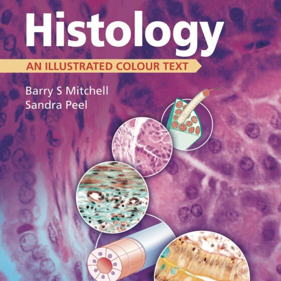
Histology An Illustrated Colour Text (Mitchell) 1 ed (2009)

From the author of the hugely popular Embryology: An Illustrated Text! A concise text, packed with high-quality illustrations, focusing on essential features – if you’re on a course where basic sciences emphasise the principle points of relevance to you, this is the perfect book for you. Volume in Illustrated Colour Text series. Same first author as Embryology volume. Clinically relevant overview ideal for medical and other health science students. Extensively illustrated with both colour photomicrographs and line drawings. Download pdf Back to the same category Note :  All files uploaded here with reserved copyrights for Medicine-21.com & Dr. Ahmed Hafez These

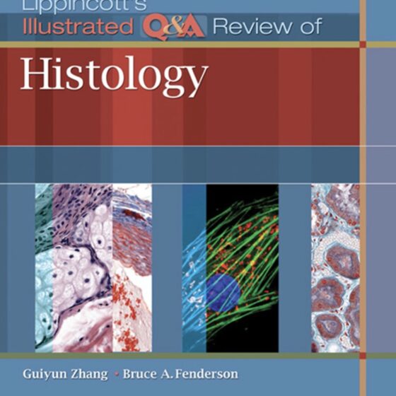
Lippincotts Illustrated QA Review of Histology (Zhang) 1 ed (2015)

Lippincott’s Illustrated Q&A Review of Histology is a resource for students engaged in histology course review and test preparation for the USMLE Step 1 and COMLEX. It contains more than 1,000 USMLE-style and content recall questions with images for approximately 40% of the questions.Features: USMLE-style multiple choice questions—each vignette is followed by a question stem that addresses a key concept in cell biology/histology Detailed explanations address incorrect answers in context Over 480 full-color images illustrate important histologic features Tissues with similar histological features are compared, providing a challenging comprehensive review Side-by-side comparisons of normal tissue and histopathology provide a bridge

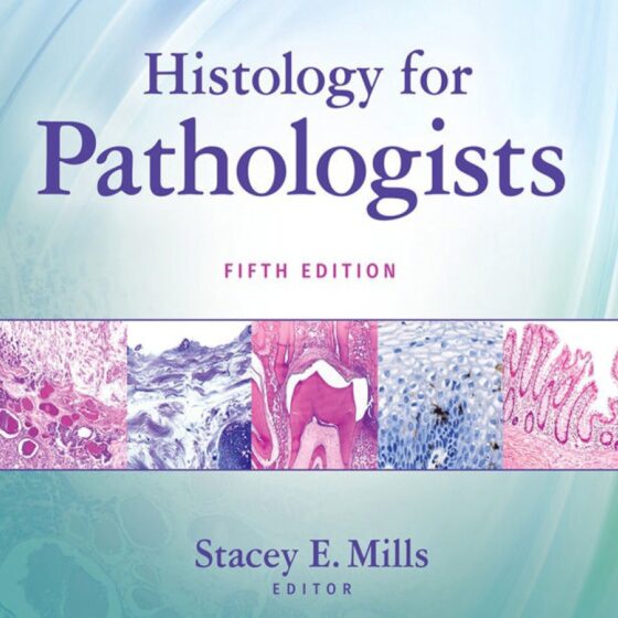
Histology for Pathologists (Mills) ed 5 (2019)

Filled with more than 1,000 images, the latest edition of this award-winning comprehensive classic—written by anatomic pathologists for anatomic pathologists—has been updated with new information on surgical principles and techniques. Like previous editions, the book is designed to bridge the gap between normal histology and pathologic alterations. Now featuring the latest developments and advances made since the previous edition five years ago. Unique among pathology textbooks in using human—as opposed to animal—tissues for discussions on histology. Essential reading for all anatomic pathologists, and particularly helpful for pathology residents throughout their training. Chapters and sections organized by biological system as well

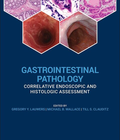
Gastrointestinal Pathology. Correlative Endoscopic and Histologic Assessment (Lauwers) 1 ed (2021)

An illustrated guide to best practices when performing and assessing biopsies for GI conditions of all kinds Accurate diagnosis of GI conditions necessarily entails both the careful taking of biopsies and the informed analysis of tissue material. With that being so, gastroenterologists and GI pathologists alike must have a solid understanding of the techniques, handling requirements, and diagnostic characteristics involved if they are to collaborate effectively. Gastrointestinal Pathology has been designed to provide a clinically focussed and richly illustrated guide to real-world scenarios faced by practicing GI specialists, offering step-by-step instruction and professional advice on the correct diagnosis of all

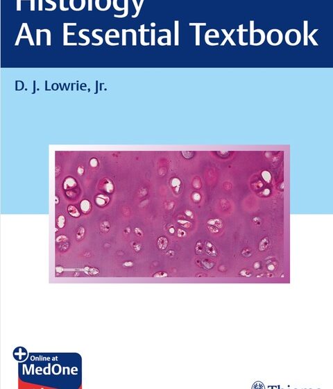
Histology. An Essential Textbook (Lowrie) 1 ed (2020)

Learn to identify histological structures and their correlated functions! Histology: An Essential Textbook is a concise, multimedia study guide for medical students who need to learn the functions and related correlations of cells, tissues, and organs of the human body. Professor D.J. Lowrie, Jr. has written a unique and practical medical histology resource based on self-directed modules originally created to replace live histology laboratory sessions. This resource will help medical students learn how to identify histological structures on slide preparations and electron micrographs. Short videos by the author, who demonstrates structures via digitized histology slides, provide additional guidance. Shorter, targeted

Color Atlas of Fetal and Neonatal Histology (Ernst) 1 ed (2011)

The first edition of Color Atlas of Fetal and Neonatal Histology was an important step in updating the histology texts available to practicing pathologists and pathology trainees who perform fetal autopsy and/or participate in research involving fetal tissues. It was a well-received volume that filled a major gap in pathology references related to normal histology and provided a comprehensive, state-of-the art review of fetal and neonatal histology.  While the basics of fetal histology have changed little in the intervening years since publication of the first edition, this successor edition provides new insights and a fresh perspective. This book contains six

Textbook of Histology (Gartner) 4 ed (2014)

Thoroughly updated to reflect all of the latest concepts and advances in the field, this concise, extensively illustrated text presents the basic science and clinical application of cellular and molecular biology. Functioning as a combined text and atlas, it is essentially two books for the price of one providing a comprehensive, visually engaging histology education from the ground up. Illustrations, tables, chapter summaries and multiple choice questions facilitate comprehension of concepts, and clinical correlations underscore the practical relevance of the material. Key Features Superb illustrations—including photomicrographs, electron micrographs, schematic diagrams and drawings—provide a visual grasp and easier retention of difficult

Outlines of Histology (Slipka) 1 ed (2017)

SUMMARY Histology is the study of tissues. In addition to the description of the basic element of tissues, a cell, this new, revised edition of general histology focuses on the description of four types of basic tissues. This book was initially intended for foreign students of stomatology, however, the text was gradually revised and adjusted to reflect the rapid development of this discipline over the past several years and to be useful for students of other medical fields as well. Download pdf Back to the same category Note :  All files uploaded here with reserved copyrights for Medicine-21.com & Dr.

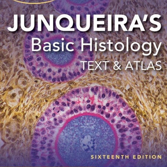
Junqueira’s Basic Histology Text and Atlas (Mescher) 15 ed (2021)

The text that has defined histology for generations―concise, clear, beautifully illustrated, and better than ever A Doody’s Core Title for 2019! For more than four decades, Junqueira’s Basic Histology has built a global reputation as the most accessible, yet comprehensive overview of human tissue structure and function available. This trusted classic delivers a well-organized and concise presentation of cell biology and histology that integrates the material with that of biochemistry, immunology, endocrinology, and physiology, and provides an excellent foundation for subsequent studies in pathology. Junqueira’s is written specifically for students of medicine and other health-related professions, as well as for

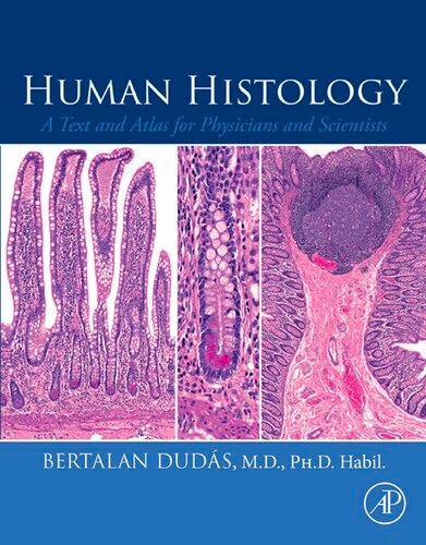
Human Histology. A Text and Atlas for Physicians and Scientists (Dudas) 1 ed (2023)

Visually engaging and easy to use, Human Histology: A Text and Atlas for Physicians and Scientists covers the normal histology of every organ in the human body. This book presents full-page high-definition photomicrographs for organs and tissues, followed by a compact and simple-to-read description of the structures identified on the micrographs, offering a clear, visual understanding of this complex subject. With over 300 outstanding images, this reference is an invaluable resource for every clinical researcher and pathologist in need of easily accessible, relatively simple but detailed enough information on normal histology of different organs systems. Due to its compact, but

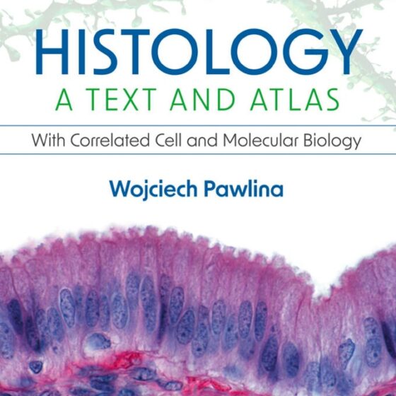
Histology. A Text and Atlas. With Correlated Cell and Molecular Biology (Pawlina) 9 ed (2023)

Combining a reader-friendly textbook and a rich, full-color atlas, Histology: A Text and Atlas: With Correlated Cell and Molecular Biology, 9th Edition, equips medical, dental, health professions, and undergraduate biology and cell biology students with a comprehensive grasp of the clinical and functional correlates of histology and a vivid understanding of the structural and functional details of cells, tissues, and organs.  The 9th Edition of this bestselling resource reflects the latest advances in cellular and molecular biology and relevant imaging techniques, accompanied by large, high-resolution illustrations and full-color photomicrographs that clarify microanatomy in vibrant detail. System chapters align conveniently with