Surgery

Varicose veins

Overview Varicose veins are dilated superficial veins commonly found on the lower limbs. Though often asymptomatic they may cause distressing symptoms and cause cosmetic upset. They are common, and prevalence increases with age and in pregnancy. Aetiology Varicose veins develop due to increased pressure in small superficial veins. In healthy veins, a one way system back to the heart is maintained by valves. This also protects small superficial veins from the increased pressures experienced within the deep compartments of the leg. Venous insufficiency may occur due to valvular incompetence resulting in raised pressures in the superficial veins and the development of varicosities. Risk

Chronic limb ischaemia

Overview Chronic limb ischaemia (CLI) refers to the development of inadequate perfusion to the lower limbs. CLI is characterised by chronic narrowing of peripheral arteries resulting in symptoms of intermittent claudication, rest pain and may threaten limb viability. It can affect the upper limbs, but lower limb disease is far more common. CLI of the lower limb will form the focus of this note. Pathogenesis Chronic limb ischaemia is most commonly the result of atherosclerosis in the arteries supplying the lower limbs. Atherosclerosis is an inflammatory process that causes narrowing of blood vessels. It involves endothelial injury, local inflammatory response and accumulation of

Aortic dissection

Definition & classification Aortic dissection refers to disruption of the medial layer of the aorta due to blood, leading to separation of the layers resulting in a true lumen and false lumen. This most commonly results from an intimal tear allowing blood to enter the intima-media space. As this false lumen fills with blood, it may propagate proximally or distally. This results in either rupture through the adventitia or re-entry to the true lumen via a second intimal tear. Classification is anatomical with two systems most commonly used, Stanford and DeBakey. Stanford: Type A: Ascending aorta is involved Type B: Ascending aorta is not involved DeBakey: Type I: Involves

Acute limb ischaemia

Overview Acute limb ischaemia (ALI) refers to a sudden decrease in blood supply resulting in ischaemic injury to the lower limbs. ALI is caused by sudden obstruction to arterial flow (venous obstruction can cause ALI but is rare) most commonly secondary to embolism or thrombosis. In the setting of complete ischaemia, necrosis results after around 6 hours. Non-viable limbs mandate amputation in around 10-15% of cases and the condition is associated with a high mortality of approximately 15-20% within one year of presentation. This mortality is related both to the condition itself and the age and co-morbid status of the patient group in which it

Abdominal aortic aneurysm

Definition Abdominal aortic aneurysm (AAA) is defined as an abnormal dilatation of the abdominal aorta with a diameter greater than 3 cm. It is a relatively common aortic pathology that results in significant amounts of morbidity and mortality. Between 1-2% of men undergoing NHS screening (over the age of 65) will be found to have a AAA. The vast majority are infrarenal (i.e below the origin of the renal arteries). Risk factors AAA may be considered a degenerative condition of the aorta though its aetiology is not fully understood. A number of risk factors have been shown to be associated with AAA: Age

Varicocele

Overview A varicocele refers to dilated veins within the pampiniform plexus. The pampiniform plexus is a network of veins found in the spermatic cord that drains the testes and epididymis. Varicoceles occur most commonly in adolescent boys and may affect up to 15% of men. The vast majority (around 90%) occur on the left side. It is strongly associated with infertility and as such varicoceles are seen in around 40% of men presenting with infertility. Aetiology Varicoceles occur due to dilation of the pampiniform plexus in the scrotum. The pampiniform plexus is a network of veins found in the spermatic cord that

Testicular torsion

Overview Testicular torsion is a urological emergency, that without intervention leads to ischaemia and loss of the testis. Testicular torsion is a rare but serious condition. Successful treatment requires prompt recognition, referral and surgical intervention. It exhibits a bimodal distribution with peaks in the neonatal period and around puberty. Differential Diagnosis Whilst other diagnoses should be considered, suspicion of testicular torsion requires urgent testicular exploration Trauma Epididymo-orchitis Torsion of the epididymal appendage Testicular tumour (an association between testicular cancer and torsion has been described) Hydrocele Inguinal hernia Systemic diseases (e.g. Henoch-Schönlein purpura, Mumps) Types Testicular torsion may be intravaginal or extravaginal. Extravaginal torsion occurs in-utero and in neonates.

Testicular cancer

Overview Testicular cancer is responsible for 1% of all new cancers in men. It typically presents with a unilateral testicular mass. Incidence appears to be increasing, with approximately 3-10 cases / 100,000 men each year in the Western world. The vast majority of testicular cancers are germ-cell tumours (95%). Overall prognosis, following appropriate therapy, is good. As with all cancers, optimal management requires a multi-disciplinary approach with GPs, specialist nurses, urologists and oncologists all essential. Figures from Cancer Research UK (last accessed Nov 2021). Epidemiology There are approximately 2,300 cases of testicular cancer in the UK each year. It is the 18th most common cancer affecting

Renal colic

Introduction Renal colic classically refers to acute severe loin pain that occurs secondary to a urinary stone. Urinary stones, also termed urolithiasis, refer to stone formation anywhere within the urinary tract. They may be asymptomatic or cause acute loin-to-groin pain due to ureteric obstruction. Urinary stones are extremely common, men are affected up to three times more than women. The peak incidence of symptomatic urinary stones is between 40-60 years in males and late 20’s in females. Aetiology Renal colic is caused by obstruction to urinary flow within the ureter that occurs secondary to urinary stones. The majority of renal stones, approximately 80%, are composed of

Renal cell carcinoma

Overview Renal cell carcinoma is the most common kidney cancer in adults. It accounts for around 80-85% of kidney cancers. They may be found incidentally on abdominal imaging, present symptomatically (e.g. haematuria, loin pain, loin mass, fever) or with features of paraneoplastic syndromes. In those with disease spread it may be local, particularly involving invasion of the renal vein and inferior vena cava (IVC) or distant (commonly the lungs). Management is highly dependent on the stage at diagnosis and patient-based factors. Epidemiology  In 2017, kidney cancers (overall) were the 7th most common cancer in the UK. There are around 13,000 cases of

Prostate cancer

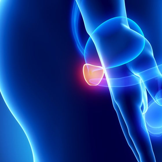
Overview Prostate cancer is the most common malignancy affecting men in the UK. Incidence increases with advancing age and men of Black ethnicity are more commonly affected. It can affect men, trans women, non-binary (assigned male sex at birth) and some intersex patients. Localised disease is commonly asymptomatic but symptoms often develop in locally advanced and metastatic disease. Management is dependent on a multitude of factors but may involve active surveillance, androgen deprivation therapies, radiotherapy, chemotherapy and surgical intervention. Epidemiology 1 in 6 men in the UK will be diagnosed with prostate cancer in their lifetime. In the UK it is the

Hydrocele

Overview Hydrocele refers to a collection of serous fluid between the parietal and visceral layers of the tunica vaginalis. Patients present with (typically) a non-tender scrotal swelling. Hydroceles may be associated with (communicating hydrocele) or without (non-communicating hydrocele) a patent processus vaginalis. Treatment depends on the underlying aetiology, age and symptomatic burden of the hydrocele. NOTE: In rare cases a similar condition may be seen in women where fluid accumulates in the canal of Nuck. Types There are two major types of hydrocele: communicating and non-communicating. Communicating These occur due to the failure of normal closure of the processus vaginalis. This allows the passage of

Chronic prostatitis

Overview Chronic prostatitis is characterised by > 3 months of urogenital pain, often associated with LUTS or sexual dysfunction. Chronic prostatitis may be categorised as: Chronic prostatitis/chronic pelvic pain syndrome: clinical features of chronic prostatitis in the absence of an identifiable bacterial infection. Chronic bacterial prostatitis: relatively uncommon, accounting for around 10% of patients with chronic prostatitis. Aetiology The aetiology of non-bacterial associated chronic prostatitis is poorly understood. Chronic prostatitis/chronic pelvic pain syndrome The aetiology remains poorly understood. Infective and inflammatory triggers may be implicated. There are suggestions that some may have a neuropathic component. Chronic bacterial prostatitis This may develop following an episode

Bladder cancer

Overview Bladder cancer is a common malignancy, in 2017 it was the 11th most common cancer in the UK. In the UK, transitional cell carcinoma is by far the most common cause (around 90%). Advancing age and smoking are significant risk factors for its development. More men than women are affected, though this appears largely due to exposure to relevant risk factors. It classically presents with painless haematuria. Management depends on staging and can involve trans-urethral resection of bladder tumour (TURBT), immunotherapy, chemotherapy, radiotherapy and cystectomy. Epidemiology There are approximately 10,300 cases of bladder cancer in the UK each year. In 2017,

Benign prostatic hyperplasia

Overview Benign prostatic hyperplasia (BPH) is characterised by hyperplasia resulting in lower urinary tract symptoms (LUTS). In BPH, prostatic hyperplasia (increased cell proliferation leading to enlargement) leads to urinary frequency, incomplete emptying, dribbling, hesitancy and nocturia. It may also be complicated by acute or chronic urinary retention. You may come across the term benign prostatic enlargement (BPE). This refers to a clinical finding on digital rectal examination (DRE), BPH refers to a histological diagnosis. There is increasing recognition that LUTS in males are often not secondary to BPH. An open mind should be had with regards to the underlying aetiology in

Acute epididymo-orchitis

Overview Acute epididymo-orchitis is a common cause of testicular pain and swelling. It is caused by acute inflammation of the epididymis (epididymitis) that may affect the testicle (orchitis). Most commonly the aetiology is infectious through the spread of pathogens from the urethra or bladder. It may be related to urinary tract infections or sexually transmitted infections. Testicular torsion must be considered in anybody presenting with acute testicular pain, particularly in children, adolescents and younger men. If there is any diagnostic uncertainty, refer to urology / paediatric surgery. Aetiology Epididymo-orchitis is most commonly caused by sexually transmitted infections or urinary pathogens. Sexually transmitted organisms tend

Acute bacterial prostatitis

Introduction Acute bacterial prostatitis refers to a an infection involving the prostate that may cause significant systemic upset. It tends to present with urinary symptoms, lower back or pelvic pain +/- symptoms of systemic infection. Urinary pathogens are often implicated, commonly Escherichia coli. Less commonly sexually transmitted infections are isolated as the cause. Aetiology E.coli is the most commonly isolated pathogen in acute bacterial prostatitis. Pseudomonas aeruginosa, Klebsiella, Enterococcus and Proteus may all be causes. Sexually transmitted infections, Chlamydia trachomatis and Neisseria gonorrhoea, are less commonly isolated. There are a number of specific scenarios to consider: Recent urethral or prostatic instrumentation (e.g. urethral catheterisation, cystoscopy or transrectal prostate biopsy): rarely acute

Skin tension lines

Overview Skin tension lines reflect internal tension within the skin. Skin tension lines essentially refer to the direction of maximal skin tension at different body sites. They reflect internal tension within the skin due to connective tissue (e.g. collagen) within the dermis. There are different types of skin tension lines. The two most widely recognised are ‘Langer’s lines’ (also known as cleavage lines) and relaxed skin tension lines. Aligning surgical incisions or excisions with these lines produces less tension, optimal scar formation, and ultimately a better cosmetic result. These lines can be drawn on a diagram of the human body. Body

Necrotising infections

Overview Necrotising soft tissue infections (NSTIs) are a collection of severe infections characterised by rapidly progressive soft tissue inflammation and necrosis. Specific examples of NSTIs include: Necrotising fasciitis – a term popularized in the 1950s to emphasise the constant feature of necrotic fascia with spread of infection along fascial planes. Gas gangrene – soft tissue infection, typically caused by Clostridium perfringens, results in myonecrosis and gas formation within tissues. Fournier’s gangrene – refers specifically to necrotising fasciitis of the perineum and scrotum. NSTIs are usually rapidly progressive, resulting in extensive tissue destruction. They are associated with high morbidity and mortality. Accordingly, prompt diagnosis and treatment are essential.

Melanoma

Summary Melanoma is a cancerous growth of melanocytes. NB – This article is in reference to cutaneous melanoma only. Melanoma is common. In the UK, it accounts for approximately 4% of all new cancers. It results from a combination of environmental and genetic factors. Features suspicious of melanoma can be remembered using the mnemonic ‘ABCDE’: Asymmetry Border (irregular) Colour alterations Diameter > 6mm Evolving lesion Patients with a suspicious lesion are referred for a tissue biopsy. In most instances, this constitutes an excisional biopsy with a 2 mm margin. If diagnostic of melanoma, the Breslow thickness, presence of ulceration and the mitotic index are used to histologically stratify the disease. Staging

Ganglion

Overview A ganglion cyst is a fluid-filled swelling the occurs over a joint or tendon sheath. Ganglions are fluid-filled swellings known as cysts that are commonly observed in the hand or wrist. They usually occur over a joint or tendon sheath. They can occur at all ages but usually occur in the second to fourth decades. They are a very common cause of swelling in the hand and wrist. Aetiology & pathophysiology Ganglions contain mucinous, gelatinous fluid. Ganglions are thought to arise from the herniation of connective tissue around key musculoskeletal structures such as tendon sheaths, ligaments, joint capsules, and bursae. This

de Quervain tendinopathy

Overview de Quervain tendinopathy is a common cause of pain in the wrist. de Quervain tendinopathy is a common cause of wrist pain and affects the abductor pollicis longus (APL) and extensor pollicis brevis (EPB) tendons. The cause is unknown but thought to be due to overuse or repetitive movements. It is classically described in women who are postpartum from repeatedly picking up their baby. The condition is typically self-limiting. Aetiology & pathophysiology The exact cause of de Quervain tendinopathy is unknown. Traditionally, de Quervain tendinopathy was thought to be due to repetitive activities that maintain the thumb in extension and abduction

Carpal tunnel syndrome

Overview Carpal tunnel syndrome is a median nerve neuropathy due to compression as it passes through the carpal tunnel in the wrist. Carpal tunnel syndrome (CTS) is due to compression of the median nerve as it passes through the carpal tunnel in the wrist leading to median nerve neuropathy. It is considered the most common upper limb mononeuropathy. CTS is common and usually presents with paraesthesia and/or sensory loss of the first three fingers (thumb, index finger, middle finger), lateral half of the fourth finger (ring finger). As the condition progresses, there is loss of motor function with hand weakness, wasting

Burns

Summary Burns result from damage to skin & deeper tissues caused by external sources or substances. Burns are a major cause of injury and death worldwide. They can have devastating physical and psychological effects on an individual, that can lead to chronic disability. They may be caused by thermal, chemical, frictional or electrical injury. Mortality is increased with large burns, with increasing age and with associated inhalational injury. Effective management necessitates a multi-disciplinary approach. Aetiology Most burns are thermal injures due to scalds, contact & flame burns. Burns may be thermal, chemical, frictional or electrical. Thermal injuries are the most common and include scalds, contact and flame burns. Epidemiology

Basal cell carcinoma

Summary Basal-cell carcinoma (BCC) is a slow-growing, locally invasive, malignant epidermal (basal layer) skin tumour. Typically, a slow-growing skin lesion (over months / years) which commonly occurs on sun-exposed areas of the body. Eighty percent occur on the head and neck. BCC is the commonest form of skin cancer. It is 4-5x more common than squamous cell carcinoma (SCC). They generally affect middle-aged/elderly individuals, unless there is a genetic susceptibility. BCCs are locally destructive and rarely metastasize. Clinical examination typically demonstrates a flesh- or pink-colored lesion with rolled edges, ulceration and telangiectasia (small blood vessels). Treatment is usually by surgical excision, electrodesiccation and curettage (EDC), cryotherapy, or Moh’s micrographic surgery. Rarely, radiotherapy is used.

Wrist fractures

Introduction Fractures of the distal radius are common orthopaedic injuries. Wrist fracture is a term that can refer to any fracture affecting the distal radius, ulna or carpal bones. However, generally, it is used interchangeably to describe a fracture of the distal radius and any accompanied ulna fracture. Isolated fractures of carpal bones tend to be considered separately. This note will focus on fractures of the distal radius (e.g. Colles’ and Smith’s fractures) Fractures of the distal radius exhibit a bimodal distribution; they are seen in the young following high-energy injuries and as fragility fractures in the elderly. Treatment aims to

Tibial plateau fractures

Introduction Tibial plateau fractures are due to high-energy trauma in the young and low-energy falls in the elderly. The tibial plateau refers to the proximal articular surface of the tibia, which forms the knee joint with the femur. Tibial plateau fractures have a bimodal distribution, classically presenting in males in their 40s and females in their 70s. The incidence of these fractures is around 10.3 per 100,000 annually. Aetiology Tibial plateau fractures occur secondary to traumatic injuries Mechanisms Fracture of the tibial plateau can arise from a valgus force, which describes an outside force pushing the knee inwards along a coronal plane.

Septic arthritis

Overview Septic arthritis is an infection of one or more joints. Bacterial infection is by far the most common cause of septic arthritis with staphylococcus aureus most frequently isolated. It normally presents with pain and swelling of the affected joint with signs of systemic infection. The condition may be categorised as: Native joint infection: infection affecting a native joint, management typically requires joint drainage (where appropriate) and antimicrobial therapy. Prosthetic joint infection (PJI): a serious complication of prosthetic joint replacement. Surgical intervention in addition to antimicrobial therapy is commonly required. Septic arthritis requires prompt recognition and management. Untreated it can lead to articular destruction

Osteomyelitis

Overview Osteomyelitis refers to infection of the bone. It is a serious infection that benefits from prompt recognition and treatment. Risk factors include diabetes, old age, peripheral vascular disease and immunocompromise. It may occur for many reasons including open fractures, skin ulcers, surgery (and prosthesis) and haematogenous spread of bacteria. Staphylococcus aureus is the most commonly identified infecting organism. Treatment involves long courses of antibiotics (normally a minimum of 4-6 weeks) and at times surgical debridement. The focus of this note will be osteomyelitis in adult patients. Causative organisms Staphylococcus aureus is the most common cause of osteomyelitis. Staphylococcus aureus: A gram-positive cocci. Includes

Hip fractures

Introduction A hip fracture is a bony injury of the proximal femur typically occurring in the elderly. Older patients are more likely to have gait unsteadiness and reduced bone mineral density, predisposing to fracture. It is a common serious injury in older people, costing the NHS and social care £1 billion per year. Basic anatomy The proximal femur consists of a head, neck, trochanters (greater and lesser) & shaft. It is the largest bone in the human body. The inter-trochanteric line lies on the anterior surface of the femoral neck, running between the trochanters. It demarcates the inferior attachments of the hip capsule. Hip capsule The capsule of the hip is attached

Ankle fractures

Overview Ankle fractures are common lower limb fractures often occurring due to low-energy torsional trauma. They affect women more than men, most commonly those aged 30-60. Ankle fractures account for around 9% of fractures presenting to accident and emergency, representing a significant portion of the trauma workload. Treatment involves restoration of normal anatomical alignment. This may involve conservative management with a walking boot or cast, or involve surgical fixation. Attention must be payed to the risk of venous thromboembolism (VTE) and prophylaxis given where appropriate. Anatomy The ankle is a hinge joint, formed by the malleoli & talus. It is reinforced medially and

Subarachnoid haemorrhage

Definition Subarachnoid haemorrhage (SAH) is bleeding in the subarachnoid space between the arachnoid and pia mater meningeal layers. Aetiology SAH can be a life-threatening emergency and it is estimated that 10-15% of patients die before they reach hospital. SAH can be divided into traumatic or spontaneous Traumatic (tSAH): most common cause of SAH. Usually in setting of a head injury (e.g. fall, assault, road traffic collision) Spontaneous: commonly due to rupture of a cerebral aneurysm (aSAH) Traumatic Trauma is the most common cause of SAH. There is usually evidence of trauma in the clinical history (e.g. road traffic accident). An isolated SAH in

Spinal cord syndromes

Overview Several classic syndromes that have characteristic clinical features based on the area of the spinal cord that is affected. Neurological signs and symptoms can usually be located to the spinal cord based on the presentation of bilateral motor and sensory signs without head or face involvement. In some situations, a specific set of clinical features can develop due to the area within the spinal cord that is affected. These are known as the spinal cord syndromes. We discuss some of the more common ones below. Anatomy The spinal cord is part of the central nervous system (CNS) and forms the main

Radiculopathies

Overview Radiculopathy refers to symptoms or impairments related to the involvement of a spinal nerve root. The spinal nerve roots serve as the main communication between the central nervous system (i.e. the spinal cord) and the peripheral nerves. When a spinal nerve root is affected, it is known as radiculopathy. Radiculopathies may be single or multiple. When multiple nerve roots are affected it is known as polyradiculopathy. The symptoms of radiculopathy are usually characteristic because each root supplies a specific area of cutaneous tissue, known as the dermatome, and a functional group of muscles, known as a myotome. Therefore, patients typically

Extradural haematoma

Definition An extradural haematoma (EDH) is a collection of blood in the extradural space, above the dura mater. Extradural haematoma (EDH), also known as epidural haematoma, refers to a collection of blood in the extradural space between dura and skull. It is most often due to trauma and commonly associated with a skull fracture (75-95% of cases). Aetiology EDH is most commonly due to trauma from a direct head injury. EDH is often life-threatening and patients may require emergency neurosurgery depending on the patient’s neurological status and size of the lesion. It is worth noting that the majority of patients with an

Chronic subdural haematoma

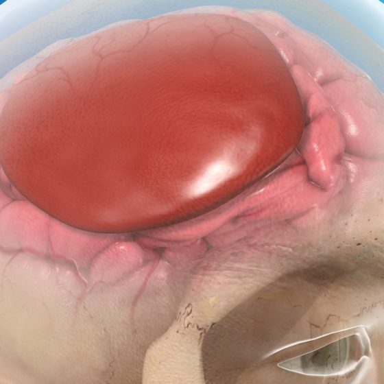
Definition Subdural haematoma is a collection of blood in the subdural space. Subdural haematoma (SDH) refers to a collection of blood in the subdural space. This is situated underneath the dura and above the arachnoid mater meningeal layers. There are several classifications of SDH: Acute (ASDH): bleeding occurring in the last 1-3 days. Chronic (CSDH): blood that has usually been present for > 3 weeks. Subacute: bleeding that occurs between 4 days and 2-3 weeks. Acute on chronic: chronic haematoma that may expand secondary to recurrent bleeding. Aetiology The most common cause of CSDH is trauma. The most common cause of CSDH is trauma in

Cauda equina syndrome

Overview Cauda equina syndrome is due to compression of the collection of nerves distal to the terminal part of the spinal cord known as the cauda equina. The cauda equina, which is Latin for ‘horses tail’, refers to the collection of spinal nerves that lie within the subarachnoid space distal to the last part of the spinal cord known as the conus medullaris. Compression of the cauda equina leads to a syndrome characterised by lower limb weakness, bladder and bowel dysfunction, and abnormal perianal sensation. It is considered a neurosurgical emergency. While not a true spinal cord lesion, cauda equina syndrome is usually

Brain tumours

Overview Brain tumour is a generic and broad term for a wide range of pathologies. A brain tumour is a broad term for an intracranial tumour, or mass, affecting structures such as the meninges, brain, glands, neurovascular structures and/or bone. Other words that may be used to describe an intracranial tumour include a mass, growth, space-occupying lesion (SOL) or central nervous system (CNS) lesion. The term ‘brain tumour’ itself is very generic and should generally be avoided as the management and prognostication for each type differ considerably. Where possible, it is important to use precise terminology. The clinical presentation, management, and prognostication

Acute subdural haematoma

Definition Subdural haematoma is a collection of blood in the subdural space. Subdural haematoma (SDH) refers to a collection of blood in the subdural space. This is situated underneath the dura and above the arachnoid mater meningeal layers. There are several classifications of SDH: Acute (ASDH): bleeding occurring in the last 1-3 days. Chronic (CSDH). blood that has usually been present for > 3 weeks. Subacute: bleeding that occurs between 4 days and 2-3 weeks Acute on chronic: chronic haematoma that may expand secondary to recurrent bleeding Aetiology The most common cause of ASDH is trauma. ASDH is often a life-threatening emergency and historically

Pancreatic adenocarcinoma

Overview Pancreatic adenocarcinoma is the most common form of pancreatic cancer and a major cause of cancer-related death. They arise from the ductal epithelium as the result of accumulated genetic mutations. Adenocarcinomas are the most common pancreatic neoplasm, accounting for around 85% of cases. Asymptomatic through much of its development, sadly this cancer is commonly diagnosed at a late stage when the chance of cure is faint. When caught early, surgery may offer a chance at curative therapy. Epidemiology There are approximately 10,500 cases of pancreatic cancer each year in the UK. Overall it is the tenth most common cancer in the UK,

Chronic pancreatitis

Overview Chronic pancreatitis refers to chronic, irreversible, inflammation and/or fibrosis of the pancreas. Chronic pancreatitis is traditionally considered as chronic, irreversible, inflammation and/or fibrosis of the pancreas. It is essentially a fibroinflammatory syndrome (evidence of fibrosis and inflammation) that occurs in patients with genetic and/or environmental risk factors for pancreatic injury. In the Western world, the leading cause of chronic pancreatitis is alcohol. Injury leads to structural and/or functional changes that include atrophy, calcification, strictures, exocrine dysfunction, endocrine dysfunction, and even increased risk of pancreatic cancer. Management centres on pain control and endoscopic or surgical treatment of complications. Exocrine and endocrine

Acute pancreatitis

Introduction Acute pancreatitis refers to an acute inflammatory process involving the pancreas. Pancreatitis occurs due to the uncontrolled release of activated pancreatic enzymes within the pancreas resulting in autodigestion. Patients may suffer a spectrum of disease from mild abdominal discomfort to multi-organ failure. Pancreatitis has an annual incidence of 13-45 cases per 100,000, with trends showing that it is becoming more common. It tends to occur more in men and is commonly secondary to gallstones or alcohol misuse. Aetiology Alcohol misuse and gallstones are the most common causes of acute pancreatitis. Alcohol misuse and gallstones are responsible for upwards of 75% of cases of

Peritonitis

Overview Peritonitis refers to inflammation of the peritoneum, which is the lining of the abdomen. Peritonitis is a really important clinical sign that refers to inflammation of the lining of the abdomen. The peritoneum is a serous membrane that essentially lines the abdominal cavity. It is composed of two layers and a potential space: Parietal peritoneum: lines the abdominal cavity. Boundaries are the pelvis inferiorly, retroperitoneal space posteriorly, diaphragm superiorly, and abdominal wall anteriorly Visceral peritoneum: wraps around the visceral (internal) organs located within the intraperitoneal space Peritoneal cavity: a potential space located between the parietal and visceral peritoneum. It contains

Perianal abscess

Overview Perianal abscesses are one of the most common complaints on the general surgery take. Perianal abscess present with an acute and tender perianal swelling. Systemic upset – with fevers and malaise – may be present. Management is typically with incision and drainage, though conservative management with antibiotics may be trialed. Risk factors There are a number of risk factors associated with perianal abscesses. Fistula-in-ano Inflammatory bowel disease Diabetes mellitus   Immunosuppression Clinical features Symptoms are typically pain and swelling in the peri-anal region. Most patients are systemically well at presentation, but a significant minority may present with features of systemic infection or

Inguinal hernias

Definition & classification A hernia refers to an organ or part of an organ that protrudes outside the walls of its usual cavity. Hernias may be described as: Reducible: the hernia may be completely returned into its original cavity. Irreducible: the hernia cannot be completely reduced, typically secondary to adhesions between the hernia and hernial sac (incarcerated). Strangulated: constriction of the hernia results in impaired circulation. These hernias represent a surgical emergency. Inguinal hernias refer to protrusions in the inguinal or scrotal region. They are a common surgical pathology, responsible for over 60,000 procedures in England in 2011/12. Men are nine times more likely to be affected than women. There

Haemorrhoids

Overview Haemorrhoids refer to abnormally swollen vascular cushions that are located in the anal canal. Haemorrhoids are essentially a cluster of vascular, smooth muscle, and connective tissue that lies along the anal canal in three columns. These are often referred to as mucosal anal cushions or haemorrhoidal cushions. These cushions are actually normal anorectal structures that are found universally in healthy individuals. In clinical practice, we use the term ‘haemorrhoids’ to refer to the symptomatic enlargement and displacement of the normal haemorrhoidal cushions that can lead to perineal irritation, anal itching, fecal soiling, and most commonly painless rectal bleeding. Haemorrhoids are extremely

Diverticulitis

Overview Diverticulitis is characterised by acute inflammation and infection of diverticula. Diverticulitis is a common presentation on any surgical take. It typically presents with abdominal pain and fevers. Terminology is key to understanding diverticulitis and its associated diseases: Diverticula: Sac like protrusion of the colonic mucosa through the muscular wall. Diverticulosis: The presence of asymptomatic diverticula. Diverticular disease: Symptomatic diverticula (e.g. abdominal pain) in the absence of inflammation. Also used as an umbrella term. Diverticulitis: Symptomatic acute inflammation and infection of diverticula. Diverticulosis most commonly affects the sigmoid colon but may occur anywhere in the gastrointestinal tract. Right sided colonic disease is more common in those of Asian

Colorectal cancer

Introduction Colorectal cancer (CRC) is the fourth most common malignancy in the UK and a major cause of morbidity and mortality. It refers to malignancies that arise from the beginning of the colon, the caecum, through to the end of the rectum. It may be found in a myriad of ways including with screening, incidentally on imaging or endoscopy, or following presentation with change in bowel habit, iron deficiency anaemia or bowel obstruction. Management depends on staging, patient factors and patient wishes. Treatment modalities include surgical resection, metastasectomy, chemotherapy and radiotherapy. Epidemiology In the UK there are around 42,300 cases of colorectal cancer

Colonic ischaemia

Introduction Colonic ischaemia refers to insufficient blood supply to the large bowel. Intestinal ischaemia occurs when blood flow to the intestines (small and large bowel) is reduced and thereby insufficient for the needs of the intestines. The cause of insufficient blood flow varies and can be occlusive (thrombosis, embolus) or non-occlusive (vasoconstriction, hypoperfusion). The terminology surrounding intestinal ischaemia can be confusing at times with certain terms used interchangeably. For the purposes of this note we define the following terminology (in line with the general consensus): Colonic ischaemia: refers to ischaemia affecting the colon (the focus of this note). Mesenteric ischaemia: this

Cholelithiasis

Overview Cholelithiasis (gallstones) refers to the development of a solid deposit or ‘stone’ within the gallbladder. Though largely asymptomatic in a significant proportion of patients they become problematic. In the UK around 60,000 cholecystectomies are performed each year. The terminology around gallstones can be confusing so is worth reviewing: Cholelithiasis: refers to gallstones – solid deposits that develop in the gallbladder. Choledocholithiasis: refers to gallstones within the biliary tree. Biliary colic: refers to a self-limiting pain in the RUQ/epigastrum associated with gallstones. Acute cholecystitis: refers to the acute inflammation of the gallbladder, most commonly caused by gallstones. Acute cholangitis: refers to infection of the biliary tree, commonly

Bowel obstruction

Definition & classification Bowel obstruction refers to complete or partial disruption of the normal flow of gastrointestinal content. It may occur in the small or large intestines, and is secondary to mechanical obstruction and/or peristaltic failure (non-mechanical). Classifying bowel obstruction depends on the location, segments of intestines involved, underlying aetiology and whether blood flow is compromised, which could lead to ischaemia and perforation. Complete obstruction: no fluid or gas is able to pass beyond the site of obstruction. Partial/incomplete obstruction: some fluid or gas is able to pass beyond the site of obstruction. Mechanical obstruction: physical blockage to the flow of gastrointestinal content.

Biliary colic

Overview Biliary colic refers to a pain in the RUQ/epigastrium caused by gallstones. It is the most common symptomatic manifestation of cholelithiasis (gallstones) affecting around 10-20% of patients. Though termed a ‘colic’ the pain is normally constant lasting from 30 minutes to 6 hours. The pain occurs when a stone impacts against the cystic duct during contraction of the gallbladder with increased pressures in the gallbladder itself. Biliary colic is generally considered an indication for elective laparoscopic cholecystectomy after appropriate investigation. See our Cholelithiasis notes for more about the aetiology, types and risk factors. Gallstones Cholelithiasis (gallstones) refers to the development of a solid

Appendicitis

Introduction Appendicitis may be defined as inflammation of the appendix. Acute appendicitis is a common surgical pathology that typically presents with acute abdominal pain. There are upwards of 50,000 cases in the UK each year. Appendicitis has a slight male preponderance and is uncommon at the extremes of age. The majority of cases occur in those aged 15-59 years old. Anatomy The appendix is a short appendage, normally 5-10 cm long, that opens onto the caecum. The appendix may also be referred to as the vermiform (to resemble a worm) appendix. It is a blind-ended tube that arises at the posteromedial aspect

Anterior abdominal wall

Introduction The abdominal wall refers to the layers of anterolateral structures including skin, muscles, nerves, vessels and connective tissues between the thorax and pelvis. The muscles of the abdominal wall have multiple functions. They act to protect and contain the abdominal viscera. They are able to contract, increasing intra-abdominal pressure, aiding expiration, coughing and vomiting. Together they help with truncal movements, particularly against resistance. They are organised into layers with the rectus abdominis found centrally and the (from superficial to deep) external oblique, internal oblique and transversus abdominis found anterolaterally. Surface anatomy The anterior abdominal wall may be divided into nine regions. The nine regions of the abdomen

Anal fissure

Overview An anal fissure refers to a tear in the lining of the anus or anal canal. An anal fissure is one of the most common anorectal disorders that refers to a tear in the lining of the anal canal. This classically leads to pain, particularly on defaecation, and can lead to PR bleeding. They may be acute or chronic: Acute: present for < 6 weeks Chronic: present for > 6 weeks Anal fissures are common with a peak incidence between 15-40 years old, although they can occur at any age and have an equal sex prevalence. Aetiology Anal fissures are most

Acute mesenteric ischaemia

Introduction Mesenteric ischaemia refers to insufficient blood supply to the small intestines leading to ischaemic and inflammatory changes. The terminology surrounding intestinal ischaemia can be confusing at times with certain terms used interchangeably. For the purposes of this note we define the following terminology (in line with the general consensus): Colonic ischaemia: refers to ischaemia affecting the colon. Mesenteric ischaemia: this term tends to be reserved to describe ischaemia affecting the small intestines. Mesenteric ischaemia can be divided into two major categories: acute and chronic. Acute mesenteric ischaemia (AMI) results from the acute insufficiency of blood supply to the small intestines. It

Acute cholecystitis

Overview Acute cholecystitis refers to inflammation of the gallbladder most commonly occurring due to impacted gallstones. After biliary colic, acute cholecystitis is the second most common complication of gallstones (cholelithiasis) affecting an estimated 0.3-0.4% of patients with asymptomatic gallstones each year. Relatively rarely acute cholecystitis occurs in the absence of gallstones (acalculous cholecystitis). Management aims to treat the infection and symptoms (antibiotics, fluids, analgesia) and prevent recurrence (laparoscopic cholecystectomy – ‘hot’ or interval). See our Cholelithiasis notes for more about the aetiology, types and risk factors. Calculous cholecystitis Cholelithiasis (gallstones) are by far the most common cause of acute cholecystitis. Cholelithiasis Gallstones affect up

Acute cholangitis

Overview Acute cholangitis refers to infection of the biliary tree characteristically resulting in pain, jaundice and fevers. Acute cholangitis almost always occurs due to bacterial infection secondary to biliary obstruction. The terms acute and ascending cholangitis can be used interchangeably. Biliary obstruction is often secondary to choledocholithiasis (gallstones in the biliary tree) or biliary strictures (both benign and malignant). Management involves antibiotics, supportive care and urgent decompression of the obstructed biliary system. Epidemiology Acute cholangitis is a relatively uncommon condition. The exact incidence is unknown. The median presenting age is 50-60, affecting men and women equally. There appears to be greater incidence in

Abdominal incisions

Overview A surgical incision refers to a cut made through the skin to access deeper tissue or facilitate an operation. An abdominal incision refers to a surgical cut made anywhere on the abdomen. This enables access to deeper tissue to facilitate an operation by gaining access to the intra-abdominal or intra-pelvic cavities. There are a number of characteristic incisions that are completed on the abdomen to facilitate open surgery. Many of these have eponymous names. Knowledge of abdominal incisions is important for exams. Kocher This is a subcostal incision that is completed to gain access to the upper abdomen. A Kocher (subcostal) incision

Tonsillitis

Overview Tonsillitis refers to the acute inflammation of the palatine tonsils secondary to infection. Acute tonsilitis is very common, especially in children. It is most frequently viral and associated with an upper respiratory tract infection (URTI). It may also be caused by bacteria (e.g. Group A Streptococci). Management typically involves reassurance, fluids and analgaesia. Where a bacterial cause is suspected, antibiotics +/- steroids are given. The most common complication of acute tonsillitis is a peritonsillar abscess (see Quinsy). Recurrent severe tonsillitis results in considerable morbidity and time lost from school or work. Clinical features Clinical examination typically demonstrates enlarged and erythematous tonsils. Exudate is more uncommon and typically suggests

Thyroid cancer

Introduction Thyroid cancer accounts for around 1% of new malignancies in the UK each year. Thyroid cancer refers to a group of malignant conditions affecting the thyroid gland. They most commonly present with a thyroid lump and are diagnosed following triple assessment. Surgical resection (thyroidectomy and hemithyroidectomy) is the mainstay of treatment. Prognosis is related to the type, stage and age at diagnosis. This note will focus primarily on the diagnosis and management of papillary and follicular thyroid cancer (together often referred to as differentiated thyroid cancers). Epidemiology There are approximately 3,900 diagnoses of thyroid cancer in the UK each year. It is

Thyroid anatomy

Overview The thyroid is located in the mid-line of the neck, anterior to the trachea and inferior to the larynx. It is found within the pre-tracheal fascia, one of the fascial compartments of the neck. It is a thin fascia in the anterior part of the neck and is composed of two parts: Muscular part: contains the infrahyoid muscles Visceral part: contains the thyroid, trachea and oesophagus The thyroid gland is comprised of two lateral lobes connected by a central isthmus. It is surrounded by a fibrous capsule and located at the level of vertebrae C5-T1. The pyramidal lobe, an embryological

Ramsay Hunt syndrome

Overview Ramsay hunt syndrome refers to the reactivation of herpes zoster in the geniculate ganglion. The terminology can be confusing. Technically it is Ramsay Hunt syndrome type II (type I is a form of cerebellar degeneration). It can also be referred to as herpes zoster oticus. It is characterised by a facial nerve palsy associated with a vesicular rash affecting the ipsilateral ear, hard palate and anterior two-thirds of the tongue. Treatment is with antivirals, steroids (+/- gastro-protection) and appropriate eye care.   Facial nerve The facial nerve is divided into five branches, which collectively control the muscles of facial expression. The

Quinsy

Overview Quinsy, also termed peritonsillar abscess, is a collection of pus in the peritonsillar space. There are two types of infection that affect the peritonsillar regions: Peritonsillar cellulitis: refers to infection of the peritonsillar tissue in the absence of a discrete collection of pus (abscess). Peritonsillar abscess (quinsy): refers to a discrete collection of pus in the peritonsillar region. Quinsy occurs most commonly as a complication of bacterial tonsillitis. Unlike simple tonsillitis, it mandates surgical intervention to drain the collection. Group A beta-haemolytic streptococcus and haemophilus influenzae are commonly implicated. Severe complications are rare but can include sepsis, the spread of the infection through the deep

Pharyngeal pouch

Overview A pharyngeal pouch, also known as Zenker’s diverticulum, is a sac-like out-pouching that occurs within the hypopharynx. A Zenker diverticulum (ZD), more commonly known as a pharyngeal pouch, is a herniation of the posterior pharyngeal wall that occurs at an area of weakness within the inferior pharyngeal constrictor. It typically occurs in older adults and can lead to symptoms of dysphagia, regurgitation, and more seriously, aspiration pneumonia. Pharyngeal pouch seen at endoscopy with a transparent distal attachment. Evidence of previous endoscopic stapling Epidemiology A pharyngeal pouch is rare before the age of 40. A pharyngeal pouch is a relatively rare condition

Ménière’s disease

Introduction Ménière’s disease is a disorder of the inner ear that manifests itself with attacks of vertigo, tinnitus and hearing loss. The majority of cases of Ménière’s disease are idiopathic but the condition is thought to be related to abnormal production and absorption of endolymph (the fluid that fills portions of the labyrinthine system). Where an underlying cause is identified it is referred to as Ménière’s syndrome. The disease is characterised by episodes of vertigo, tinnitus and fluctuating hearing loss. As the condition develops, symptoms, in particular tinnitus and hearing loss, may become permanent and progressively worse. Management involves supportive care during acute

Malignant otitis externa

Overview Malignant otitis externa is a serious condition where infection spreads from the external auditory canal to the skull base. It most commonly affects the elderly, diabetic or immunocompromised. Pseudomonas aeruginosa is by far the most common causative pathogen. Treatment is primarily with intravenous antibiotics and supportive care. Risk factors The majority of cases of malignant otitis externa are associated with diabetes or glucose intolerance. Diabetes Old age Immunocompromise (e.g. HIV) Aetiology Pseudomonas aeruginosa is the most common cause of malignant otitis externa. Pseudomonas aeruginosa is a gram-negative rod. It normally causes infections in patients with deficient immune systems or poor glycemic control. Less commonly other pathogens are

Epistaxis

Introduction Epistaxis refers to bleeding from the blood vessels within the nasal mucosa. Nosebleeds are common. The vast majority resolve spontaneously or with basic first aid. However, it remains a relatively common cause of presentation to A&E departments, with up to 6% of the UK population seeking medical attention due to epistaxis at some point in time. On occasion, epistaxis requires intervention (e.g. nasal cautery or nasal packing) typically performed by an A&E or ENT doctor. In certain settings (discussed in more detail below), epistaxis should trigger an outpatient referral to ENT to exclude an underlying causative lesion (e.g. malignancy). Aetiology

Chronic rhinosinusitis

Overview Chronic rhinosinusitis refers to inflammation of the nasal cavities and paranasal sinuses lasting > 12 weeks. Chronic rhinosinusitis is broadly defined as inflammation of the nasal cavities and paranasal sinuses that lasts for longer than 12 weeks. However, it is actually a very heterogenous condition that is difficult to fully define and may be part of a wider systemic illness (e.g. granulomatosis with polyangiitis). There are various subtypes of chronic rhinosinusitis although the cardinal features remain the same, which include nasal congestion, mucopurulent nasal discharge, and facial pain or pressure. Treatment aims to reduce symptom burden and improve quality of

BPPV

Overview Benign paroxysmal positional vertigo (BPPV) is an inner ear disorder characterised by episodes of positional vertigo. BPPV is the most commonly encountered cause of vertigo seen in clinical practice. It is caused by otoconia (loose debris composed of calcium carbonate) within the semilunar canals of the inner ear. Attacks are triggered by head movements that result in movement of the otoconia, abnormal motion of endolymph and the feeling of vertigo. A careful history and examination are required to exclude other causes of vertigo. ‘Manoeuvres’ (explained in detail below) are used in both the diagnosis and treatment of BPPV. The Dix-Hallpike

Allergic rhinitis

Overview Allergic rhinitis is a common condition secondary to IgE-mediated inflammation of the nasal mucosa. Allergic rhinitis is a common condition characterised by sneezing, runny nose (i.e. rhinorrhoea), nasal obstruction and itching. It is more colloquially known as ‘Hayfever’. These symptoms are brought about by IgE-mediated inflammation secondary to antigens known as ‘allergens’ that can include grass, tree pollen, or house dust mites among many others. The diagnosis of allergic rhinitis is clinical and management aims to dampen the immune response to allergens principally with anti-histamines and intranasal glucocorticoids. It may be a very disabling condition and is commonly associated with

Acute rhinosinusitis

Overview Acute rhinosinusitis refers to acute inflammation of the nose and paranasal sinuses. Acute rhinosinusitis is a common condition that is usually caused by a viral pathogen. It leads to typical features of nasal congestion, nasal discharge, and facial pressure/pain that is worse on bending forward. These symptoms completely resolve within 4 weeks. Similar to an upper respiratory tract infection, symptoms will improve without intervention and antibiotics are rarely required. Terminology Rhinosinusitis is a better term than simply ‘sinusitis’ because inflammation of the nasal cavities almost always accompanies sinusitis. Sinusitis versus rhinosinusitis Sinusitis: symptomatic inflammation of the paranasal sinuses Rhinosinusitis: symptomatic inflammation

Acute otitis media

Overview Acute otitis media (AOM) refers to inflammation of the middle ear with effusion and clinical features of a middle ear infection. Normally AOM is a self-limiting condition, commonly occurring in children, though adults can also be affected. In general symptoms settle after 3 days, but can remain for a week. Bacteria or viruses are normally implicated – differentiating the two is challenging. The majority of cases do not require antibiotic therapy and will settle with time. Though rare, serious complications such as mastoiditis, meningitis, intracranial abscess, sinus thrombosis or facial nerve paralysis may occur. AOM may become persistent or recurrent. NICE

Acute otitis externa

Overview Otitis externa refers to inflammation of the external auditory canal. Though the inflammation can have many causes (e.g. allergic, dermatologic), infection, in particular by bacteria, is the most common cause. It may be categorised (definition from NICE CKS) as: Acute: lasts 3 weeks or less Chronic: lasts 3 months or longer Malignant otitis externa (MOE) is a rare life-threatening condition where the infection spreads to the surrounding bone, it is covered here. Epidemiology Up to 10% of people will experience acute otitis externa in their lifetime. Though otitis externa (both acute and chronic) can occur at any age, it is more common in children and

Acoustic neuroma

Overview An acoustic neuroma is a benign Schwann-cell derived tumour, which commonly arises from the eight cranial nerve. An acoustic neuroma, also known as a vestibular schwannoma, is a benign intracranial tumour that is derived from Schwann cells that are one of the major supporting nerve cells in the peripheral nervous system. It is typically a slow growing tumour that arises from the eighth cranial nerve supplying the inner ear. This cranial nerve, also known as the vestibulocochlear nerve, has important functions in both hearing and balance. An acoustic neuroma is typically unilateral and leads to unilateral sensorineural hearing loss (bilateral

Lactational mastitis

Introduction Lactational mastitis is a relatively common condition affecting post-partum women. Also termed puerperal mastitis, it can affect up 1/3 of women in the post-partum setting (although many studies put this figure closer to 10%). It can be complicated by the development of a breast abscess. These terms may be defined as: Mastitis: refers to inflammation of the breast tissue. Mastitis may be lactational (occurring in lactating women) or non-lactational. It can also be categorised as infectious or non-infectious. Breast abscess: refers to a localised collection of pus within the breast. Again these may be lactational (occurring in lactating women) or

Fibroadenoma

Introduction Fibroadenomas are the most common benign tumour of the breast. Traditionally referred to as ‘breast mice’, these discrete, mobile lumps often occur in younger women. It is important to distinguish and exclude breast cancer, as such patients should be referred for a triple assessment in line with NICE guidance. As a general rule, 50% regress spontaneously, 25% will remain unchanged and 25% will get bigger. Epidemiology Fibroadenomas most commonly occur in women aged 14 to 35. These benign tumours are typically seen in younger patients and often regress after menopause. There is up to a 10% lifetime incidence of fibroadenomas in women. Types Fibroadenoma may be split

Breast cancer

Introduction Breast cancer is the most common malignancy affecting women in the UK. It may be diagnosed during screening or patients may present with a breast (or axillary) lump. Pain, skin and nipple changes may also prompt presentation. On occasion, patients will present with symptoms of metastatic spread. Management is holistic, with input from members of the multi-disciplinary team (MDT) and centred around the individual patient’s thoughts and wishes. Breast cancer can occur in anyone. It can affect women, trans-women, trans-men, men and non-binary individuals. In men, it is less common and is not within the top 20 cancers affecting men in the

Abdominal aortic aneurysm

Definition Abdominal aortic aneurysm (AAA) is defined as an abnormal dilatation of the abdominal aorta with a diameter greater than 3 cm. It is a relatively common aortic pathology that results in significant amounts of morbidity and mortality. Between 1-2% of men undergoing NHS screening (over the age of 65) will be found to have a AAA. The vast majority are infrarenal (i.e below the origin of the renal arteries). Risk factors AAA may be considered a degenerative condition of the aorta though its aetiology is not fully understood. A number of risk factors have been shown to be associated

Acute bacterial prostatitis 

Introduction Acute bacterial prostatitis refers to a an infection involving the prostate that may cause significant systemic upset. It tends to present with urinary symptoms, lower back or pelvic pain +/- symptoms of systemic infection. Urinary pathogens are often implicated, commonly Escherichia coli. Less commonly sexually transmitted infections are isolated as the cause.     Aetiology E.coli is the most commonly isolated pathogen in acute bacterial prostatitis. Pseudomonas aeruginosa, Klebsiella, Enterococcus and Proteus may all be causes. Sexually transmitted infections, Chlamydia trachomatis and Neisseria gonorrhoea, are less commonly isolated. There are a number of specific scenarios to consider: Recent urethral

Basal cell carcinoma

Summary Basal-cell carcinoma (BCC) is a slow-growing, locally invasive, malignant epidermal (basal layer) skin tumour. Typically, a slow-growing skin lesion (over months / years) which commonly occurs on sun-exposed areas of the body. Eighty percent occur on the head and neck. BCC is the commonest form of skin cancer. It is 4-5x more common than squamous cell carcinoma (SCC). They generally affect middle-aged/elderly individuals, unless there is a genetic susceptibility. BCCs are locally destructive and rarely metastasize. Clinical examination typically demonstrates a flesh- or pink-colored lesion with rolled edges, ulceration and telangiectasia (small blood vessels). Treatment is usually by surgical

Ankle fractures

Overview Ankle fractures are common lower limb fractures often occurring due to low-energy torsional trauma. They affect women more than men, most commonly those aged 30-60. Ankle fractures account for around 9% of fractures presenting to accident and emergency, representing a significant portion of the trauma workload. Treatment involves restoration of normal anatomical alignment. This may involve conservative management with a walking boot or cast, or involve surgical fixation. Attention must be payed to the risk of venous thromboembolism (VTE) and prophylaxis given where appropriate. Anatomy The ankle is a hinge joint, formed by the malleoli & talus. It is

Acute subdural haematoma

Definition Subdural haematoma is a collection of blood in the subdural space. Subdural haematoma (SDH) refers to a collection of blood in the subdural space. This is situated underneath the dura and above the arachnoid mater meningeal layers. There are several classifications of SDH: Acute (ASDH): bleeding occurring in the last 1-3 days. Chronic (CSDH). blood that has usually been present for > 3 weeks. Subacute: bleeding that occurs between 4 days and 2-3 weeks Acute on chronic: chronic haematoma that may expand secondary to recurrent bleeding     Aetiology The most common cause of ASDH is trauma. ASDH is

Acute pancreatitis

Introduction Acute pancreatitis refers to an acute inflammatory process involving the pancreas. Pancreatitis occurs due to the uncontrolled release of activated pancreatic enzymes within the pancreas resulting in autodigestion. Patients may suffer a spectrum of disease from mild abdominal discomfort to multi-organ failure. Pancreatitis has an annual incidence of 13-45 cases per 100,000, with trends showing that it is becoming more common. It tends to occur more in men and is commonly secondary to gallstones or alcohol misuse.     Aetiology Alcohol misuse and gallstones are the most common causes of acute pancreatitis. Alcohol misuse and gallstones are responsible for

Abdominal incisions

Overview A surgical incision refers to a cut made through the skin to access deeper tissue or facilitate an operation. An abdominal incision refers to a surgical cut made anywhere on the abdomen. This enables access to deeper tissue to facilitate an operation by gaining access to the intra-abdominal or intra-pelvic cavities. There are a number of characteristic incisions that are completed on the abdomen to facilitate open surgery. Many of these have eponymous names. Knowledge of abdominal incisions is important for exams. Kocher This is a subcostal incision that is completed to gain access to the upper abdomen. A

Acoustic neuroma

Overview An acoustic neuroma is a benign Schwann-cell derived tumour, which commonly arises from the eight cranial nerve. An acoustic neuroma, also known as a vestibular schwannoma, is a benign intracranial tumour that is derived from Schwann cells that are one of the major supporting nerve cells in the peripheral nervous system. It is typically a slow growing tumour that arises from the eighth cranial nerve supplying the inner ear. This cranial nerve, also known as the vestibulocochlear nerve, has important functions in both hearing and balance. An acoustic neuroma is typically unilateral and leads to unilateral sensorineural hearing loss

Breast cancer

Introduction Breast cancer is the most common malignancy affecting women in the UK. It may be diagnosed during screening or patients may present with a breast (or axillary) lump. Pain, skin and nipple changes may also prompt presentation. On occasion, patients will present with symptoms of metastatic spread. Management is holistic, with input from members of the multi-disciplinary team (MDT) and centred around the individual patient’s thoughts and wishes. Breast cancer can occur in anyone. It can affect women, trans-women, trans-men, men and non-binary individuals. In men, it is less common and is not within the top 20 cancers affecting