Dermatology

Genetic Skin Disorders

Neurofibromatosis Type 1 (NF1) (von Recklinghausen disease) This is a complex multi-system disorder, caused by loss of protein Neurofibromin needed in many cells– An autosomal dominant condition which is caused by a mutation/deletion of NF-1 gene on Chromosome 17 Skin features:>5 Café au lait marks >1.5cm diameter (uniformly pigmented brown macules)– Due to a collection of pigment producing melanocytes in the epidermis of the skin– Freckles found in the axilla and groin region– Peripheral neurofibromas Other Organs:– Spine –> Scoliosis– Eye –> Lisch nodules (dome shaped gelatinous masses on iris surface)– Endocrine –> Pheochromocytoma (adrenaline secreting tumour of the chromaffin

Drug Reactions

Urticaria + Angioedema This is a type 1 hypersensitivity reaction which gives a local release of histamine– This increases the permeability of small capillaries and venules resulting in fluid shift to the extracellular space and pruritus, causing swelling of tissues. Symptoms:– Urticaria –> Swelling of the superficial dermis– Presents as itchy red wheals which appear all over the body – Angioedema –> deeper swelling of the dermis– Presents as a swelling of tongue and lips Management:– Urticaria –> Antihistamines– Angioedema –> Corticosteroids   Hereditary angioedema This is an autosomal dominant condition which occurs due to a deficiency in C1-INH (Complement 1 inhibitor)–

Dermis and Epidermal Tumours

Benign Lesions Seborrheic keratosis This is a benign squamous cell proliferation, common in elderly– Sudden onset of many lesions suggests GI carcinoma, known as the Leser-Trelat sign Appearance– Raised brown/black plaques on extremities/face– Coin like, “stuck on” appearance that can be very variable ManagementCompletely benign but can be removed if suspicion of cancer   Dermatofibroma This is an overgrowth of fibrous tissue in the dermis– Made up of fibroblasts, it is thought to be a reactive process or a neoplasm– It is completely benign and does not turn into cancer Appearance– Single dermal firm nodule found usually on the legs

Skin Infections

Bacterial Infections Acne Vulgaris This is a condition where there is increased sebum production due to increased androgens– This causes excess keratin production which block follicles making comedones-Anaerobic bacteria Propionibacterium acnes colonise plugs giving inflammation Appearance– Red spots with pustules on face, cheeks, neck and upper trunk N.B. Can become disseminated around the body with fever, malaise.– This is known as acne fulminans and needs to be treated in hospital.   Management:– 1st line is single topical creams –> Benzoyl peroxide (antimicrobial action) or Vitamin A derivative–  If not successful –> try multiple topical agents together– If still uncontrolled –> oral antibiotics – tetracyclines (unless pregnant

Inflammatory Dermatological Conditions

Acne Rosacea        This is a chronic skin disease of unknown cause in adults which characteristically affects the nose, cheeks and forehead, most often between the ages of 30-60. – It is more common in white people with blue eyes, made worse by sunlight Appearance– Starts with flushing before giving persistent redness– Gives pustule formation and prominent blood vessels (telangiectasia)– Can lead to rhinophyma – bulbous nose due to chronic inflammation   Management– Reduce the factors which cause flushing such as sun exposure– Tetracycline antibiotics for 6-12 weeks –> doxycycline, minocycline– If resistant –> co-trimoxazole or metronidazole– If

Genetic Skin Disorders

Neurofibromatosis Type 1 (NF1) (von Recklinghausen disease) This is a complex multi-system disorder, caused by loss of protein Neurofibromin needed in many cells– An autosomal dominant condition which is caused by a mutation/deletion of NF-1 gene on Chromosome 17 Skin features:>5 Café au lait marks >1.5cm diameter (uniformly pigmented brown macules)– Due to a collection of pigment producing melanocytes in the epidermis of the skin– Freckles found in the axilla and groin region– Peripheral neurofibromas Other Organs:– Spine –> Scoliosis– Eye –> Lisch nodules (dome shaped gelatinous masses on iris surface)– Endocrine –> Pheochromocytoma (adrenaline secreting tumour of the chromaffin

Disorders of Melanocytes

These conditions are marked by an intrinsic problem with melanocytes, the pigment producing cells. Vitiligo This is an acquired autoimmune condition which leads to the destruction of melanocytes causing depigmentation of the skin (leukoderma).– In people with darker skin, it can be very apparent and psychologically scarring– It is more common in India and the onset is usually in youths before the age of 30. Associations:Type 1 diabetes, Addison’s disease, thyroid disease– Connective tissue disorders –> SLE, rheumatoid arthritis Appearance:Well-defined patches of depigmentation (on face, neck fingers and toes)– Koebner phenomenon occurs –> development of lesions on sites of injury–

Autoimmune Conditions

Bullous pemphigoid This is an autoimmune skin condition causing blisters under the epidermis.– Due to a type II hypersensitivity reaction which gives destruction of hemi- desmosomes between basal cells and basal membrane.– IgG antibody is directed against BP180 and BP 230 of basement membrane – More common in elderly patients >70 years but there is no obvious cause  Appearance– Starts as a non-specific rash a month before the blisters– Then itchy blisters form around the skin folds.– Blisters are sup-epidermal and quite tense (do not rupture very easily) and heal without scarring – Blisters contains clear or cloudy yellowish fluid,

Eczema

Eczema (Dermatitis) This is the general term which describes a condition which causes general inflammation of the skin.   Atopic Eczema (Atopic dermatitis) This is the most common type of eczema and most common inflammatory disease of early childhood– It affects about 20% of children under 5 and 2% of adults– It is an immunologically mediated inflammatory skin reaction, mediated by increased Th2 cells– This promotes pathogen specific IgE antibodies (type I hypersensitivity) increasing eosinophils and mast cells Cause: Underpinned by a genetic defect in skin barrier function– Associated with a loss of function variant of protein filaggrin.– This leads to

Autoimmune Conditions

Bullous pemphigoid This is an autoimmune skin condition causing blisters under the epidermis.– Due to a type II hypersensitivity reaction which gives destruction of hemi- desmosomes between basal cells and basal membrane.– IgG antibody is directed against BP180 and BP 230 of basement membrane – More common in elderly patients >70 years but there is no obvious cause  Appearance– Starts as a non-specific rash a month before the blisters– Then itchy blisters form around the skin folds.– Blisters are sup-epidermal and quite tense (do not rupture very easily) and heal without scarring – Blisters contains clear or cloudy yellowish fluid,

Dermis and Epidermal Tumours

Benign Lesions Seborrheic keratosis This is a benign squamous cell proliferation, common in elderly– Sudden onset of many lesions suggests GI carcinoma, known as the Leser-Trelat sign Appearance– Raised brown/black plaques on extremities/face– Coin like, “stuck on” appearance that can be very variable ManagementCompletely benign but can be removed if suspicion of cancer   Dermatofibroma This is an overgrowth of fibrous tissue in the dermis– Made up of fibroblasts, it is thought to be a reactive process or a neoplasm– It is completely benign and does not turn into cancer Appearance– Single dermal firm nodule found usually on the legs

Skin Infections

Bacterial Infections Acne Vulgaris This is a condition where there is increased sebum production due to increased androgens– This causes excess keratin production which block follicles making comedones-Anaerobic bacteria Propionibacterium acnes colonise plugs giving inflammation Appearance– Red spots with pustules on face, cheeks, neck and upper trunk N.B. Can become disseminated around the body with fever, malaise.– This is known as acne fulminans and needs to be treated in hospital.   Management:– 1st line is single topical creams –> Benzoyl peroxide (antimicrobial action) or Vitamin A derivative–  If not successful –> try multiple topical agents together– If still uncontrolled –> oral antibiotics – tetracyclines (unless pregnant

Birth Marks

Children often present with a variety of birthmarks – these can be asymptomatic or develop into diseases. They can be categorised by their colour.   Blue birthmarks   Mongolian Blue spots These are congenital marks, which occur mainly in Asian/African children– Colour occurs as dermal melanocytes become interrupted in their migration from neural crest to epidermis in utero. They are completely asymptomatic Appearance – Blue-grey macules, usually lumbosacral    Brown birthmarks   Congenital Melanocytic naevi These are congenital marks which occur due to congenital proliferation of melanocytes Appearance– Present as a brown plaque which can be hairy.– Size ranges Small/medium/ Large/

Drug Reactions

Urticaria + Angioedema This is a type 1 hypersensitivity reaction which gives a local release of histamine– This increases the permeability of small capillaries and venules resulting in fluid shift to the extracellular space and pruritus, causing swelling of tissues. Symptoms:– Urticaria –> Swelling of the superficial dermis– Presents as itchy red wheals which appear all over the body – Angioedema –> deeper swelling of the dermis– Presents as a swelling of tongue and lips Management:– Urticaria –> Antihistamines– Angioedema –> Corticosteroids   Hereditary angioedema This is an autosomal dominant condition which occurs due to a deficiency in C1-INH (Complement 1 inhibitor)–

Systemic Skin Conditions

There are many skin conditions which are associated with an underlying systemic pathology. Diabetic Skin Conditions   Diabetic dermopathy This is the presence of atrophic brown patches on shins, “shin spots”– It occurs due to changes in the microvasculature and leakage of blood into the skin – Occurs in about a third of diabetic patients   Necrobiosis Lipiodica These are yellow/brown waxy plaques that are slow growing and can ulcerate– They are also seen in rheumatoid arthritis– Lesions occur usually on the patient’s shins due to collagen degeneration– Appears as hardened, raised area of skin with yellowish tint Treatment– PUVA

Genetic Skin Disorders

Neurofibromatosis Type 1 (NF1) (von Recklinghausen disease) This is a complex multi-system disorder, caused by loss of protein Neurofibromin needed in many cells– An autosomal dominant condition which is caused by a mutation/deletion of NF-1 gene on Chromosome 17 Skin features:>5 Café au lait marks >1.5cm diameter (uniformly pigmented brown macules)– Due to a collection of pigment producing melanocytes in the epidermis of the skin– Freckles found in the axilla and groin region– Peripheral neurofibromas Other Organs:– Spine –> Scoliosis– Eye –> Lisch nodules (dome shaped gelatinous masses on iris surface)– Endocrine –> Pheochromocytoma (adrenaline secreting tumour of the chromaffin

Disorders of Melanocytes

These conditions are marked by an intrinsic problem with melanocytes, the pigment producing cells. Vitiligo This is an acquired autoimmune condition which leads to the destruction of melanocytes causing depigmentation of the skin (leukoderma).– In people with darker skin, it can be very apparent and psychologically scarring– It is more common in India and the onset is usually in youths before the age of 30. Associations:Type 1 diabetes, Addison’s disease, thyroid disease– Connective tissue disorders –> SLE, rheumatoid arthritis Appearance:Well-defined patches of depigmentation (on face, neck fingers and toes)– Koebner phenomenon occurs –> development of lesions on sites of injury–

Dermis and Epidermal Tumours

Benign Lesions Seborrheic keratosis This is a benign squamous cell proliferation, common in elderly– Sudden onset of many lesions suggests GI carcinoma, known as the Leser-Trelat sign Appearance– Raised brown/black plaques on extremities/face– Coin like, “stuck on” appearance that can be very variable ManagementCompletely benign but can be removed if suspicion of cancer   Dermatofibroma This is an overgrowth of fibrous tissue in the dermis– Made up of fibroblasts, it is thought to be a reactive process or a neoplasm– It is completely benign and does not turn into cancer Appearance– Single dermal firm nodule found usually on the legs

Traumatic Conditions

Burns injury In a burn, there is local response with progressive tissue loss and release of inflammatory cytokines– Loss of capillary membrane integrity leads to fluid leading into interstitial space, leading to hypovolaemic shock– There is increased risk of bacterial infections (S. Aureus), acute peptic stress ulcers and lung injury   Before treating the burn, it is essential to measure the extent and depth of the burnExtent – Measured by Wallace’s Rule of Nines, divided body into 11 sections each measuring 9% surface area– Used to generate burn measurement quantified by the total body surface area (TBSA)   Depth – This is measured by the depth

Autoimmune Conditions

Bullous pemphigoid This is an autoimmune skin condition causing blisters under the epidermis.– Due to a type II hypersensitivity reaction which gives destruction of hemi- desmosomes between basal cells and basal membrane.– IgG antibody is directed against BP180 and BP 230 of basement membrane – More common in elderly patients >70 years but there is no obvious cause  Appearance– Starts as a non-specific rash a month before the blisters– Then itchy blisters form around the skin folds.– Blisters are sup-epidermal and quite tense (do not rupture very easily) and heal without scarring – Blisters contains clear or cloudy yellowish fluid,

Skin Infections

Bacterial Infections Acne Vulgaris This is a condition where there is increased sebum production due to increased androgens– This causes excess keratin production which block follicles making comedones-Anaerobic bacteria Propionibacterium acnes colonise plugs giving inflammation Appearance– Red spots with pustules on face, cheeks, neck and upper trunk N.B. Can become disseminated around the body with fever, malaise.– This is known as acne fulminans and needs to be treated in hospital.   Management:– 1st line is single topical creams –> Benzoyl peroxide (antimicrobial action) or Vitamin A derivative–  If not successful –> try multiple topical agents together– If still uncontrolled –> oral antibiotics – tetracyclines (unless pregnant

Inflammatory Dermatological Conditions

Acne Rosacea        This is a chronic skin disease of unknown cause in adults which characteristically affects the nose, cheeks and forehead, most often between the ages of 30-60. – It is more common in white people with blue eyes, made worse by sunlight Appearance– Starts with flushing before giving persistent redness– Gives pustule formation and prominent blood vessels (telangiectasia)– Can lead to rhinophyma – bulbous nose due to chronic inflammation   Management– Reduce the factors which cause flushing such as sun exposure– Tetracycline antibiotics for 6-12 weeks –> doxycycline, minocycline– If resistant –> co-trimoxazole or metronidazole– If

Eczema

Eczema (Dermatitis) This is the general term which describes a condition which causes general inflammation of the skin.   Atopic Eczema (Atopic dermatitis) This is the most common type of eczema and most common inflammatory disease of early childhood– It affects about 20% of children under 5 and 2% of adults– It is an immunologically mediated inflammatory skin reaction, mediated by increased Th2 cells– This promotes pathogen specific IgE antibodies (type I hypersensitivity) increasing eosinophils and mast cells Cause: Underpinned by a genetic defect in skin barrier function– Associated with a loss of function variant of protein filaggrin.– This leads to

Basic Principles

The skin functions as a barrier – made of the dermis and epidermis: The epidermis can be split into 4 main layers:1. Stratum corneum – composed of dead keratinocytes2. Stratum granulosum – has granules in keratinocytes3. Stratum spinosum – this layer is characterized by desmosomes between keratinocytes4. Stratum basalis – regenerative stem cell layer   When taking an assessment of the skin, the history is very important. i) Nature of the problem ii) Duration of the problem iii) Treatments so far iv) Allergies or Sensitivities:– Including atopy (eczema, asthma, allergy)   v) Sun exposure:– Skin response to sun– Can be

Vitiligo

Overview Vitiligo is an acquired depigmentation disorder due to the selective loss of melanocytes. Vitiligo is a common, chronic skin condition that results in depigmentation due to the loss of cells known as melanocytes that are important in the production and distribution of the pigment melanin. It causes well-circumscribed areas of depigmentation leading to large areas of white skin. Vitiligo can affect any race, but the condition is most striking in people with darker skin that can have a significant impact on quality of life and mental health. In most cases, it is a clinical diagnosis based on the characteristic finding

Psoriasis

Introduction Psoriasis is a common, chronic, inflammatory papulosquamous disorder typically characterised by well-demarcated, scaly plaques and a relapsing-remitting course. Psoriasis is a common skin disorder that affects an estimated 1-3% of the world’s population. There are a number of distinct clinical patterns, the most common being chronic plaque psoriasis which accounts for 80-90% of cases. It is frequently associated with systemic diseases (e.g. psoriatic arthritis) and can also impact individuals’ self-esteem and mental wellbeing. Treatment tends to be with topical therapies (e.g. emollients, steroids, vitamin D analogues) with phototherapy and systemic therapies reserved for more severe or treatment-resistant disease. Epidemiology It is

Impetigo

Overview Impetigo is a common superficial bacterial skin infection that is highly contagious. Impetigo is considered the most common bacterial infection in children but can occur at any age. It relates to a superficial infection in the upper layer of the skin, known as the epidermis. The most commonly implicated bacteria as Staphylococcus aureus and Group A Streptococcus. Topical antibiotics are usually sufficient to treat the condition. Aetiology & pathophysiology Impetigo is commonly caused by Staphylococcus aureus, which is a gram-positive coccus. Impetigo is usually caused by microorganisms that are commonly found on the skin. Bacteria are able to enter through breaks in the skin

Erythema nodosum

Introduction Erythema nodosum (EN) is a panniculitis that presents with tender raised nodules that typically develop over the anterior shins. It is thought to occur due to a delayed hypersensitivity reaction that results in a panniculitis (inflammation of the subcutaneous fat). It is idiopathic in 30-50% of cases with the remainder occurring secondary to a wide variety of triggers. EN typically self-resolves over a few weeks though NSAIDs (in the absence of contraindications) are often given for symptomatic relief and may aid resolution. It is important to try and establish any underlying disorder that may exist. Epidemiology EN most commonly presents in women aged

Erythema multiforme

Overview Erythema multiforme is an immune-mediated reaction that results in characteristic target lesions on the skin. Erythema multiforme (EM) is a skin hypersensitivity reaction that commonly occurs secondary to an infection or drugs. A hypersensitivity reaction is an exaggerated immune reaction that occurs in response to an antigen or allergen. EM is characterised by the presence of target lesions that are usually < 3 cm in size. These lesions have three striking features: A central blister or dusky red area A surrounding pale ring of oedema An erythematous halo in the periphery Epidemiology EM most commonly occurs in young adults (20-40 years old).

Atopic eczema

Introduction Atopic eczema (atopic dermatitis) is a common inflammatory skin condition that typically presents early in life. Atopic eczema normally presents in the first few years of life and normally follows a relapsing-remitting course. It is characterised by a dry, cracked and itchy rash that may follow a number of patterns of distribution. Episodic flares are followed by periods of remission, though the interval and length of episodes vary widely and some patients develop chronic disease without remission. Management tends to follow a stepped approach that involves emollients and additional agents such as topical steroids, antihistamines and oral steroids where needed. Complications

Acne vulgaris

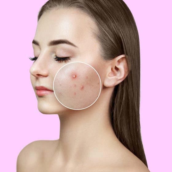
Introduction Acne vulgaris is a common chronic dermatological condition resulting from inflammation of the pilosebaceous unit. Acne vulgaris is extremely common in adolescence affecting up to 95% of young people. Though in most cases it is relatively mild, up to 1/3 have moderate or severe disease. It results in comedones and inflammatory lesions that typically affect the face, chest and back. Beyond (and secondary to) its dermatological manifestations it can have a profound impact on self-esteem and mental wellbeing. Most cases respond to topical and/or oral therapies (e.g. antibiotics and retinoids). Specialist dermatological and mental health support can be required. Epidemiology 20-35% of

Acne vulgaris

Introduction Acne vulgaris is a common chronic dermatological condition resulting from inflammation of the pilosebaceous unit. Acne vulgaris is extremely common in adolescence affecting up to 95% of young people. Though in most cases it is relatively mild, up to 1/3 have moderate or severe disease. It results in comedones and inflammatory lesions that typically affect the face, chest and back. Beyond (and secondary to) its dermatological manifestations it can have a profound impact on self-esteem and mental wellbeing. Most cases respond to topical and/or oral therapies (e.g. antibiotics and retinoids). Specialist dermatological and mental health support can be required.