Clinical

Psychiatric History

The psychiatric history is probably one of the most difficult histories to conduct, as the patient may present with no physical symptoms at all, just a general sense of feeling low. As a result, they may not give you much information at all and not be willing to engage with your questions. ‌ The biggest mistake that students make when taking a psychiatric history for depression is losing their structure. It is important to remember that we still want to find out about the presenting complaint – feelings of lethargy, poor sleep, loss of appetite are all symptoms and we

Paediatric History

This is a history taking template for a young child where you will be taking the history from the parent.  Now that you are familiar with taking basic histories, there are some situations where we have to amend our general structure. Keep in mind, whether it be children, mental health patients or other specific conditions, we are still very much interested in finding out the presenting complaint, ICE and the past medical, family, drug and social history. The only difference in these histories is that we must also ask some additional questions to screen for problems specific to this group

Vomiting

The vomiting history is similar to cough – it is a symptom which has an onset, timing etc. but also we need details about the physical content of the vomit. So what are some differentials for vomiting? Neurological Tumour Raised ICP Idiopathic intracranial hypertension Hydrocephalus Bleed   Infection Gastroenteritis Infective colitis Malignancy Gastric cancer GI cancer   GI Pancreatitis Bowel obstruction Oesophageal varices Peptic ulcers Alcohol/gastritis It is very important to screen the abdominal system right from top to bottom. Notice how vomiting with constipation is more likely to suggest bowel obstruction, compared to with diarrhoea (more likely food poisoning). Unexplained vomiting

Urinary Frequency

This history will usually refer to a middle-aged/elderly male complaining or urinary symptoms. The main purpose of the history will be to distinguish whether this condition is benign (BHP) or something more sinister, such as prostate/bladder cancer. In addition, for a younger patient, we might also be concerned with a STI. So what are some common conditions which might cause urinary frequency? Urinary tract Benign prostatic hyperplasia Prostate cancer Bladder cancer Over-active bladder Infection UTI STI Looking at these differentials, the systems we have to explore are the urinary system and possible sexually transmitted infections. Again when going through the symptoms, we should enquire

Syncope

This history refers to patients who have experienced a loss of consciousness on more than one occasion. What are some common conditions which might cause this? Raised intracranial pressure Bleed Tumour Hydrocephalus   Neurological Epilepsy Cardiovascular Palpitations (AF) Vasovagal reflex Postural hypotension Looking at these differentials, the systems we are most interested in exploring during our specific systems review will be the neurological and cardiovascular systems. Remember, common things are common. Someone presenting with syncope is much more likely to have some form of vasovagal reflex, or a side effect of their blood pressure medication than epilepsy – but this is a diagnosis we do not

Rectal Bleeding

Rectal bleeding is a serious symptom which should ring bells. If we take the anatomical approach the main conditions which can cause this are bowel cancer, diverticulitis, IBD but also more benign conditions like haemorrhoids and an anal fissure. Therefore, in our GI specific systems review, we will need to work through the GI tract. Again, in any female of child bearing age (14 to 60) we should always query pregnancy and gynaecological conditions.   Introduction Good afternoon, my name is _______ and I am one of the student doctors here. Is it _______(patient’s name)? I just wanted to ask

Palpitations

As a medical student, this is actually one of the simplest histories to take. The reason is that apart from the heart, there are few things which can cause palpitations. Nevertheless, what we do have to concentrate on is taking a really detailed account of the presenting complaint – specifically when do the palpitations start, what brings them on, are they there all the time etc. In addition, we have to screen for cardiovascular and breathing symptoms, as these are connected to the heart. So what specific systems should we screen for?‌ Cardiovascular–> Chest pain, loss of consciousness Respiratory –> Breathlessness

Joint Pain

Joint pain is one of the most common presentations you will see. Again we are interested in the onset, timing etc. A key aspect for joint pain is working out whether this is an inflammatory or non-inflammatory problem. The difference in prognosis, treatment of osteoarthritis vs rheumatoid is substantial. So an important symptom to explore is STIFFNESS. Is it worse in the morning? How long does it last? So what are the differentials for joint pain? Trauma Sports injury Fall   Non-inflammatory Osteoarthritis   Other Chronic fatigue syndrome Fibromyalgia Inflammatory Rheumatoid arthritis Rheumatoid variants Gout Pseud-gout Bursitis Septic arthritis Therefore,

Headache

When considering a headache, what are some common conditions which might cause this? Raised intracranial pressure Bleed Tumour Idiopathic intracranial hypertension Hydrocephalus   Infection Sinusitis Meningitis Encephalitis Brain abscess  Trauma Extradural/subdural hematoma   Neurological Epilepsy Migraine Tension headache   Infarction Ischaemic stroke Therefore, when conducting the specific systems review, we will need to ask questions that cover all of these differentials and allows us to narrow down (if not rule out) the most serious causes. This will most cover the head and neck and the nervous system   Introduction Good afternoon, my name is _______ and I am one of the student

Cough

Taking a history for a cough, vomiting, diarrhoea, and blood in the urine all fall into a group. Here you have a type of symptom which requires exploration not only about the onset, timeline, associated symptoms, but also about the physical content of the substance. A history which covers only one aspect will be inadequate and will mean that you miss out key information. Therefore, in these histories, do not forget to ask about the “S (specific questions)” in OATES. ‌ When considering a cough, what are some common conditions which might cause this? Chest infection Viral infection Pneumonia Fungus  

Chest Pain

Chest pain is one of the most common reasons why patients present into A&E. So what are some of the differentials to consider? Cardiovascular MI Aortic dissection Palpitations Aneurysm Heart failure Pericarditis GI Gastro-oesophageal reflux Respiratory Asthma Pulmonary embolism Chest infection   MSK Pulled muscle Costochondritis Therefore, when conducting the specific systems review, we will need to ask questions that cover all of these systems, which will allow us to narrow down (if not rule out) the most serious causes. In addition, during the presenting complain, as you gain more experience, you will naturally learn to ask more specific questions,

Abdominal Pain

With these different histories, whilst the overall structure will be identical to before, pay close attention to the presenting complaint and systems review sections – as this is what will differ.‌ For the presenting complaint, I have included the most common symptoms a patient is initially likely to tell you. Remember to screen fully, and only after you have fully screened for all the symptoms, you can follow SOCRATES or OATES to explore each one in detail. If there are symptoms listed in the example presenting complaints that the patient does not say, remember to ask about these in the

FAQs

It is important to note that the example history before is just a model, and so it might not work in 100% of situations. However, it is a template which should always give you a base with which to work from and not panic in stressful situations. Below are some common queries which students have and how to get round them.   1. What if the patient mentions their family history or something different during a different part of the history? Whilst you might have this model template in your mind, the patient will not. They are just going to

History Taking – Explained

Now that you have read a general template, let’s go through each aspect trying to understand exactly why we ask these specific questions, and what information we hope to gain from them.   Introduction Good afternoon, my name is _______ and I am one of the student doctors here. Is it _______(patient’s name) To begin within, it is essential that you introduce yourself to the patient and clearly explain what level you are i.e. whether you are a medical student, junior doctor, consultant. By doing this, the patient will be able to probably gauge your level of experience and knowledge

History Taking – A General Template

Below is a general template for taking a history. In a nutshell, we should aim to introduce ourselves politely and start with an open question thus allowing the patient to describe in their own words what the problem is. After this we can work through the different categories obtaining information to gain a full perspective of the patient’s illness and obtaining the most likely differentials. Do not worry if you do not understand why we are asking all these questions, it will be explained in much more detail on the next page.   Introduction Good afternoon, my name is _______

History Taking Principles

History taking is one of the most important skills required by a doctor. Many clinicians argue that 70% of the diagnosis can be found in the history, with follow up investigations, imaging and special tests only really required to answer initial doubts.‌ Each question in your history should be asked with a purpose. Let us imagine that someone has presented with vomiting. What are some differentials in this scenario? Gastrointestinal Infective colitis Pancreatitis Biliary conditions Bowel Obstruction Achalasia   CardioRespiratory Myocardial infarction   Neurological Cerebrovascular accident Extra/sub dural haematoma Raised ICP Brain tumour   Non-organic Drugs Alcohol Allergies   By

Study designs

Overview The reporting of medical findings originally focussed on the description of individual patients with an unusual presentation. This would be a novel or rarely used method of treatment, or an unexpected outcome. Some reports described several such cases treated by the same practitioner. The 20th century saw the development of larger scale studies that involved (for example) the collection of the views of individual patients, the inspection of patient medical records, following patients over time using standardised record keeping, or performing comparisons of patients receiving different treatments. Recent years have seen the development of medical literature databases on the Internet, facilitating

Sampling

Rationale for sampling A population is a complete group of individuals such as all the residents of a country. Involving the whole population is unrealistic in terms of cost and staff time. Sampling is preferable and often the only way forward. Individuals who are reasonably easy to contact are selected making the study viable. Remember that due to random fluctuation information gained from different samples will not be identical even with the same method of selection. Also, some of the individuals selected will not be willing and/ or able to be involved leading to non-response bias. Random sampling A sampling

Hypothesis testing

Investigating a population Deductions about the characteristics of a population can be made using a representative sample drawn from the population. This procedure starts with a statement regarding the population and information from the sample is used to decide whether or not there is enough evidence to conclude that this statement might be false. The null hypothesis The initial statement about the population (the null hypothesis) is very specific. For the case of a single sample it needs to describe the population, introduce the characteristic under study, include a statistical measure of interest (e.g. the mean) and propose an assumed value for this measure.

Diagnostic tests

Introduction Diagnostic testing is based on the phenomenon that components of blood and other body fluids are found at different concentrations in individuals with a particular disease relative to people without the disease. For instance, fasting blood glucose concentrations are raised in those who suffer from diabetes. Blood glucose concentration can therefore be used as a method for identifying undetected cases of diabetes in the population. In men, prostate specific antigen (PSA) levels in blood are higher on average in those who have prostate cancer and can be used to detect and manage progression of the disease. In these notes,

Data presentation

Summary Data can be binary, nominal, ordered, discrete quantitative, or continuous quantitative. Graphs include bar-charts, pie-charts, histograms, box-and-whisker plots and scatter diagrams. The type of graph used should be chosen according to the type of data being displayed. Be aware that graphs may be presented in misleading ways. Furthermore, graphs can be symmetrical, positively skewed or negatively skewed. Types of average include the mean, median, and mode. Measures of spread include the range, interquartile range, variance, and standard deviation. Means and measures of spread should be chosen according to the type of data being summarised and some measures are susceptible

Statins

Introduction Statins have revolutionised the management of hypercholesterolaemia. Statins have become essential in the management of patients with, or at risk, of cardiovascular disease including coronary artery disease (CAD), cerebrovascular disease and peripheral vascular disease (PVD). Indications Statins were primarily created to treat hypercholesterolaemia, which is an independent risk factor for cardiovascular disease. Statins are indicated in both primary and secondary prevention of cardiovascular disease for many conditions. Primary prevention Cardiovascular risk assessment score ≥10%* and ≤ 84 years old (risk/benefit if >84) Cardiovascular risk assessment score ≥10%* and type 2 diabetes mellitus (T2DM) Type 1 diabetes mellitus (T1DM) and additional criteria (age, kidney disease, duration) Chronic kidney disease (CKD)

Oxygen

Overview Oxygen should be regarded as a drug that is prescribed for patients with hypoxaemia (low blood oxygen concentration). Oxygen is the most commonly used drug in emergency situations. It can be a life-saving drug to prevent severe hypoxaemia that refers to a low arterial oxygen concentration. However, used inappropriately oxygen can have serious or fatal consequences. The headline point is that oxygen should be used to treat hypoxaemia and maintain a patients’ saturations in the target range. This should be 94-98% or 88-92% in patients at risk of type 2 respiratory failure. When prescribing and administering oxygen there are several

Metformin

Summary Metformin is considered the first-line hypoglycaemic agent for the treatment of type 2 diabetes mellitus. Metformin has been one of most prescribed medications worldwide over the last decade. The typical starting dose for adults is 500 mg daily, which can be increased to a maximum dose of 2 g daily. The most common adverse-effect is gastrointestinal upset (i.e. nausea, abdominal pain, diarrhoea), although rarely metformin can result in lactic acidosis. Origin Derived from galegine, which is a natural protein found in the plant Galega officinalis. Metformin was first synthesised around the 1920s and then used clinically in the 1950s. It is now

Bronchodilators

Introduction Airway resistance occurs as frictional forces oppose the flow of air through the conducting airways. Normal flow is laminar, the flow is ordered and quicker in the centre. As airways divide and become narrow increasingly turbulent flow occurs. Poiseuille’s equation describes resistance (if flow is laminar) with resistance (R), length (L), radius (r) and viscosity (η). π and 8 are constants: Resistance (R) = 8 x L x η / π x r4 Bronchomotor tone Bronchomotor tone controls the ease with which air is conducted through airways. It exhibits a circadian rhythm where tone is greatest in the early morning. We note from the

Antihypertensives

Overview The anti-hypertensives are an important & broad group of medications. Hypertension is a very common condition that represents a significant source of morbidity and mortality. It is a major risk factor for MI, stroke and chronic kidney disease. Hypertension is managed in a step-wise fashion according to the NICE guidelines (see our Hypertension notes for more). Several groups of drugs, by varying mechanisms, are used to reduce blood pressure. Regulation Blood pressure is maintained by a number of physiological reflexes that respond to acute and chronic changes. Blood pressure = cardiac output x peripheral vascular resistance Numerous interconnected systems contribute to the regulation of blood pressure.

Antiarrhythmics

Vaughan-William’s classification Anti-arrhythmics are drugs that modify cardiac conduction, they are used to treat arrhythmias and are classified according to the Vaughan-William’s system. This classification divides these drugs into four classes according to their effects on cardiac action potential. Anti-arrhythmics have complex actions and classes may overlap. It is important to note, that this classification system has become increasingly inadequate with improved understandings of drug mechanisms and development of new antiarrhythmics. Arrhythmogenesis To understand the action of the antiarrhythmics it helps to understand how arrhythmias may develop. Arrhythmias are disorders of rate and rhythm of the heart, which arise due to either abnormal generation or

Pleural fluid

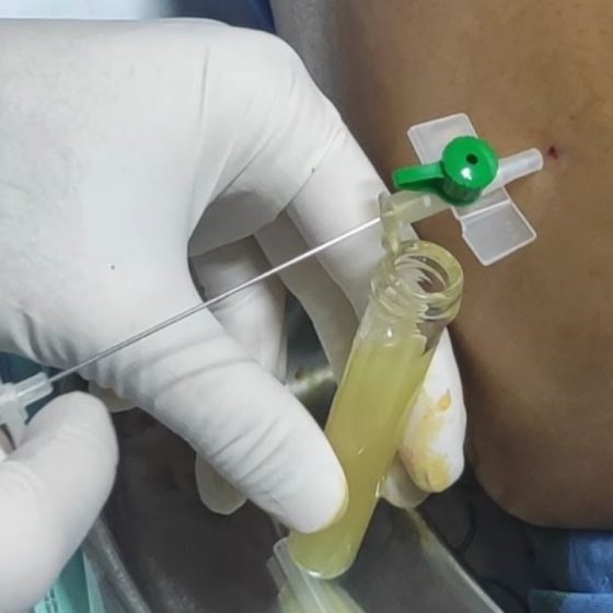
Overview Analysis of pleural fluid is important for the workup of a pleural effusion. A pleural effusion is the most common manifestation of pleural disease and it may occur from a wide range of aetiologies. It refers to an abnormal collection of fluid with the pleural space. Pleural fluid may be aspirated and analysed to help determine the underlying cause. Pleural effusions may be a manifestation of numerous conditions that requires a careful history, clinical examination, imaging (e.g. chest x-ray), and pleural fluid analysis to determine the cause. Sometimes the cause may be obvious (e.g. heart failure) and pleural aspiration is

Blood bottles

Overview We give a practical guide to the use of blood bottles in clinical practice. In modern medicine, most hospitals and GP practices are set up with computer software that automatically prints blood labels following a request. This process automatically assigns a test to a blood bottle and reduces the need to handwrite patient information. The required blood bottle on these labels is differentiated by the coloured top (e.g. lavender or gold). Occasionally, when the test is specialist or there is ‘computer downtime’ knowledge of the appropriate blood bottle is required, but you can always ask! We outline a practical guide to

Ascitic fluid

Overview Ascites refers to the presence of pathological fluid within the abdominal cavity. In men, no fluid should be present. In women, up to 20 mls may be considered normal depending on the timing of their menstrual cycle. Ascites is most commonly associated with liver disease. In this context, ascites develops due to portal hypertension. This refers to increased pressure within the portal venous system that drains blood from the gastrointestinal tract to the liver. Other causes of ascites are broadly due to local infiltration (e.g. tumour) of the peritoneal lining, inflammation or infection. Aetiology Ascites can be broadly be divided

Respiratory history

Introduction The respiratory history should focus on key system-specific symptoms related to the respiratory system. A respiratory history focuses the consultation on the respiratory system. This is usually because a patient presents with a respiratory problem such as shortness of breath or wheeze. The idea of a system-specific history is to explore key factors that are relevant to the affected system during the consultation. In respiratory disease, this may include pertinent past medical history (e.g. asthma/COPD), inhaler use, smoking history, occupational exposures, and even childhood illnesses. History of presenting complaint Breathlessness is a major symptom of respiratory disease. Shortness of breath or

Musculoskeletal history

Overview Musculoskeletal disorders are often multi-system that requires a wide range of questioning in the history. A musculoskeletal (MSK) history focuses primarily on presentations affecting the musculoskeletal system including bones, joints, tendons, ligaments, and muscles. Remember that many musculoskeletal disorders are multi-system meaning a wide range of inquiry is required during the consultation to determine the involvement of other organ systems. Presentations may be acute and obvious (e.g. fractured bone after fall) or more chronic with vague extending over months to years. Be patient and listen to the patient. Finally, it is essential to ask what the functional and psychological impact of

Gastrointestinal history

Overview The gastrointestinal (GI) history should focus on key system-specific symptoms related to the GI tract. The gastrointestinal tract runs all the way from mouth to anus. Pathology can occur anywhere along the tract from oesophagus to the intestines to the bile ducts. Therefore, the GI history is a system-specific history that needs to focus on the different elements that can affect each part. Any GI history should focus on the presenting symptom of the patient (e.g. diarrhoea or dysphagia) and then proceed to ask more broad questions related to other parts of the GI tract History of presenting complaint Isolate

Cardiovascular history

Introduction The cardiovascular history should focus on key system-specific symptoms related to the heart and cardiovascular system. A cardiovascular history should focus the consultation on the cardiovascular system. This is usually because a patient presents with a cardiovascular problem such as chest pain or palpitations. The idea of a system-specific history is to explore key factors that are relevant to the affected system during the consultation. In cardiovascular disease, this may include cardiovascular risk factors, medications (i.e. that prevent or provoke cardiovascular disease), lifestyle factors (e.g. obesity, smoking), and relevant family history. History of presenting complaint Chest pain can be described using the SOCRATES mnemonic.

Breast history

Overview The breast history should focus of system-specific questions relating to disease of the breast. A breast history focuses the consultation on key clinical features that affect the breasts. The hallmark of breast disease is a breast lump and you should feel confident in exploring the features of a breast lump in full detail. It is important within your history to determine any systemic features that may indicate an underlying malignancy (e.g. fever, weight loss, loss of appetite) and to tease out any significant risk factors for breast disease. For example, mastitis is more likely to occur if the patient is

Basic history

Introduction The art of medicine is to determine why a patient has sought help. The key skills to help establish the underlying cause of a patients’ symptoms (the diagnosis) is based on talking to the patient (the history), examining the patient (the examination) and requesting tests like bloods and x-rays (the investigations). The information gathered from the history and examination is used to form a hypothesis of the possible underlying diagnosis. Investigations can then be used to either confirm or refute this diagnosis. Some diagnoses can be made just by talking to a patient, while others are reliant on a specific test. As a medical student, student nurse, physician associate, or allied

9. Reporting an ECG

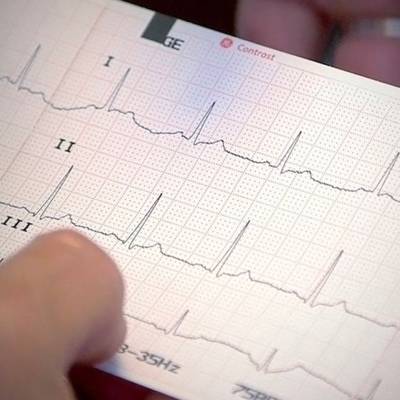
Introduction It is important to have a good structure for analysing and subsequently reporting an ECG. When reporting an ECG, you should use the same structured approach every time. This is particularly important when first learning about ECGs. A structured approach allows you to systematically assess the ECG in order and not forget any key elements. Once you improve, you’ll start to develop pattern recognition for classic ECG features.  We propose the following structure for analysing and reporting an ECG: Confirm correct patient details Rate Rhythm Cardiac axis P waves, Q waves & QRS complexes ST segments & T waves  QT interval Putting it all together This

8. ST segment & T waves

Introduction It is important we analyse each aspect of the ECG morphology including P wave, QRS complex, ST segment and T wave. Abnormalities of the P wave, QRS complex, ST segment and T wave can tell us a lot about the patient. Analysing these parts of the ECG should always be taken in context of the rate, rhythm and clinical status of the patient.  Certain changes to the ECG morphology are classical of an underlying pathology. For example, ST elevation is the characteristic feature of an acute ST-elevation myocardial infarction (STEMI). Other changes are non-specific and can be suggestive of multiple pathologies. For

7. P wave, QRS & QT interval

Introduction It is important we analyse each aspect of the ECG morphology including P wave, QRS complex, ST segment and T wave. Abnormalities of the P wave, QRS complex, ST segment and T wave can tell us a lot about the patient. Analysing these parts of the ECG should always be taken in context of the rate, rhythm and clinical status of the patient.  Certain changes to the ECG morphology are classical of an underlying pathology. For example, ST elevation is the characteristic feature of an acute ST-elevation myocardial infarction (STEMI). Other changes are non-specific and can be suggestive of multiple pathologies. For

6. Tachycardias

Introduction Tachycardia refers to an abnormally fast heart rate. Tachycardia is usually defined as an abnormally fast heart rate greater than 100 bpm. We refer to all the abnormally fast heart rhythms as tachyarrhythmias. When a tachyarrhythmia occurs intermittently, we call it paroxysmal. Tachyarrhythmias may develop due to an ectopic foci of electrical activity within the atria, atrioventricular node (AVN) or ventricles. The aetiology of these ectopic foci can get quite complex, but is broadly due to problems with impulse conduction (i.e. transmission of electrical activity) or impulse formation (i.e. generation of electrical activity). Impulse conduction problems: caused by conduction blocks (discussed in our Bradycardia notes) and formation of reentrant

5. Bradycardias

Introduction Bradycardia refers to an abnormally slow heart rate. Bradycardia is usually defined an abnormally slow heart rate less than 60 bpm. We refer to all the abnormally slow heart rhythms as bradyarrhythmias. Bradyarrhythmias may develop due to a variety of intrinsic or extrinsic factors. Intrinsic Within the heart, a slow rate may occur due to failure to initiate, or transmit, electrical activity. Failure to initiate electrical activity can cause another part of the heart to take over as the primary pacemaker. This is called an escape rhythm. For example, if the sinoatrial note (SAN) fails to undergo spontaneous depolarisation the atrioventricular node (AVN) may initiate

2. Rate & rhythm

Introduction Rate and rhythm are the first things to assess when analysing an ECG. The rate refers to the frequency of electrical activity. It correlates with muscular contraction and therefore heart rate. Normal electrical activity in the absence of contraction is termed ‘pulseless electrical activity’. This rhythm is not compatible with life and can be seen in cardiac arrest. The rhythm refers to the area of the heart that is controlling electrical activity. In other words, the part of the heart that is initiating electrical activity, which then spreads throughout the heart. Due to spontaneous depolarisation, different parts of the heart can initiate electrical

1. How to read an ECG

Introduction Conduction through the heart is dependent on pacemaker cells, which are organised into key structures. The heart is a dual pump that sits at the centre of the cardiovascular system. It is composed of both contractile cells and autorhythmic cells (also known as pacemaker cells). Approximately 1% of cardiac tissue is composed pacemaker cells, which are organised into key structures and can undergo spontaneous depolarisation. Depolarisation refers to the electrical changes that occur within a muscle to allow it to contract. The heart is essentially one big muscle that can contract by itself. We can detect these electrical changes, which are associated with

Shock

Introduction Shock describes circulatory failure resulting in inadequate tissue perfusion and insufficient delivery of oxygen. Shock is a broad and at times unhelpful term. It refers to any cause of circulatory failure that results in inadequate oxygen delivery to tissues. There are many causes with different underlying pathophysiological processes, normally they are divided into four categories: Hypovolaemic Distributive Cardiogenic Obstructive It should be noted the term ‘shock’ is unliked by many in the medical field. The Royal College of Emergency Medicine describe it as a ‘somewhat lazy shorthand’ preferring clinicians take a more granular approach to their description of the patients clinical state that

Multi-organ dysfunction syndrome

Overview Multi-organ dysfunction refers to progressive organ dysfunction whereby homeostasis cannot be maintained without intervention. Multiple organ dysfunction syndrome (MODS), also known as multi-organ failure (MOF), is a severe clinical syndrome that is seen in critically ill patients. It is characterised by progressive organ dysfunction with a failure to maintain homeostasis without intervention. MODS is commonplace in the intensive treatment unit (ITU) setting and usually present at the time of ITU admission and/or at the time of death. MODS is broadly defined as primary or secondary: Primary: direct, identifiable injury (e.g. liver failure due to a hepatotoxic agent) Secondary: no direct

JVP

Introduction Examination of the jugular venous pressure provides an estimate of the central venous pressure. Assessment of the jugular venous pressure (JVP) is a normal component of the cardiovascular examination. It is commonly used to help determine a patients’ fluid balance (i.e. do they have too much or too little fluid in the body). This is because the JVP can be used as an indirect marker of central venous pressure (CVP), which is a measure of pressure within the vena cava. The term jugular venous pulse is often used synonymously with jugular venous pressure. To determine the JVP, we have to visualise the

Fluid prescribing

Overview Prescribing intravenous fluids is one of the cornerstone tasks of junior doctors & prescribers. Many patients who are admitted to hospital require intravenous fluids. The task of prescribing intravenous fluids typically falls under the realm of the junior doctor. Becoming proficient at prescribing fluids is one of the key skills during the first year of clinical practice. The indication for fluids can be: Shock (inadequate tissue perfusion) Excess fluid losses (e.g. diarrhoea) Inadequate intake (e.g. nil by mouth) Replacing electrolytes (e.g. potassium) The National Institute of Clinical Excellence (NICE) produced guidelines on the administration of intravenous fluids in both adults and children. However, it is difficult to learn how to

Confirmation of death

Overview Confirmation of death is an important part of healthcare practice. The confirmation of death is an essential part of practice within both the community and hospital setting. In 2008, the Academy of Royal Medical Colleges issued a generalised code of practice for the confirmation of death. Death may occur from: Irreversible cessation of brain-stem function (i.e. brain-stem death) Following cessation of cardiorespiratory function (including failed resuscitation) The confirmation of brain-stem death (i.e. the heart and lungs can only be maintained by artificial ventilation) is advanced and has to be carried out by at least two medical practitioners (minimum one consultant) who have been registered

Respiratory alkalosis

Overview Respiratory alkalosis is characterised by a pH > 7.45 and low partial pressure of arterial carbon dioxide (< 4 kPa). Respiratory alkalosis primarily occurs as a result of increased clearance of carbon dioxide from the lungs. It is commonly seen as a compensatory response to metabolic acidosis. Alternatively, it can occur as a primary abnormality (e.g. pulmonary embolism). It is characterised by alkalosis (pH > 7.45) and a low PaCO2 (< 4 kPa). The principle mechanism of respiratory alkalosis, regardless of whether it is compensatory or primary, is hyperventilation. This refers to an increase in alveolar ventilation, which leads to clearance or ‘blowing off’

Respiratory acidosis

Overview Respiratory acidosis is characterised by a pH < 7.35 and raised partial pressure of arterial carbon dioxide (>6 kPa). Respiratory acidosis primarily occurs as a result of inefficient clearance of carbon dioxide from the lungs. It may be acute or chronic. It is characterised by acidosis (pH < 7.35) and a high PaCO2 (elevated above the upper limit of the reference range). This is commonly 6 kPa. The increase in carbon dioxide may occur due to several mechanisms: Abnormal alveolar ventilation (lungs unable to clear carbon dioxide) Increased carbon dioxide production (systemic illnesses increase levels and ‘overwhelm’ lungs) Increased carbon dioxide intake (e.g. rebreathing gas containing

pH control

Overview Within the body, normal pH is 7.35-7.45. The pH refers to the acidity or alkalinity of the blood. Tight control of pH is needed for normal metabolic function. Within the body, normal arterial pH is 7.35-7.45. This is determined by the concentration of hydrogen ions. Precise regulation of pH requires alteration of the free hydrogen ion concentration within bodily fluids. The actual concentration of hydrogen ions is extremely small (~40 nano moles per litre). Therefore, pH is an easier way to express the concentration of hydrogen ions. pH is a logarithmic scale. Inverse relationship The numerical value of pH is inversely proportional

Metabolic alkalosis

Overview Metabolic alkalosis is characterised by a pH > 7.45 and raised plasma bicarbonate level (> 26 mmol/L). There are numerous causes of metabolic alkalosis, which are broadly divided based on the volume status (i.e. fluid replete or replete) of the patient. It is characterised by alkalosis (pH > 7.45) and a high plasma bicarbonate level (> 26 mmol/L). For metabolic alkalosis to develop, there needs to be an ‘initiating event’ and then ‘maintenance of alkalosis’. In some cases, these two factors are the same physiological process. Initiating event Metabolic alkalosis, put simply, can result from a loss of hydrogen ions or gain of bicarbonate. Hydrogen ion loss: usually

Metabolic acidosis

Overview Metabolic acidosis is characterised by a pH < 7.35 and reduced plasma bicarbonate level (< 22 mmol/L). Metabolic acidosis is one of the most common acid-base abnormalities seen in clinical practice. It is characterised by acidosis (pH < 7.35) and a low plasma bicarbonate level (< 22 mmol/L). Metabolic acidosis, put simply, can be from a primary increase in hydrogen ions (e.g. addition of strong acid) or reduction in bicarbonate concentration: Addition of acid (e.g. diabetic ketoacidosis or methanol ingestion) Loss of bicarbonate (e.g. diarrhoea or renal loss) Determining the cause There are numerous causes of metabolic acidosis. These are broadly differentiated based on the anion gap. The

ABG interpretation

Overview Interpretation of an arterial blood gas is an essential skill required by all doctors and most healthcare professionals. In simplistic terms, an arterial blood gas (ABG) tells us about three main things: Oxygenation: measurement of oxygen within the blood. Ventilation: process of respiratory function (i.e. breathing). Acid-base balance: the control of pH. To enable us to interpret oxygenation, respiratory function and acid-base balance, an ABG analyser gives us key bits of information. This includes the pH, partial pressure of oxygen, partial pressure of carbon dioxide and calculated bicarbonate. Key components pH (normal range 7.35-7.45): refers to the acidity or alkalinity

Data presentation

Summary Data can be binary, nominal, ordered, discrete quantitative, or continuous quantitative. Graphs include bar-charts, pie-charts, histograms, box-and-whisker plots and scatter diagrams. The type of graph used should be chosen according to the type of data being displayed. Be aware that graphs may be presented in misleading ways. Furthermore, graphs can be symmetrical, positively skewed or negatively skewed. Types of average include the mean, median, and mode. Measures of spread include the range, interquartile range, variance, and standard deviation. Means and measures of spread should be chosen according to the type of data being summarised and some measures are susceptible

Antiarrhythmics

Vaughan-William’s classification Anti-arrhythmics are drugs that modify cardiac conduction, they are used to treat arrhythmias and are classified according to the Vaughan-William’s system. This classification divides these drugs into four classes according to their effects on cardiac action potential. Anti-arrhythmics have complex actions and classes may overlap. It is important to note, that this classification system has become increasingly inadequate with improved understandings of drug mechanisms and development of new antiarrhythmics.     Arrhythmogenesis To understand the action of the antiarrhythmics it helps to understand how arrhythmias may develop. Arrhythmias are disorders of rate and rhythm of the heart, which

Ascitic fluid

Overview Ascites refers to the presence of pathological fluid within the abdominal cavity. In men, no fluid should be present. In women, up to 20 mls may be considered normal depending on the timing of their menstrual cycle. Ascites is most commonly associated with liver disease. In this context, ascites develops due to portal hypertension. This refers to increased pressure within the portal venous system that drains blood from the gastrointestinal tract to the liver. Other causes of ascites are broadly due to local infiltration (e.g. tumour) of the peritoneal lining, inflammation or infection.     Aetiology Ascites can be

Basic history

Introduction The art of medicine is to determine why a patient has sought help. The key skills to help establish the underlying cause of a patients’ symptoms (the diagnosis) is based on talking to the patient (the history), examining the patient (the examination) and requesting testslike bloods and x-rays (the investigations). The information gathered from the history and examination is used to form a hypothesis of the possible underlying diagnosis. Investigations can then be used to either confirm or refute this diagnosis. Some diagnoses can be made just by talking to a patient, while others are reliant on a specific

How to read an ECG

Introduction Conduction through the heart is dependent on pacemaker cells, which are organised into key structures. The heart is a dual pump that sits at the centre of the cardiovascular system. It is composed of both contractile cells and autorhythmic cells (also known as pacemaker cells). Approximately 1% of cardiac tissue is composed pacemaker cells, which are organised into key structures and can undergo spontaneous depolarisation. Depolarisation refers to the electrical changes that occur within a muscle to allow it to contract. The heart is essentially one big muscle that can contract by itself. We can detect these electrical changes,

Confirmation of death

Overview Confirmation of death is an important part of healthcare practice. The confirmation of death is an essential part of practice within both the community and hospital setting. In 2008, the Academy of Royal Medical Colleges issued a generalised code of practice for the confirmation of death. Death may occur from: Irreversible cessation of brain-stem function (i.e. brain-stem death) Following cessation of cardiorespiratory function (including failed resuscitation) The confirmation of brain-stem death (i.e. the heart and lungs can only be maintained by artificial ventilation) is advanced and has to be carried out by at least two medical practitioners (minimum one

ABG interpretation

Overview Interpretation of an arterial blood gas is an essential skill required by all doctors and most healthcare professionals. In simplistic terms, an arterial blood gas (ABG) tells us about three main things: Oxygenation: measurement of oxygen within the blood. Ventilation: process of respiratory function (i.e. breathing). Acid-base balance: the control of pH. To enable us to interpret oxygenation, respiratory function and acid-base balance, an ABG analyser gives us key bits of information. This includes the pH, partial pressure of oxygen, partial pressure of carbon dioxide and calculated bicarbonate. Key components pH (normal range 7.35-7.45): refers to the acidity or