Day: April 20, 2024

Infective Endocarditis

This refers to inflammation of the endocardium that lines the surface of heart valves. It can lead to vegetations on the valve surface that can destroy the valve. In addition, it can lead to septic emboli formation leading to other complications. Causes Staphylococcus aureus This is the most common cause of IE which is usually seen in IV drug abusers It is a high virulence organism that destroys valves, most commonly the tricuspid valve Risk factors for this bacterium include skin breaches (dermatitis, IV lines), kidney failure and diabetes   Viridans Streptococci This is a group of low-virulence bacteria that

Heart Valve Conditions

Rheumatic fever This is a complication after being infected with group A β-haemolytic streptococcus. Antibodies directed against the streptococcus pyogenes M-antigen cross react with myosin and smooth muscle. This leads to a systemic condition which occurs in acute attacks, taking about three months to recover. It can also cause chronic inflammation leading to permanent damage to heart valves. It typically causes stenosis of valves (usually mitral) with a “fish mouth” appearance. Symptoms History of previous streptococcal infection (e.g., pharyngitis) Infective signs – fever and arthralgia Cardiac symptoms (e.g., mitral stenosis) may occur after multiple attacks of RF   Key tests

Heart axis deviation

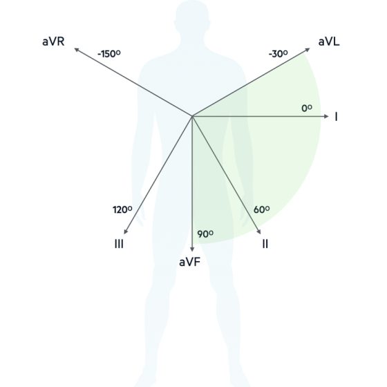
 Main ECG waves Atrial depolarization (P) axis : it is the general direction in which atrial depolarization conducts Ventricular depolarization (QRS) axis : it is the general direction in which depolarization conducts Ventricular repolarization (T) axis : it is the general direction in which ventricular repolarization conducts Rt : RightLt : Leftmv : Milli voltaVR : Augmented Voltage on Right armaVL : Augmented Voltage on Left armaVF : Augmented Voltage on FootRA : Right AtriumLA : Left AtriumRV : Right VentricleLV : Left VentricleLI : Lead 1LII : Lead 2LIII : Lead 3 Definition Of Mean QRS Axis : The

Heart Failure

This is a clinical syndrome consisting of symptoms (e.g., breathlessness, oedema, fatigue) that occurs due to abnormalities in cardiac structure or function, causing inadequate cardiac output or raised intracardiac pressures. Usually, it is due to myocardial dysfunction, but can be due to other causes like valvular disease, pericardial disease or arrhythmias.   Systolic failure This is an inability for the ventricle to contract properly, decreasing cardiac output. In this case, the ejection fraction (EF) is < 40%. It occurs due to conditions which weaken the heart muscle reducing contractility, e.g., ischaemic cardiomyopathy, dilated cardiomyopathy and myocarditis.   Diastolic failure Refers

Ischaemic Heart Disease

Angina This is the term which is used to describe symptomatic chest pain which occurs due to myocardial ischaemia. In the heart, the coronary circulation fills during diastole. Due to atherosclerosis, over time, the coronary arteries become occluded impeding blood flow to the muscle. It means areas of myocardium become ischaemic, especially in times of greater oxygen demand, which leads to chest pain. To counteract this, the body increases sympathetic stimulation which aims to increase cardiac output. But this worsens the issue as it increases O2 demand further. Hence, when treating angina, the main goals are increasing coronary filling by vasodilation,

Introduction to ECG

Clinical values of ECG By the end of this course you will learn how to detect many diseases by ECG which is important, easy & inexpensive clinical test as following : Heart rate : Normal, bradycardia or tachycardia How to detect Heart axis and measure α angle Hypertrophy of both aria & ventricles on both sides right and left Myocardial Infarction, type, localization & stage Arrhythmia types, localization & stages Heart blocks, types & stages unusual syndromes of arrhythmia Features of Time voltage chart The P-QRS-T sequence is usually recorded on special ECG graph paper that is divided into grid-like