Day: April 5, 2024

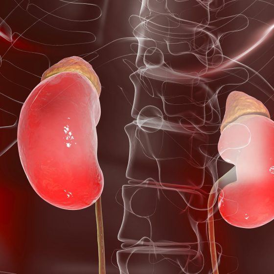
Treatment for phaeochromocytomas

The main treatment for phaeochromocytoma is surgery. Other treatments include internal radiotherapy, external radiotherapy and chemotherapy. Your treatment depends on different factors including: the size of the tumour whether it has spread your general health and fitness A team of doctors and other professionals discuss the best treatment and care for you. They are called a multidisciplinary team (MDT). The team usually includes a: specialist surgeon doctor specialising in hormone disorders (endocrinologist) cancer doctor (oncologist) doctor specialising in the diseases of tissues or cells (pathologist) doctor specialised in reading scans (radiologist) clinical nurse specialist (CNS) You may need to travel

Treatment for pseudomyxoma peritonei

The main treatments for pseudomyxoma peritonei (PMP) are surgery and chemotherapy. Your treatment depends on the size of the cancer and your general health. You might not start treatment straight away. Your doctor closely monitors your cancer in case you need treatment in the future. This is called watch and wait.  If you need treatment you might have: surgery combined with chemotherapy into the tummy (abdomen) surgery to remove as much cancer as possible (debulking surgery) chemotherapy Watch and wait Your doctor might decide to closely monitor your cancer if it’s small and slow growing and you don’t currently need

Treating retinoblastoma

Retinoblastoma is rare and so your child will go to a specialist centre for treatment. There are different treatments such as freezing and laser therapy, chemotherapy, radiotherapy and surgery. The treatment your child has depends on several things including the size of the cancer and if it is in one or both eyes. Treatment centres There are 2 specialist retinoblastoma centres in England and they are: The Royal London Hospital Birmingham Children’s Hospital These 2 centres serve all parts of the UK including Wales, Scotland and Northern Ireland. They have teams of specialists who know a lot about retinoblastoma and

Treatment for small bowel cancer

Your treatment will depend on what type of small bowel cancer you have. This page is about the treatment you might have if you have small bowel adenocarcinoma. This is the most common type of small bowel cancer.  Treatment for other types of small bowel cancer, such as neuroendocrine tumours and lymphoma will be different. Deciding which treatment you need Your doctor will talk to you about the treatment they suggest. They will explain its benefits and the possible side effects. Your treatment will depend on: where your cancer is how big it is and whether it has spread (the stage) the type

Gonorrhoea

Overview Gonorrhoea is a common sexually transmitted infection caused by the bacterium Neisseria gonorrhoeae. Gonorrhoea is a common sexually transmitted infection (STI) that is transmitted by direct contact with infected secretions. It is caused by the gram negative organism Neisseria gonorrhoeae. Gonorrhoea causes infection of mucous membranes, most notably the urethra, endocervix, rectum, pharynx and conjunctiva. Rarely, it can cause disseminated infection. Epidemiology Gonorrhoea is most commonly seen among patients 15-24 years old. Rates of gonorrhoea have been increasing with 70,936 cases in the UK in 2019, which is a 26% increase from 2018. This accounts for around 15% of all new cases of STIs. Chlamydia

Chlamydia

Overview Chlamydia is a common sexually transmitted infection caused by the bacterium Chlamydia trachomatis. Chlamydia is considered the most common treatable bacterial sexually transmitted infection (STI). It is caused by the obligate intracellular bacterium Chlamydia trachomatis, which is also responsible for Lymphogranuloma venereum (LGV) and ocular infections. The phrase ‘chlamydia’ is generally reserved for urogenital infections by C. trachomatis that is caused by certain serovars of the organism (D-K). Serovars are a way of groups organisms into species or subspecies based on the surface antigens. The majority of chlamydia infections are asymptomatic (50% of males and ~70% of females). Untreated, chlamydia can cause significant long-term morbidity including

Chancroid

Overview Chancroid is a sexually transmitted infection caused by Haemophilus ducreyi. Chancroid is a sexually transmitted infection (STI) caused by the organism Haemophilus ducreyi. It is highly contagious and characterised by multiple, deep painful ulcers. It is one of the sexually transmitted causes of genital ulcers. Traditionally, chancroid was very common, but with more attention paid to STI transmission with the rise in HIV cases in the 1980’s, the prevalence of chancroid has been falling. Herpes simplex is now the most common genital ulcerative disease. Genital ulcers The major causes of genital ulcers include: Syphilis: characterised by a single painless ulcer (chancre) Herpes

Bacterial vaginosis

Overview Bacterial vaginosis refers to an overgrowth of predominantly anaerobic microorganisms in the vaginal flora. Bacterial vaginosis (BV) refers to a condition where there is disruption in the normal vaginal flora. There is an excess of anaerobic microorganisms and loss of the normal lactobacilli, which leads to a vaginal odour and increased discharge. The condition is a not a sexually transmitted infection (STI) but it occurs more commonly in sexually-active women. Epidemiology BV is the most common cause of vaginal discharge in women of childbearing age. Almost one third of women between the ages of 14-49 years old have evidence of BV. It is highest amongst

Anogenital warts

Overview Anogenital warts are caused by human papillomavirus. Classical anogenital warts cause cauliflower-like lesions known as condylomata, which are typically painless and occur on moist surfaces. They are caused by human papillomavirus (HPV) of which there are > 100 known genotypes. It is estimated that > 90% of anogenital warts occur secondary to HPV genotypes 6 and 11. Epidemiology Anogenital warts are very common, but most infections do not result in visible lesions and self-resolve. The estimated annual prevalence of anogenital warts is 0.15% in the adult population of the developed world. In the UK, it is estimated that 130,000 cases of anogenital warts are

Anogenital herpes

Overview Anogenital herpes is caused by herpes simplex virus 1 & 2 and leads to painful ulcerated genital lesions. Anogenital herpes is characterised by multiple, painful, ulcerated genital lesions. They arise as small vesicles that progress to pustules, which subsequently ulcerate and crust over. Anogenital herpes can be caused by herpes simplex virus 1 (HSV1) or herpes simplex virus 2 (HSV2), which are both part of the larger family of human herpesviruses (HHVs). This family also includes varicella-zoster virus, Epstein-Barr virus and cytomegalovirus. Epidemiology Worldwide there are over 23 million cases of HSV infection per year. HSV-1 is usually acquired in childhood and often

Rhabdomyolysis

Overview Rhabdomyolysis refers to a clinical syndrome characterised by skeletal muscle necrosis. Rhabdomyolysis is a condition that develops due to skeletal muscle necrosis and the release of toxic intracellular contents including myoglobin and electrolytes. It is characterised by markedly elevated levels of creatinine kinase (CK). Rhabdomyolysis is a potentially life-threatening condition. The full clinical syndrome is characterised by evidence of myoglobinuria from muscle breakdown and acute tubular necrosis (ATN) causing acute kidney injury (AKI). It is a common condition in the adult population that occurs from a variety of causes including trauma and prolonged immobilisation (e.g. following a fall in the

Refeeding syndrome

Overview Refeeding syndrome is an adverse clinical and biochemical response to feeding in severely malnourished patients. Refeeding syndrome (RFS) is generally defined as potentially fatal shifts in fluids and electrolytes that can occur in severely malnourished patients undergoing feeding. Throughout human history, it has been described following famines or in prisoners of war who start eating again after long periods of starvation. The overzealous reintroduction of diet leads to profound electrolyte and fluid derangements that can cause cardiac arrhythmias or cardiac and respiratory failure. Epidemiology It is difficult to estimate the true incidence of RFS due to varied definitions and at times lack of

Hypokalaemia

Introduction Potassium is an essential body cation, which has a normal plasma concentration of 3.5-5.5 mmol/L. Hypokalaemia is one of the most common electrolyte abnormalities within the hospital setting. It is defined as a plasma potassium concentration < 3.5 mmol/L. Hypokalaemia can be a potentially life-threatening condition leading to dangerous cardiac arrhythmias, although most cases are mild and patients are asymptomatic. Hypokalaemia can be further divided as follows: Mild: 3.0-3.4 mmol/L Moderate: 2.5-2.9 mmol/L Severe: < 2.5 mmol/L or symptomatic Epidemiology Hypokalaemia is a common electrolyte abnormality in secondary care affecting up to 20% of inpatients. Potassium concentration is highly variable depending on age, sex,

Hypophosphataemia

Overview Hypophosphataemia is defined as a serum phosphate concentration < 0.8 mmol/L. The normal serum concentration of phosphate is 0.8-1.5 mmol/L. Hypophosphataemia is a common electrolyte abnormality that is estimated to affect up to 5% of patients admitted to hospital. In certain populations, hypophosphataemia is common such as those who are malnourished or suffering from refeeding syndrome. In alcoholics, it may be seen in up to 50%. In patients with paracetamol overdose, it is a good prognostic marker that signifies hepatic recovery. Phosphate physiology The human body contains approximately 1000 g of phosphate of which 80-90% is found in bone. Phosphate is

Hyponatraemia

Definition & classification Hyponatraemia is a common medical problem defined as a low plasma sodium (Na+) concentration less than 135 mmol/L. The normal range for serum sodium is 135-145 mmol/L. Hyponatraemia can be classified according to the severity of the biochemical value or the rate of onset. Biochemical severity Mild hyponatraemia: serum Na+ 130-135 mmol/L. Moderate hyponatraemia: serum Na+ 125-129 mmol/L. Severe hyponatraemia: serum Na+ < 125 mmol/L. Rate of onset Acute: duration < 48 hours. Chronic: duration > 48 hours Epidemiology Hyponatraemia is a common electrolyte disturbance encountered in primary and secondary care. The incidence and prevalence of hyponatraemia is variable and dependent on the population studied.

Hypomagnesaemia

Overview Hypomagnesaemia is defined as a serum magnesium concentration < 0.7 mmol/L. Hypomagnesaemia is a very common electrolyte disturbance that can be seen in up to 65% of intensive care unit patients. Magnesium is commonly lost in the gastrointestinal tract and kidneys. Even small deficits in the net magnesium balance will lead to hypomagnesaemia. This is due to an inability to rapidly exchange between serum magnesium and the bony reserve. The normal range of serum magnesium is 0.7-1.1 mmol/L. Hypomagnesaemia is defined as a serum concentration < 0.7 mmol/L, it is commonly found with other electrolyte derangement including hypokalaemia and hypocalcaemia.

Hypocalcaemia

Overview Hypocalcaemia is defined as a serum corrected calcium concentration < 2.2 mmol/L. Hypocalcaemia can lead to dangerous cardiac arrhythmias and requires urgent identification and treatment. The normal serum calcium concentration is 2.2-2.6 mmol/L and this should be corrected for albumin. Hypocalcaemia is a common electrolyte abnormality with a wide range of causes. It is commonly seen in chronic kidney disease (CKD) and vitamin D deficiency. Acute symptomatic hypocalcaemia is most often seen following thyroidectomy (removal of the thyroid gland) which can disrupt the parathyroid glands that are needed for in parathyroid hormone (PTH) secretion. Acute hypocalcaemia can be further divided

Hyperphosphataemia

Overview Hyperphosphataemia is defined as a serum phosphate concentration > 1.5 mmol/L. Hyperphosphataemia is commonly observed in patients with chronic kidney disease (CKD) because regulation of dietary phosphate is primarily by renal excretion. The normal plasma concentration of phosphate is 0.8-1.5 mmol/L. Hyperphosphataemia is defined by an elevated phosphate level > 1.5 mmol/L. It typically occurs when dietary intake of phosphate exceeds renal excretion but may also occur with massive cell lysis (as phosphate is predominantly an intracellular ion). Phosphate physiology The human body contains approximately 1000 g of phosphate of which 80-90% is found in bone. Phosphate is an essential ion

Hypernatraemia

Overview Hypernatraemia is defined as a serum sodium concentration > 145 mmol/L. Hypernatraemia is commonly encountered in clinical practice. Normal serum sodium concentration is 135-145 mmol/L. Elevations above 145 mmol/L are consistent with hypernatraemia. Hypernatraemia should be considered a problem with total body water (TBW), rather than a problem with sodium homeostasis. There is typically a fall in TBW relative to sodium with failure of normal adaptive mechanisms including thirst and anti-diuretic hormone (ADH). Acute vs chronic When the differentiation between acute and chronic is unknown, patients should always be assumed to have chronic hypernatraemia due to the risk of cerebral

Hypermagnesaemia

Overview Hypermagnesaemia is defined as a serum magnesium concentration > 1.1 mmol/L. Hypermagnesaemia is an uncommon condition. It is usually only seen in the context of renal impairment and excess administration of magnesium. The normal range of serum magnesium is 0.7-1.1 mmol/L. Hypermagnesaemia is defined as a serum concentration > 1.1 mmol/L, but clinical symptoms usually only develop when the level rises > 2.0 mmol/L. Magnesium physiology Magnesium is principally absorbed from the intestines and excreted by the kidneys. Magnesium is absorbed in the intestines and excreted by the kidney in urine. Bone is the main reservoir of magnesium in the body, but exchange

Hyperlipidaemia

Overview Hyperlipidaemia broadly refers to having too many lipids (i.e. fats) in the blood. Hyperlipidaemia is a broad term that refers to high levels of one or more of total cholesterol (TC), low-density lipoprotein cholesterol (LDL-C), and triglycerides (TG). Hyperlipidaemia is very common and a major risk factor in the development of atherosclerosis and cardiovascular disease. Importantly, high levels of high-density lipoprotein cholesterol (HDL-C), commonly referred to as ‘good’ cholesterol, actually confers cardiovascular protection. Various terms for excess serum lipids including hypercholesterolaemia, dyslipidaemia and hyperlipidaemia are often used interchangeably in clinical practice but refer to specific problems with lipid levels and

Hyperkalaemia

Overview Potassium is the most abundant intracellular cation, with a normal plasma concentration of 3.5-5.5 mmol/L. Hyperkalaemia may be defined as a blood potassium level ≥ 5.5 mmol/L. It can represent a life-threatening emergency requiring urgent management. Hyperkalaemia can be further divided as follows: Mild: 5.5-5.9 mmol/L Moderate: 6.0-6.4 mmol/L Severe: ≥ 6.5 mmol/L Physiology Potassium is primarily an intracellular cation. Approximately 98% of potassium within the body is found within cells.  An adult has approximately 3,500 mmol of potassium. In the normal individual, a daily intake of 90 mmol/day is recommended by the World Health Organisation.  Cellular uptake of potassium is controlled by sodium-potassium adenosine

Hypercalcaemia

Overview Hypercalcaemia is defined as a serum corrected calcium concentration > 2.6 mmol/L. Hypercalcaemia is a common electrolyte abnormality. It occurs when the serum calcium concentration exceeds the amount by which calcium can be deposited in bone or excreted by the kidneys. Normal serum calcium levels range from 2.2-2.6 mmol/L. Corrected calcium levels > 2.6 mmol/L are defined as hypercalcaemia. Depending on the level of serum calcium, hypercalcaemia can be graded: Mild: 2.6-3.0 mmol/L Moderate: 3.0-3.5 mmol/L Severe: > 3.5 mmol/L The most common causes of hypercalcaemia are Malignant hypercalcaemia and Primary hyperparathyroidism. Calcium physiology Calcium is distributed between bone and the intra- and extra-cellular compartments.

Familial hyperlipidaemia

Overview Familial hyperlipidaemia refers to the inheritance of a single (or multiple) genetic variant(s) that lead to elevated lipid levels. Familial hyperlipidaemia is also known as primary hyperlipidaemia. It refers to the inheritance of single or multiple genetic variants that cause an abnormal elevation in serum lipids. These lipids include total cholesterol (TC), low-density lipoprotein cholesterol (LDL-C), and triglycerides (TG). If there is a single genetic variant (i.e. mutation) this is known as a monogenic disease. If there are multiple genetic variants that all contribute to the development of the condition this is known as a polygenic disease. The pattern of

Pre-assessment clinic

Overview Anaesthetic pre-assessment clinic is key to providing patients with a smooth and safe elective operation. Elective surgery refers to operations that are planned in advance to admission to hospital at a time convenient for the patient, staff and hospital. Patients undergoing elective surgery should always be pre-assessed prior to admission to hospital. This involves pre-operative assessment clinics that review the patient and their co-morbidities with reference to the planned procedure. It may be lead by nurses, PA’s, surgical juniors or anaesthetists (particularly for high-risk patients). The goal is to ensure the patient is appropriate and prepared for surgery, potentials issues

Anaesthetic pre-assessment

Introduction All patients will be reviewed by an anaesthetist prior to their surgery. This generally occurs on the morning of the operation by the anaesthetist who will be looking after the patient. It is a time for the anaesthetist to undertake a systematic review of the patient, review any investigations and arrange urgent ones if essential to proceeding with surgery safely. It is also an essential time to explain the anaesthetic approaches available and explain the relevant benefits and risks of each to allow a shared decision to be reached with informed consent. Elective vs emergency Pre-op assessment is similar in

Varicose veins

Overview Varicose veins are dilated superficial veins commonly found on the lower limbs. Though often asymptomatic they may cause distressing symptoms and cause cosmetic upset. They are common, and prevalence increases with age and in pregnancy. Aetiology Varicose veins develop due to increased pressure in small superficial veins. In healthy veins, a one way system back to the heart is maintained by valves. This also protects small superficial veins from the increased pressures experienced within the deep compartments of the leg. Venous insufficiency may occur due to valvular incompetence resulting in raised pressures in the superficial veins and the development of varicosities. Risk

Chronic limb ischaemia

Overview Chronic limb ischaemia (CLI) refers to the development of inadequate perfusion to the lower limbs. CLI is characterised by chronic narrowing of peripheral arteries resulting in symptoms of intermittent claudication, rest pain and may threaten limb viability. It can affect the upper limbs, but lower limb disease is far more common. CLI of the lower limb will form the focus of this note. Pathogenesis Chronic limb ischaemia is most commonly the result of atherosclerosis in the arteries supplying the lower limbs. Atherosclerosis is an inflammatory process that causes narrowing of blood vessels. It involves endothelial injury, local inflammatory response and accumulation of

Aortic dissection

Definition & classification Aortic dissection refers to disruption of the medial layer of the aorta due to blood, leading to separation of the layers resulting in a true lumen and false lumen. This most commonly results from an intimal tear allowing blood to enter the intima-media space. As this false lumen fills with blood, it may propagate proximally or distally. This results in either rupture through the adventitia or re-entry to the true lumen via a second intimal tear. Classification is anatomical with two systems most commonly used, Stanford and DeBakey. Stanford: Type A: Ascending aorta is involved Type B: Ascending aorta is not involved DeBakey: Type I: Involves

Acute limb ischaemia

Overview Acute limb ischaemia (ALI) refers to a sudden decrease in blood supply resulting in ischaemic injury to the lower limbs. ALI is caused by sudden obstruction to arterial flow (venous obstruction can cause ALI but is rare) most commonly secondary to embolism or thrombosis. In the setting of complete ischaemia, necrosis results after around 6 hours. Non-viable limbs mandate amputation in around 10-15% of cases and the condition is associated with a high mortality of approximately 15-20% within one year of presentation. This mortality is related both to the condition itself and the age and co-morbid status of the patient group in which it