Day: April 3, 2024

Treatment for small bowel neuroendocrine tumours

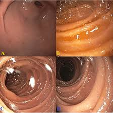
The treatment you have depends on a number of things. This includes where the cancer started, its size and whether it has spread (the stage). Surgery is the main treatment for small bowel neuroendocrine tumours. But surgery isn’t always possible. Some small bowel neuroendocrine tumours might have already started to spread when you are diagnosed. Or you might not be well enough to have it. You continue to have treatment to help your symptoms if surgery isn’t an option. Deciding which treatment you need A team of doctors and other professionals discuss the best treatment and care for you. They

Treatment by type of stomach NET

The treatment you have for a stomach neuroendocrine tumour (NET) depends on a number of things. This includes the type of NET and whether it has spread. Doctors call this the stage of the cancer. NETs grow at different rates, but they often grow very slowly. Some might not grow at all for months or years. So, you might not need treatment straight away. Your doctor might ask you to have regular tests to keep an eye on it. Which treatment do I need? A team of doctors and other professionals discuss the best treatment and care for you. They

Thymus gland cancer

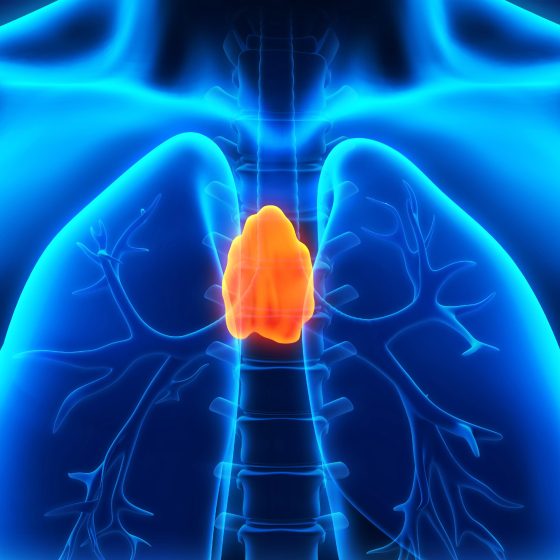
Cancer of the thymus gland is rare. Most cancers that start in the thymus gland are called thymomas. There’s also a much rarer form of thymus gland cancer called thymic carcinoma. What is the thymus gland? The thymus gland is in your chest, in between your lungs. A gland is an organ in your body that makes and releases substances such as hormones. The thymus gland is part of our lymphatic system . It’s involved in the development of a type of white blood cell called a T lymphocyte. These white blood cells are part of your immune system .

Treating Wilms tumour

Doctors plan your child’s treatment in one of the UK’s children’s cancer centres. Your child has most of their treatment in this specialist centre, but some care might take place at a hospital closer to home. Children’s cancer centres have teams of specialists who know about Wilms tumours and the best way to treat them. Wilms tumour is curable in about 9 out of 10 children (about 90%). The main treatments include: chemotherapy for almost all children surgery for all children radiotherapy for some children In the UK and many other countries, children usually start treatment without having a biopsy if the tumour looks

Treating tonsil cancer

The main treatments for tonsil cancer are surgery, radiotherapy and chemotherapy. You might have a combination of these or one treatment on its own. The tonsils are part of the oropharynx and treatment is similar to other cancers of the oropharynx. Treatment by stage Staging is a way of describing the size of cancer and how far it has grown. We have information about treatment for early and advanced cancer. Your doctor will tell you more about the stage of your cancer and the treatment you will have. There is an increase in the number of tonsil cancers that are

Treating tongue cancer

The main treatments for tongue cancer are surgery, radiotherapy and chemotherapy, either combined or on their own. Your treatment depends on how big the cancer is and whether it has spread (the stage). It also depends on which part of the tongue is affected. Treating the oral tongue The front two thirds of the tongue (oral tongue) are treated like a mouth cancer. Early stage Early cancer means your cancer is smaller than 4cm and is contained within the tongue. The most common treatment is surgery to remove the affected area. You might also have radiotherapy after surgery to try

Placental site trophoblastic tumour and epithelioid trophoblastic tumour

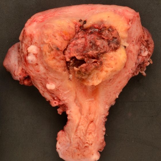
Placental site trophoblastic tumours (PSTTs) and epithelioid trophoblastic tumours (ETTs) are cancers that happen after pregnancy. They are extremely rare and are slow growing.  What are placental site trophoblastic tumours and epithelioid trophoblastic tumours? These tumours are part of a group of conditions called gestational trophoblastic disease (GTD). GTD is the name for abnormal cells or tumours that grow from the tissue that forms in the womb during pregnancy. Less than 1 in 100 (less than 1%) of GTDs are placental site trophoblastic tumours or epithelioid trophoblastic tumours. In PSTT and ETT the tumour develops from the cells that grow

Abdominal aortic aneurysm

Definition Abdominal aortic aneurysm (AAA) is defined as an abnormal dilatation of the abdominal aorta with a diameter greater than 3 cm. It is a relatively common aortic pathology that results in significant amounts of morbidity and mortality. Between 1-2% of men undergoing NHS screening (over the age of 65) will be found to have a AAA. The vast majority are infrarenal (i.e below the origin of the renal arteries). Risk factors AAA may be considered a degenerative condition of the aorta though its aetiology is not fully understood. A number of risk factors have been shown to be associated with AAA: Age

Varicocele

Overview A varicocele refers to dilated veins within the pampiniform plexus. The pampiniform plexus is a network of veins found in the spermatic cord that drains the testes and epididymis. Varicoceles occur most commonly in adolescent boys and may affect up to 15% of men. The vast majority (around 90%) occur on the left side. It is strongly associated with infertility and as such varicoceles are seen in around 40% of men presenting with infertility. Aetiology Varicoceles occur due to dilation of the pampiniform plexus in the scrotum. The pampiniform plexus is a network of veins found in the spermatic cord that

Testicular torsion

Overview Testicular torsion is a urological emergency, that without intervention leads to ischaemia and loss of the testis. Testicular torsion is a rare but serious condition. Successful treatment requires prompt recognition, referral and surgical intervention. It exhibits a bimodal distribution with peaks in the neonatal period and around puberty. Differential Diagnosis Whilst other diagnoses should be considered, suspicion of testicular torsion requires urgent testicular exploration Trauma Epididymo-orchitis Torsion of the epididymal appendage Testicular tumour (an association between testicular cancer and torsion has been described) Hydrocele Inguinal hernia Systemic diseases (e.g. Henoch-Schönlein purpura, Mumps) Types Testicular torsion may be intravaginal or extravaginal. Extravaginal torsion occurs in-utero and in neonates.

Testicular cancer

Overview Testicular cancer is responsible for 1% of all new cancers in men. It typically presents with a unilateral testicular mass. Incidence appears to be increasing, with approximately 3-10 cases / 100,000 men each year in the Western world. The vast majority of testicular cancers are germ-cell tumours (95%). Overall prognosis, following appropriate therapy, is good. As with all cancers, optimal management requires a multi-disciplinary approach with GPs, specialist nurses, urologists and oncologists all essential. Figures from Cancer Research UK (last accessed Nov 2021). Epidemiology There are approximately 2,300 cases of testicular cancer in the UK each year. It is the 18th most common cancer affecting

Renal colic

Introduction Renal colic classically refers to acute severe loin pain that occurs secondary to a urinary stone. Urinary stones, also termed urolithiasis, refer to stone formation anywhere within the urinary tract. They may be asymptomatic or cause acute loin-to-groin pain due to ureteric obstruction. Urinary stones are extremely common, men are affected up to three times more than women. The peak incidence of symptomatic urinary stones is between 40-60 years in males and late 20’s in females. Aetiology Renal colic is caused by obstruction to urinary flow within the ureter that occurs secondary to urinary stones. The majority of renal stones, approximately 80%, are composed of

Renal cell carcinoma

Overview Renal cell carcinoma is the most common kidney cancer in adults. It accounts for around 80-85% of kidney cancers. They may be found incidentally on abdominal imaging, present symptomatically (e.g. haematuria, loin pain, loin mass, fever) or with features of paraneoplastic syndromes. In those with disease spread it may be local, particularly involving invasion of the renal vein and inferior vena cava (IVC) or distant (commonly the lungs). Management is highly dependent on the stage at diagnosis and patient-based factors. Epidemiology  In 2017, kidney cancers (overall) were the 7th most common cancer in the UK. There are around 13,000 cases of

Prostate cancer

Overview Prostate cancer is the most common malignancy affecting men in the UK. Incidence increases with advancing age and men of Black ethnicity are more commonly affected. It can affect men, trans women, non-binary (assigned male sex at birth) and some intersex patients. Localised disease is commonly asymptomatic but symptoms often develop in locally advanced and metastatic disease. Management is dependent on a multitude of factors but may involve active surveillance, androgen deprivation therapies, radiotherapy, chemotherapy and surgical intervention. Epidemiology 1 in 6 men in the UK will be diagnosed with prostate cancer in their lifetime. In the UK it is the

Hydrocele

Overview Hydrocele refers to a collection of serous fluid between the parietal and visceral layers of the tunica vaginalis. Patients present with (typically) a non-tender scrotal swelling. Hydroceles may be associated with (communicating hydrocele) or without (non-communicating hydrocele) a patent processus vaginalis. Treatment depends on the underlying aetiology, age and symptomatic burden of the hydrocele. NOTE: In rare cases a similar condition may be seen in women where fluid accumulates in the canal of Nuck. Types There are two major types of hydrocele: communicating and non-communicating. Communicating These occur due to the failure of normal closure of the processus vaginalis. This allows the passage of

Chronic prostatitis

Overview Chronic prostatitis is characterised by > 3 months of urogenital pain, often associated with LUTS or sexual dysfunction. Chronic prostatitis may be categorised as: Chronic prostatitis/chronic pelvic pain syndrome: clinical features of chronic prostatitis in the absence of an identifiable bacterial infection. Chronic bacterial prostatitis: relatively uncommon, accounting for around 10% of patients with chronic prostatitis. Aetiology The aetiology of non-bacterial associated chronic prostatitis is poorly understood. Chronic prostatitis/chronic pelvic pain syndrome The aetiology remains poorly understood. Infective and inflammatory triggers may be implicated. There are suggestions that some may have a neuropathic component. Chronic bacterial prostatitis This may develop following an episode

Bladder cancer

Overview Bladder cancer is a common malignancy, in 2017 it was the 11th most common cancer in the UK. In the UK, transitional cell carcinoma is by far the most common cause (around 90%). Advancing age and smoking are significant risk factors for its development. More men than women are affected, though this appears largely due to exposure to relevant risk factors. It classically presents with painless haematuria. Management depends on staging and can involve trans-urethral resection of bladder tumour (TURBT), immunotherapy, chemotherapy, radiotherapy and cystectomy. Epidemiology There are approximately 10,300 cases of bladder cancer in the UK each year. In 2017,

Benign prostatic hyperplasia

Overview Benign prostatic hyperplasia (BPH) is characterised by hyperplasia resulting in lower urinary tract symptoms (LUTS). In BPH, prostatic hyperplasia (increased cell proliferation leading to enlargement) leads to urinary frequency, incomplete emptying, dribbling, hesitancy and nocturia. It may also be complicated by acute or chronic urinary retention. You may come across the term benign prostatic enlargement (BPE). This refers to a clinical finding on digital rectal examination (DRE), BPH refers to a histological diagnosis. There is increasing recognition that LUTS in males are often not secondary to BPH. An open mind should be had with regards to the underlying aetiology in

Acute epididymo-orchitis

Overview Acute epididymo-orchitis is a common cause of testicular pain and swelling. It is caused by acute inflammation of the epididymis (epididymitis) that may affect the testicle (orchitis). Most commonly the aetiology is infectious through the spread of pathogens from the urethra or bladder. It may be related to urinary tract infections or sexually transmitted infections. Testicular torsion must be considered in anybody presenting with acute testicular pain, particularly in children, adolescents and younger men. If there is any diagnostic uncertainty, refer to urology / paediatric surgery. Aetiology Epididymo-orchitis is most commonly caused by sexually transmitted infections or urinary pathogens. Sexually transmitted organisms tend

Acute bacterial prostatitis

Introduction Acute bacterial prostatitis refers to a an infection involving the prostate that may cause significant systemic upset. It tends to present with urinary symptoms, lower back or pelvic pain +/- symptoms of systemic infection. Urinary pathogens are often implicated, commonly Escherichia coli. Less commonly sexually transmitted infections are isolated as the cause. Aetiology E.coli is the most commonly isolated pathogen in acute bacterial prostatitis. Pseudomonas aeruginosa, Klebsiella, Enterococcus and Proteus may all be causes. Sexually transmitted infections, Chlamydia trachomatis and Neisseria gonorrhoea, are less commonly isolated. There are a number of specific scenarios to consider: Recent urethral or prostatic instrumentation (e.g. urethral catheterisation, cystoscopy or transrectal prostate biopsy): rarely acute

Skin tension lines

Overview Skin tension lines reflect internal tension within the skin. Skin tension lines essentially refer to the direction of maximal skin tension at different body sites. They reflect internal tension within the skin due to connective tissue (e.g. collagen) within the dermis. There are different types of skin tension lines. The two most widely recognised are ‘Langer’s lines’ (also known as cleavage lines) and relaxed skin tension lines. Aligning surgical incisions or excisions with these lines produces less tension, optimal scar formation, and ultimately a better cosmetic result. These lines can be drawn on a diagram of the human body. Body

Necrotising infections

Overview Necrotising soft tissue infections (NSTIs) are a collection of severe infections characterised by rapidly progressive soft tissue inflammation and necrosis. Specific examples of NSTIs include: Necrotising fasciitis – a term popularized in the 1950s to emphasise the constant feature of necrotic fascia with spread of infection along fascial planes. Gas gangrene – soft tissue infection, typically caused by Clostridium perfringens, results in myonecrosis and gas formation within tissues. Fournier’s gangrene – refers specifically to necrotising fasciitis of the perineum and scrotum. NSTIs are usually rapidly progressive, resulting in extensive tissue destruction. They are associated with high morbidity and mortality. Accordingly, prompt diagnosis and treatment are essential.

Melanoma

Summary Melanoma is a cancerous growth of melanocytes. NB – This article is in reference to cutaneous melanoma only. Melanoma is common. In the UK, it accounts for approximately 4% of all new cancers. It results from a combination of environmental and genetic factors. Features suspicious of melanoma can be remembered using the mnemonic ‘ABCDE’: Asymmetry Border (irregular) Colour alterations Diameter > 6mm Evolving lesion Patients with a suspicious lesion are referred for a tissue biopsy. In most instances, this constitutes an excisional biopsy with a 2 mm margin. If diagnostic of melanoma, the Breslow thickness, presence of ulceration and the mitotic index are used to histologically stratify the disease. Staging

Ganglion

Overview A ganglion cyst is a fluid-filled swelling the occurs over a joint or tendon sheath. Ganglions are fluid-filled swellings known as cysts that are commonly observed in the hand or wrist. They usually occur over a joint or tendon sheath. They can occur at all ages but usually occur in the second to fourth decades. They are a very common cause of swelling in the hand and wrist. Aetiology & pathophysiology Ganglions contain mucinous, gelatinous fluid. Ganglions are thought to arise from the herniation of connective tissue around key musculoskeletal structures such as tendon sheaths, ligaments, joint capsules, and bursae. This

de Quervain tendinopathy

Overview de Quervain tendinopathy is a common cause of pain in the wrist. de Quervain tendinopathy is a common cause of wrist pain and affects the abductor pollicis longus (APL) and extensor pollicis brevis (EPB) tendons. The cause is unknown but thought to be due to overuse or repetitive movements. It is classically described in women who are postpartum from repeatedly picking up their baby. The condition is typically self-limiting. Aetiology & pathophysiology The exact cause of de Quervain tendinopathy is unknown. Traditionally, de Quervain tendinopathy was thought to be due to repetitive activities that maintain the thumb in extension and abduction

Carpal tunnel syndrome

Overview Carpal tunnel syndrome is a median nerve neuropathy due to compression as it passes through the carpal tunnel in the wrist. Carpal tunnel syndrome (CTS) is due to compression of the median nerve as it passes through the carpal tunnel in the wrist leading to median nerve neuropathy. It is considered the most common upper limb mononeuropathy. CTS is common and usually presents with paraesthesia and/or sensory loss of the first three fingers (thumb, index finger, middle finger), lateral half of the fourth finger (ring finger). As the condition progresses, there is loss of motor function with hand weakness, wasting

Burns

Summary Burns result from damage to skin & deeper tissues caused by external sources or substances. Burns are a major cause of injury and death worldwide. They can have devastating physical and psychological effects on an individual, that can lead to chronic disability. They may be caused by thermal, chemical, frictional or electrical injury. Mortality is increased with large burns, with increasing age and with associated inhalational injury. Effective management necessitates a multi-disciplinary approach. Aetiology Most burns are thermal injures due to scalds, contact & flame burns. Burns may be thermal, chemical, frictional or electrical. Thermal injuries are the most common and include scalds, contact and flame burns. Epidemiology

Basal cell carcinoma

Summary Basal-cell carcinoma (BCC) is a slow-growing, locally invasive, malignant epidermal (basal layer) skin tumour. Typically, a slow-growing skin lesion (over months / years) which commonly occurs on sun-exposed areas of the body. Eighty percent occur on the head and neck. BCC is the commonest form of skin cancer. It is 4-5x more common than squamous cell carcinoma (SCC). They generally affect middle-aged/elderly individuals, unless there is a genetic susceptibility. BCCs are locally destructive and rarely metastasize. Clinical examination typically demonstrates a flesh- or pink-colored lesion with rolled edges, ulceration and telangiectasia (small blood vessels). Treatment is usually by surgical excision, electrodesiccation and curettage (EDC), cryotherapy, or Moh’s micrographic surgery. Rarely, radiotherapy is used.

Wrist fractures

Introduction Fractures of the distal radius are common orthopaedic injuries. Wrist fracture is a term that can refer to any fracture affecting the distal radius, ulna or carpal bones. However, generally, it is used interchangeably to describe a fracture of the distal radius and any accompanied ulna fracture. Isolated fractures of carpal bones tend to be considered separately. This note will focus on fractures of the distal radius (e.g. Colles’ and Smith’s fractures) Fractures of the distal radius exhibit a bimodal distribution; they are seen in the young following high-energy injuries and as fragility fractures in the elderly. Treatment aims to

Tibial plateau fractures

Introduction Tibial plateau fractures are due to high-energy trauma in the young and low-energy falls in the elderly. The tibial plateau refers to the proximal articular surface of the tibia, which forms the knee joint with the femur. Tibial plateau fractures have a bimodal distribution, classically presenting in males in their 40s and females in their 70s. The incidence of these fractures is around 10.3 per 100,000 annually. Aetiology Tibial plateau fractures occur secondary to traumatic injuries Mechanisms Fracture of the tibial plateau can arise from a valgus force, which describes an outside force pushing the knee inwards along a coronal plane.

Septic arthritis

Overview Septic arthritis is an infection of one or more joints. Bacterial infection is by far the most common cause of septic arthritis with staphylococcus aureus most frequently isolated. It normally presents with pain and swelling of the affected joint with signs of systemic infection. The condition may be categorised as: Native joint infection: infection affecting a native joint, management typically requires joint drainage (where appropriate) and antimicrobial therapy. Prosthetic joint infection (PJI): a serious complication of prosthetic joint replacement. Surgical intervention in addition to antimicrobial therapy is commonly required. Septic arthritis requires prompt recognition and management. Untreated it can lead to articular destruction

Osteomyelitis

Overview Osteomyelitis refers to infection of the bone. It is a serious infection that benefits from prompt recognition and treatment. Risk factors include diabetes, old age, peripheral vascular disease and immunocompromise. It may occur for many reasons including open fractures, skin ulcers, surgery (and prosthesis) and haematogenous spread of bacteria. Staphylococcus aureus is the most commonly identified infecting organism. Treatment involves long courses of antibiotics (normally a minimum of 4-6 weeks) and at times surgical debridement. The focus of this note will be osteomyelitis in adult patients. Causative organisms Staphylococcus aureus is the most common cause of osteomyelitis. Staphylococcus aureus: A gram-positive cocci. Includes

Hip fractures

Introduction A hip fracture is a bony injury of the proximal femur typically occurring in the elderly. Older patients are more likely to have gait unsteadiness and reduced bone mineral density, predisposing to fracture. It is a common serious injury in older people, costing the NHS and social care £1 billion per year. Basic anatomy The proximal femur consists of a head, neck, trochanters (greater and lesser) & shaft. It is the largest bone in the human body. The inter-trochanteric line lies on the anterior surface of the femoral neck, running between the trochanters. It demarcates the inferior attachments of the hip capsule. Hip capsule The capsule of the hip is attached

Ankle fractures

Overview Ankle fractures are common lower limb fractures often occurring due to low-energy torsional trauma. They affect women more than men, most commonly those aged 30-60. Ankle fractures account for around 9% of fractures presenting to accident and emergency, representing a significant portion of the trauma workload. Treatment involves restoration of normal anatomical alignment. This may involve conservative management with a walking boot or cast, or involve surgical fixation. Attention must be payed to the risk of venous thromboembolism (VTE) and prophylaxis given where appropriate. Anatomy The ankle is a hinge joint, formed by the malleoli & talus. It is reinforced medially and

Subarachnoid haemorrhage

Definition Subarachnoid haemorrhage (SAH) is bleeding in the subarachnoid space between the arachnoid and pia mater meningeal layers. Aetiology SAH can be a life-threatening emergency and it is estimated that 10-15% of patients die before they reach hospital. SAH can be divided into traumatic or spontaneous Traumatic (tSAH): most common cause of SAH. Usually in setting of a head injury (e.g. fall, assault, road traffic collision) Spontaneous: commonly due to rupture of a cerebral aneurysm (aSAH) Traumatic Trauma is the most common cause of SAH. There is usually evidence of trauma in the clinical history (e.g. road traffic accident). An isolated SAH in

Spinal cord syndromes

Overview Several classic syndromes that have characteristic clinical features based on the area of the spinal cord that is affected. Neurological signs and symptoms can usually be located to the spinal cord based on the presentation of bilateral motor and sensory signs without head or face involvement. In some situations, a specific set of clinical features can develop due to the area within the spinal cord that is affected. These are known as the spinal cord syndromes. We discuss some of the more common ones below. Anatomy The spinal cord is part of the central nervous system (CNS) and forms the main