Day: March 26, 2024

Ventilation

Ventilation Ventilation is the movement of air between the lungs and the surrounding environment. Ventilation is key to maintaining adequate arterial oxygenation and for the removal of CO2. Under normal circumstances, breathing is a passive process controlled by centres in the brainstem. These respiratory centres are specialised groups of neurones. The complex interaction of these neuronal collections are responsible for ventilation. Three such groups have been identified: the pons respiratory centre, the medullary respiratory centre and the pre-Bötzinger complex. Respiratory centres Pons respiratory centre The pons (primary) respiratory centre is composed of two centres. Pneumotaxic centres: can interact with the dorsal respiratory group (see below) to suppress inspiration.

Tuberculosis

Introduction Tuberculosis (TB), caused by bacteria of the mycobacterium tuberculosis complex, is one of the world’s most common and deadly infective diseases. TB is an infectious disease responsible for significant morbidity and mortality worldwide. There is a great deal of variety in the clinical presentation and the disease can manifest in a number of organs. It most commonly affects the lungs in what is called pulmonary TB. The disease may exist in a number of phases: Latent TB: This refers to individuals infected with TB, who suppressed the initial infection, have no active disease and are not infectious. It is thought that around

Spontaneous pneumothorax

Introduction A pneumothorax is a collection of air within the pleural space. The pleural space (or cavity) refers to the space between the parietal (lines the inner wall of the lung) and visceral pleura (lines the surface of the lung). When air enters this space it is referred to as a pneumothorax. There are two major types of pneumothorax: Spontaneous: occurring without prior trauma, normally in older patients with underlying lung disease or younger patients with apical blebs. Traumatic: occurring in the setting of trauma, may be open (direct communication between the outside air and pleural space) or closed. Aetiology Spontaneous pneumothoraces may be either primary or secondary First

Sarcoidosis

Overview Sarcoidosis is a rare multisystem granulomatous disorder of unknown aetiology. Sarcoidosis is considered a multi-system disorder that most commonly affects the lungs. The aetiology, though poorly understood, is thought to involve immune dysfunction and T-cell overactivity. Sarcoidosis can have a wide range of clinical manifestations and is able to affect most organ systems within the body. Nonetheless, sarcoidosis most frequently affects the lungs causing a form of interstitial lung disease. Epidemiology Sarcoidosis affects approximately 10-20 people per 100,000 in the UK. There is a great deal of geographic variation in the prevalence and clinical manifestations of sarcoidosis. In Finland the prevalence is approximately

Respiratory failure

Introduction Respiratory failure occurs when there is inadequate gaseous exchange in the lungs. Respiratory failure is defined by the partial pressure of oxygen and carbon dioxide in the arterial blood. Arterial samples, termed an arterial blood gas (ABG), are typically obtained with an arterial sample drawn from the radial artery (other arteries may be used) or a sample taken from an arterial line. Two types of respiratory failure are defined based upon the partial pressure of oxygen and carbon dioxide: Type 1 respiratory failure (T1RF): is characterised by hypoxaemia (PaO2 < 8 kPa) and a normal or low CO2. Type 2 respiratory failure (T2RF):

Pulmonary embolism

Overview Pulmonary embolism can be a life-threatening condition caused by occlusion within the pulmonary arteries. Venous thromboembolism Venous thromboembolism (VTE) is a term that encompasses two conditions: Pulmonary embolism (PE): acute/chronic occlusion of pulmonary arteries. Clot breaks off and travels to the lungs (emboli). Deep vein thrombosis (DVT): acute/chronic occlusion of deep vein(s). Commonly affects the lower limbs through the formation of a clot (thrombus). VTE is commonly asymptomatic, but 1-2 per 1000 people every year have symptomatic VTE. DVT accounts for two-thirds of these cases and is commonly seen with PE. It is estimated that 80% of cases of PE are associated with

Pneumonia

Definition & classification Pneumonia is the inflammation of the parenchyma of the lung. Pneumonia is a common type of infection affecting the lung tissue (i.e. parenchyma). In pneumonia, the air sacs (i.e. alveoli) become filled with microorganisms and inflammatory cells leading to poor lung function with features of cough, fever, and shortness of breath. Pneumonia can be life-threatening, particularly in frail, elderly patients or those who are immunosuppressed. Pneumonia is one of several types of respiratory infections, which include: Upper respiratory tract infection (URTI): inflammation of the mucosa of the nostrils, nasal cavity, mouth, throat (i.e. pharynx), and larynx. Colloquially known as

Pleural effusions

Overview A pleural effusion refers to an abnormal collection of fluid within the pleural space. A pleural effusion is the most common manifestation of pleural disease and it may occur from a wide range of aetiologies. It refers to an abnormal collection of fluid within the pleural space. Pleural effusions are most commonly detected on chest x-ray. They are seen as blunting of the costophrenic angle (where the diaphragm meets the ribs). This may progress to form a dense white area with a meniscus (curve formed by the upper layer of fluid). Large right-sided pleural effusion Pleural effusion is a broad

Muscles of respiration

Mechanics of respiration The basics of ventilation are simple. To inspire, the intrathoracic pressure must be below that of atmospheric pressure. The reverse must be true in order to expire. There are two mechanisms by which we may change the intrathoracic volume (and therefore pressure): Movement of the diaphragm: the diaphragm is the main muscle of respiration. When it contracts its domes descend expanding the thoracic cavity. Movement of the ribcage: The action of various muscles can either move the ribcage up and outward or down and inward. The muscles of the neck, thorax and abdomen all contribute by causing changes

Lung function tests

Introduction Lung function tests aid the diagnosis of numerous respiratory conditions. Ventilation is the process by which air (or other gases) is moved in and out of the lungs to facilitate gas exchange. Lung function tests allow us to measure the volume and flow of inhaled gases. An understanding of these tests and what they show, both in health and disease, aids clinicians in the diagnosis of respiratory (and related) disease. Volumes The volume of air during inspiration and expiration, at different levels of exertion, can be categorised into lung volumes. These volumes are highly dependent on height, weight, gender, age

Lung cancer

Introduction Lung cancer is a common malignant tumour, with around 48,500 cases diagnosed in the UK each year. It is the third most common malignancy in the UK and is the leading cause of cancer-related death. Smoking is the most important aetiological factor, implicated in upwards of 80% of cases. It is categorised by the underlying cell type: Small-cell lung cancer (SCLC) Non-small cell lung cancer (NSCLC): Adenocarcinoma Squamous cell carcinoma Large cell Management depends on the subtype, stage at diagnosis and patient co-morbidities. Broadly the options include chemotherapy, radiotherapy and surgical resection. Figures from Cancer Research UK (last accessed Nov 2021). Aetiology Smoking is by far

Idiopathic pulmonary fibrosis

Introduction Idiopathic pulmonary fibrosis (IPF) is a chronic condition characterised by progressive fibrosis of unknown aetiology with typical imaging and histological findings. Interstitial lung disease Interstitial lung disease (ILD) refers to a family of conditions with shared characteristics of interstitial inflammation, fibrosis and/or cellular changes in the absence of infection or malignancy. In most cases interstitial fibrosis is found and commonly related to sarcoidosis, chronic hypersensitivity pneumonitis, autoimmune disease or in cases with an unknown aetiology, idiopathic interstitial pneumonia. Idiopathic pulmonary fibrosis IPF is the most common form of idiopathic interstitial pneumonia. It occurs more commonly with advancing age and is rare

Cystic fibrosis

Overview Cystic fibrosis (CF) is an autosomal recessive multi-system disease predominantly characterised by respiratory features. It is caused by mutations to the cystic fibrosis transmembrane conductance regulator (CFTR) gene found on chromosome 7. This encodes a chloride channel and abnormalities have wide-ranging effects. Since the introduction of newborn screening for CF in 2007, the vast majority of patients are diagnosed in the first two months of life. Others are diagnosed following characteristic presentations (e.g. failure to thrive, recurrent chest infections, steatorrhea) As treatment improves, we are seeing significant improvements in life-expectancy, based on data from the 2019 CF Registry Report the median predicted survival

Bronchiectasis

Introduction Bronchiectasis describes the irreversible and abnormal dilatation of the airways. It normally results from the inflammatory destruction of the elastic and muscular components of the airways. This leads to abnormally dilated airways, persistent sputum production with ineffectual clearance and recurrent chest infections. Bronchiectasis commonly occurs secondary to cystic fibrosis though this is often considered and covered separately. Epidemiology In 2012 it was estimated that 210,000 people in the UK had bronchiectasis. The figures for prevalence and incidence vary widely by source as the condition often goes unrecognised or undiagnosed. Data from the British Lung Foundation indicates females are affected more than males. In 2012 they

Asthma (adults)

Introduction Asthma is a common chronic inflammatory disorder of the airways. Approximately, 12% of the UK population have a diagnosis of asthma and 5.4 million are receiving treatment for the condition. Clinically it presents with classical features including cough, wheeze, chest tightness, and shortness of breath. It can present acutely as an ‘exacerbation of asthma’, which may be life-threatening. It is characterised by: Reversible airflow limitation Airway hyperresponsiveness Inflammation of the bronchi Over the last decade, the approach to the diagnosis and management of asthma has been changing rapidly. There are currently two broad guidelines for the management of asthma (NICE

Bowel problems and sex after radiotherapy for prostate cancer 

Radiotherapy uses radiation to kill cancer cells. It is a common treatment for prostate cancer. Radiotherapy to the prostate gland can affect your sex life in several ways. For example, daily treatments for several weeks can make you very tired. You might not feel like having sex. Other side effects can affect you when having anal sex. Side effects include: loose poo or diarrhoea irritation and pain around the back passage and anus leaking poo How diarrhoea affects your sex life You might get diarrhoea during radiotherapy and for some time afterwards. This can be unpleasant and tiring. It may put you

Less interest in sex (libido)

Prostate cancer and its treatments can cause a loss of interest in sex. This is also called a low libido or sex drive. Your sex drive might be lower because of a number of reasons. These include: tiredness anxiety about having prostate cancer  loss of confidence and self esteem  It may also be a side effect of hormone therapy.  How hormone therapy causes a loss of libido Hormone therapy blocks or lowers the amount of testosterone in the body. This can cause less interest in sex.  Different hormone therapy drugs can cause different side effects. So your doctor might suggest changing

Infertility after prostate cancer treatment 

You might not be able to have children after prostate cancer treatment. This can be difficult to cope with. Understanding why your fertility can be affected and what can help may help you to cope with these changes. How prostate cancer surgery affects your fertility A common treatment for prostate cancer is surgery to remove the prostate gland. This is a radical prostatectomy.  After a radical prostatectomy, you will not be able to have children by sexual intercourse. If you want to have children, your doctor might suggest collecting sperm before surgery. The sperm is frozen and stored until you

Sex and erection problems after treatment for prostate cancer

Prostate cancer treatments can affect your sex life. Some treatments can damage the nerves that you need to get an erection. Other treatments can affect the levels of hormones needed to get and maintain an erection.   Even if you can’t get a full erection, you can still have a sex life. There are several treatments that may help you but not everyone chooses to have treatment. Treatments might include: drug treatments such as tablets and creams vacuum pumps implants Sex therapy and counselling can also help. Speak to your doctor or specialist nurse if this is something you think might help.  

Surgery to remove your testicles (orchidectomy)

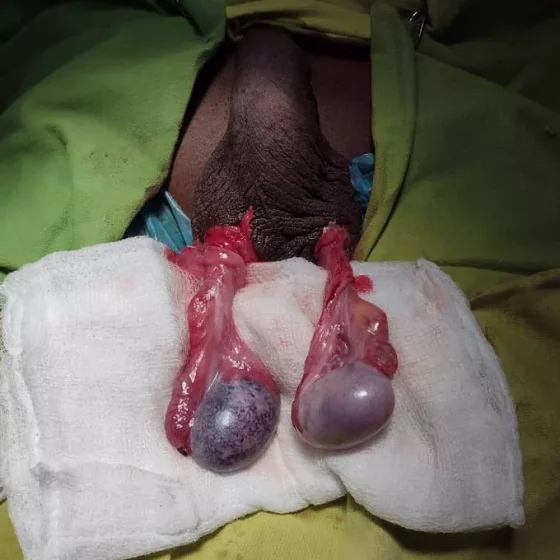
You might have an operation to remove your testicles (orchidectomy). The testicles produce testosterone, which can help prostate cancer grow. So removing the testicles can help to control the growth of prostate cancer. After the removal of the testicles, the level of testosterone in the blood falls quickly.   Having an orchidectomy doesn’t cure prostate cancer. But it can control the cancer and reduce your symptoms. Removing your testicles is a type of hormone therapy for prostate cancer. It is not a common treatment. You’re more likely to have injections or tablets to reduce the level of testosterone in your blood. Some

What is hormone therapy for prostate cancer?

Hormone therapy is a possible treatment for prostate cancer. Sometimes it is also called anti hormone therapy. What is hormone therapy? Hormones occur naturally in your body. They control the growth and activity of normal cells. Testosterone is a male hormone mainly made by the testicles. Prostate cancer usually depends on testosterone to grow. Hormone therapy blocks or lowers the amount of testosterone in the body. Hormone therapy on its own doesn’t cure prostate cancer. But it can lower the risk of an early prostate cancer coming back when you have it with other treatments. Hormone therapy is also a possible

Problems with sleeping

Hormone therapy for prostate cancer can change the levels of sex hormones in your body. This can cause different side effects including difficulty sleeping (insomnia).  What is insomnia? Insomnia is when you have difficulty falling asleep or staying asleep at night. Does hormone therapy for prostate cancer cause insomnia? Hormone therapy can cause side effects such as hot flushes and anxiety. This can make it difficult to sleep. What are the best treatments for insomnia?   Relaxation techniques such as meditation can help. Your doctor might also suggest medicines if you’re finding it very difficult to sleep. Difficulty sleeping and

Hot flushes in men 

Hormone therapy for prostate cancer can change the levels of sex hormones in your body. This can cause hot flushes.  This page is about hot flushes in men with prostate cancer. But some of the tips on how to cope with hot flushes are also applicable to men who have hot flushes for other reasons.  What causes hot flushes in men? Some prostate cancer treatments lower the amount of sex hormones in the body. These lower levels of hormones can cause hot flushes. What do they feel like? They usually start as a feeling of warmth in your neck and

Male sex hormones and cancer

Hormones are natural substances made by the glands and organs of your body. Treatment for prostate cancer can lower the amount of sex hormones in your body. This can affect you in different ways.   What is the male sex hormone? Testosterone is the main sex hormone in men. Where is testosterone made? Most testosterone is made by the testicles. Small amounts are also made in a gland above the kidney. What are the symptoms of low testosterone? Symptoms of low testosterone include hot flushes and changes in memory or mood. Cancer treatments and hormones Hormones act as messengers and carry signals from

Urinary tract infections

Definition & classification Urinary tract infections (UTIs) refer to an infection of any part of the urinary system from kidney to the urethra. UTIs are generally defined as the presence of characteristic symptoms (e.g. dysuria, frequency) and significant bacteriuria (presence of bacteria in urine). Significant bacteriuria is defined as > 105 colony forming units (CFU)/ml. In the absence of symptoms, this level of bacteriuria is termed asymptomatic bacteriuria. UTIs can be further categorised depending on the location of infection (e.g. upper or lower) or the presence of co-morbidities (e.g. complicated or uncomplicated). Upper UTI: infection of the kidney (pyelonephritis) Lower UTI: infection of the bladder (cystitis) and

Tubulointerstitial nephritis

Overview Tubulointerstitial nephritis refers to a primary insult to the renal tubules and interstitium. Tubulointerstitial nephritis (TIN) refers to inflammation of the renal tubules and interstitium that is most commonly the result of a hypersensitivity reaction to a medication (e.g. penicillin). This leads to acute inflammation with or without acute kidney injury (AKI). Tubulointerstitial nephritis may be acute or chronic: Acute TIN: develops over days or months. Majority of cases (~85%) due to drugs or infections. May cause AKI to variable severity. Chronic TIN: develops over years. Continued tubulointerstitial insults lead to chronic inflammation and eventually fibrosis with decline in renal function. Most often

Nephrotic syndrome

Overview Nephrotic syndrome is broadly defined as a triad of heavy proteinuria > 3.5 g/day, hypoalbuminaemia, and oedema. The nephrotic syndrome describes a very classic presentation of glomerular disease that is characterised by a triad of: ‘Nephrotic-range’ proteinuria (> 3.5 g/day) Hypoalbuminaemia (< 35 g/L) Oedema (e.g. peripheral, periorbital) In addition, hyperlipidaemia, increased risk of venous/arterial thrombosis and higher risk of infection is typical of the syndrome. This is because of loss of important proteins (e.g. immunoglobulins, albumin) through the glomeruli. Nephrotic syndrome is one of the hallmarks of glomerular disease. For more general information see our notes on Glomerular disease. Aetiology The cause of

Nephritic syndrome

Overview Nephritic syndrome is classically described as the presence of haematuria, variable proteinuria, renal impairment, and hypertension. Nephritic syndrome is also known as glomerulonephritis or simply ‘nephritis’. It describes the presence of an inflammatory process within the glomeruli. The nephritic syndrome describes a very classic presentation of glomerular disease that is characterised by Haematuria Variable proteinuria (may be nephrotic range) Renal impairment Hypertension Glomerulonephritis is a more encompassing term than nephritic syndrome because it reflects the variable clinical presentation of glomerular inflammation. Some patients may have minimal haematuria and proteinuria whereas others develop rapidly progressive renal impairment. Aetiology The cause of nephritic syndrome is

Microscopic polyangiitis

Overview Microscopic polyangiitis (MPA) is a small vessel vasculitis. Microscopic polyangiitis (MPA) is one of the anti-neutrophil cytoplasmic antibody (ANCA) associated vasculitides (AAV) that can present with rapidly progressive renal impairment and other systemic manifestations. In the UK, the incidence is estimated at 2 cases per 100,000 people. It is more common among older Caucasians with an equal sex distribution. ANCA associated vasculitis AAV is an umbrella term for three conditions: Microscopic polyangiitis (MPA) Granulomatosis with polyangiitis (GPA): previously known as Wegener’s granulomatosis Eosinophilic granulomatosis with polyangiitis (EGPA): previously known as Churg-Strauss syndrome These conditions are all small vessel vasculitides that

IgA nephropathy

Overview IgA nephropathy is the most common primary chronic glomerular disease worldwide. It commonly presents with macroscopic haematuria or asymptomatically with microscopic haematuria, proteinuria +/- hypertension picked up incidentally or during investigation for renal impairment. The condition remains a leading caused of chronic kidney disease and renal failure. The diagnosis can be confirmed with a renal biopsy. There is no targeted therapy but angiotensin-converting enzyme (ACE) inhibitors or angiotensin-II receptor blockers (ARB) may be used for proteinuria and hypertension. In those with ongoing severe disease immunosuppressive therapies may be considered. Epidemiology IgA nephropathy predominantly affects children and young adults. Though the disease occurs at

Granulomatosis with polyangiitis

Overview Granulomatosis with polyangiitis (GPA) is a small vessel vasculitis. Granulomatosis with polyangiitis (GPA) is one of the anti-neutrophil cytoplasmic antibody (ANCA) associated vasculitides (AAV) that can present with rapidly progressive renal impairment and other systemic manifestations. In the UK, the incidence is estimated at 10.2 cases per 1,000,000 people. It is slightly more common in men (1.5:1 male to female ratio) and typically presents in middle-age (35-55 years old). ANCA associated vasculitis AAV is an umbrella term for three conditions: Microscopic polyangiitis (MPA) Granulomatosis with polyangiitis (GPA): previously known as Wegener’s granulomatosis Eosinophilic granulomatosis with polyangiitis (EGPA): previously known as

Glomerular disease

Overview The ‘glomerulopathies’ are a constellation of diseases characterised by injury to renal glomeruli. Glomerular disease (also known as ‘glomerulonephritides’ or ‘glomerulopathies’) refers to a group of conditions that cause damage to the renal glomeruli. The glomeruli represent the site of filtration (i.e. where blood is filtered) within the kidneys. Glomerular diseases are an important cause of both acute kidney injury (AKI) and chronic kidney disease (CKD). Glomerular diseases are traditionally difficult to learn. This is because of confusing terminology that combines clinical and histopathological terms, the wide variety of clinical presentations and significant overlap between conditions. Two commonly discussed clinical manifestations

Chronic kidney disease

Overview Chronic kidney disease (CKD) can be defined by the presence of kidney damage or reduced kidney function for three or more months. Reduced kidney function is suggested by a reduction in the glomerular filtration rate (GFR) or by evidence of kidney damage which can be characterised by the presence of one or more of the following pathological markers: Albuminuria (e.g. albumin:creatinine ratio > 3 mg/mmol or > 30 mg/g) Urinary sediment abnormalities (e.g. white cell or red cell casts) Radiological abnormalities (e.g. polycystic kidneys) Pathological abnormalities (e.g. seen on renal biopsy) History of kidney transplantation CKD is a common condition with a progressive nature. As CKD progresses

Anti-GBM disease

Overview Anti-glomerular basement membrane (Anti-GBM) disease is a rare small-vessel vasculitis. Anti-glomerular basement membrane (Anti-GBM) disease is a small vessel vasculitis with an annual incidence of one case per million population. It is characterised by the formation of anti-GBM antibodies that are directed against collagen in the kidneys and lungs. This can lead to life-threatening glomerulonephritis and/or pulmonary haemorrhage. Anti-GBM disease is a relatively new term that encompasses the clinical spectrum of both renal and pulmonary manifestations. The condition is associated with two older terms named after Ernest Goodpasture, who initially discovered the condition in 1919: Goodpasture syndrome: refers to clinically

Alport’s syndrome

Overview Alport’s syndrome is a rare hereditary nephropathy occurring due to mutations to genes encoding type IV collagen. Alport’s may demonstrate different inheritance patterns though it is most commonly X-linked due to mutations of COL4A5 on the X chromosome. The disease affects the glomerular basement membrane (GBM) leading to nephritis. Other features may include sensorineural hearing loss and ocular defects. Genetics Alport’s may arise from a number of mutations to genes responsible for type IV collagen. It may occur due to mutations to alpha-3, alpha-4 and alpha-5 chains of type IV collagen. Type IV collagen is important to basement membranes and mutations lead to

ADPKD

Overview Autosomal dominant polycystic kidney disease is a common genetic disorder characterised by multiple renal cysts. Autosomal dominant polycystic kidney disease (ADPKD) is due to inheritance of the abnormal genes PKD1 or PKD2 that is thought to occur in around 1 in 1000 live births. The disease is characterised by development of multiple renal cyst and progressive renal impairment, which leads to end-stage renal disease (ESRD) in around 50% of patients by 60 years of age. It accounts for up to 10% of patients with ESRD. ADPKD is generally a condition of adults and rarely presents in childhood. The estimated median age of developing ESRD is in 6th

Acute kidney injury

Overview Acute kidney injury (AKI) is generally defined as a sudden decline in renal function over hours or days. AKI is a common medical condition affecting up to 15% of emergency hospital admissions and the mortality associated with severe AKI can be up to 30-40%. A decline in renal function can lead to dysregulation of fluid balance, acid-base homeostasis and electrolytes. AKI has largely replaced the term ‘acute renal failure’. This change in nomenclature reflects the significance that small decrements in renal function do not lead to overt renal failure, but do have a clinical impact on morbidity and mortality. Classification