Description
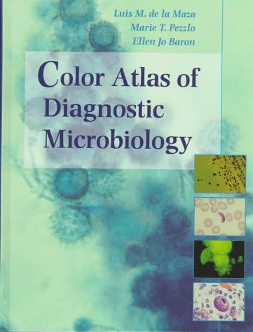
Description of color atlas of diagnostic microbiology PDF
Authors Of Color Atlas Of Diagnostic Microbiology PDF
This book is written by the coordination of three authors,
Luis M. de la Maza, M.D.,Ph.D.
He is the professor of the Department of Pathology, Director of Medical Microbiology at the University Of California, Irvine Medical Centre Orange located in California.
Marie T. Pezzlo, M.A., F(AAM)
He is the Administrative Director of the Department Of Pathology at the University Of California, Irvine Medical Centre Orange located in California.
Ellen Jo Baron, Ph.D., F(AAM)
He is the Adjunct Administrative Professor of Medicine at the University Of California in Los Angeles and Clinical Associative Professor Of Molecular Biology and Immunology at the University Of Southern California in Los Angeles.
Content Of Microbiology Colored Atlas
Diagnostic Microbiology is the ability to recognize the characteristics of the microorganism both microscopically and macroscopically furthermore it is a visual science and to find colored images related to Microbiology are not conveniently available. The book Color Atlas Of Diagnostic Microbiology is designed to seek to the situation.
The book contains a total of 18 chapters with a total of 223 pages.
-
- Laboratory safety: This chapter contains the laboratory safety guidelines which are mandated by OSHA (Occupational Safety And Health Act, created in 1970) and illustrated with the pictorial presentation. It contains all the information from handling equipment to the introduction to disinfectants, waste disposal, and Sterilization.
- Specimen collection: This chapter is based on the pros and cons of the specimen collection furthermore it defines the different methods to collect specimens for the specific microorganisms.
-
- Specimen Processing: As in the second chapter we have learned about collecting specimens for the test and now this chapter is based on How to process a specimen using different techniques including specimen preparation selection and inoculation of media.
- Gram stain: The gram staining is used for classifying microorganisms based on their morphology. In terms of gram staining Microorganism is divided into two types; Gram-Negative and Gram-positive.
- Micrococcuceae: The three genres of the family micrococcuceae are the common cause of the infection found in the human body. In this chapter different tests on the Staphylococcus, micrococcus, and Stomatococcus are given with the help of different images on the test performance.
- Sterptococcaeceae: This chapter is about the family streptococcaceae and its related genera which includes nine genera tested in the clinical laboratory. There is more than one detailed image for every test taken for the research.
- Aerobic Gram-positive Bacilli And Actinomyces spp: This chapter is based on the test of the common genera of aerobic gram-positive bacteria to isolate them. The chapter is defined through the pixels free images of every test on the given genera performed through the test tubes and agar plates.
- Enterobacteriaceae: The chapter includes the most common isolated bacteria in the clinical laboratory with the different steps of isolation of them.
- Other Gram-negative Bacteria Microorganism: In this chapter colored images of isolation is given of the bacteria not included in Enterobacteriaceae.
- Anaerobic Bacteria: are those bacteria that are unable to multiply in the presence of atmospheric oxygen. The chapter includes the study of these bacteria to research them in detail.
- Mycobacteria: it is based on the different staining techniques used to research the characteristic of Mycobacterium Tuberculosis.
- Microbial Pathogens Isolated And/ Or Identified By Tissue Culture Or Other Special Methods: This includes the tests based on the tissue culture of the different microbial pathogen with the macroscopic images of every tissue.
- Antimicrobial Susceptibility test: The most interesting chapter of the book is this one because in addition to performing the different tests it contains the preparation of inoculum and the disk diffusion test with an illustration of every step through images
- Mycology: It is the study of fungi with their genetic and biochemical properties. The chapter includes the images for every category of fungi to understand all the phenomena involved in mycology.
- Parasitology: it is the study of different parasites and the diseases caused by them. The book Color Atlas Of Diagnostic Microbiology PDF includes a visual description of the different parasites.
- Virology: the chapter is based on the different viruses with the laboratory research on them along with the pictures of different viruses taken through microscopy.
- Immunoserology: It includes the test systems used in identifying the causative organisms of the infectious disease.
- Molecular Techniques: Some techniques are used to make the feasibility of the test are called molecular techniques.which elucidate the tests performed.
Features Of Color Atlas Of Diagnostic Microbiology PDF
The book Color Atlas Of Diagnostic Microbiology contains 753 full-colored images including specimen collection, handling, Transport devices, Lab safety, and different microscopic and macroscopic images of the microbes in the human body.
- The microscopic images were taken with the Nikon FX-35A Camera from Zeiss Universal Microscope equipped with Zeiss and Olympus lenses.
- The microscopic images were obtained using the Nikon EL Camera with a Micro-Nikkor 55mm f/3.5 Lens and a Contex RTS Camera with a Carl Zeiss A-Planar 60mm f/2.8 lens.
- Kodachrome 25 professional films, Velvia and Provia Fujichrome Films were used for most of the images of the book.
- The Electron Micrographs were taken with a Philip EM400 Electron Microscope and Electron Image Film SO-163.
- In addition to the book content, it contains the index on the last pages of the book from Page no. 212 to 223 that contain different terminology with their page numbers to find the respective term easily in the book.







Reviews
There are no reviews yet.